

Introduction
Composting is a process of microbial aerobic decomposition under controlled conditions that allow thermophilic microbes to generate high temperatures. The end product is stable and free of pathogens and viable weed seeds and thus ready to be used in plant cultivation (Martin and Brathwaite, Reference Martin and Brathwaite2012). The type of feedstock, the compost formulation and the composting process, system and management methods have all been reported to affect compost maturity and quality. Compost can be used as an organic amendment or organic substrate (Rynk, Reference Rynk1992; Haug, Reference Haug1993; Sullivan and Miller, Reference Sullivan, Miller, Stoffella and Kahn2000). One of the unique properties of compost is its ability to suppress pathogens, and this ability has been widely linked to living microorganisms in the compost (Bonanomi et al., Reference Bonanomi, Antignani, Capodilupo and Scala2010). Studies have shown that heating or autoclaving eliminates the suppressive capacity of compost, but that this capacity can be recovered by mixing heated or sterilized composts with natural composts (Hoitink and Fahy, Reference Hoitink and Fahy1986). These results support the idea that the microbial community in composts mediates their suppressive capacity. In addition, physico-chemical properties such as nitrogen, pH, the C/N ratio, heat, moisture and the degree of compost maturity have also been linked to suppression activity (Hoitink and Grebus, Reference Hoitink, Grebus, Hayes and Wilson1997). Several studies have focused on the use of different biological parameters (e.g., the microbial population, diversity, activity and function) in compost to develop disease-suppressive composts. After an extensive data review, Bonanomi et al. (Reference Bonanomi, Antignani, Capodilupo and Scala2010) concluded that fluorescein diacetate hydrolysis, basal respiration, microbial biomass, total cultivable bacteria and fluorescent Pseudomonas and Trichoderma populations provide the best predictions of disease suppression capacity.
The development of high-throughput molecular tools has made it possible to characterize the taxonomic, phylogenetic and functional diversity of soil/compost microbial communities to find new predictors of the suppressive capacity of composts. Sequencing has been used to explore the structure and composition of compost microbial communities (de Gannes et al., Reference de Gannes, Eudoxie and Hickey2013; Neher et al., Reference Neher, Weicht, Bates, Leff and Fierer2013) and the suppressive capabilities of a given compost (Yu et al., Reference Yu, Sinkkonen, Hui, Kurola, Kukkonen, Parikka, Vestberg and Romantschuk2015; Blaya et al., Reference Blaya, Marhuenda, Pascual and Ros2016). However, genomic analysis reveals the total abundance of microbial populations and not just the active populations (Bastida et al., Reference Bastida, Torres, Moreno, Baldrian, Ondoño, Ruiz-Navarro, Hernandez, Richnow, Starke, García and Jehmlich2016). Microbial populations can be in sporulated and dormant forms. Moreover, researchers have recently discovered that up to 50% of the microbial nucleic acid sequences in environmental samples can correspond to dead and non-active biomass (Carini et al., Reference Carini, Marsden, Leff, Morgan, Strickland and Fierer2016). In contrast to genomic analysis, which focuses on phylogenetic data, meta-proteomics was defined by Wilmes and Bond (Reference Wilmes and Bond2006) as the large-scale characterization of the entire protein complement of environmental microbiota at a given point of time. Proteins that have been synthesized by microorganisms at the time of sampling reflect the actual functionality with respect to metabolic reactions and regulatory cascades, and give more direct information about microbial functionality than genes (Wilmes and Bond, Reference Wilmes and Bond2006). Microbial proteins have now been extracted from different environmental matrices, such as soil samples (Chourey et al., Reference Chourey, Jansson, VerBerkmoes, Shah, Chavarria, Tom, Brodie and Hettich2010; Bastida et al., Reference Bastida, Hernández and Garcia2014, Reference Bastida, Selevsek, Torres, Hernandez and Garcia2015), leaf litter (Schneider et al., Reference Schneider, Keblinger, Schmid, Sterflinger-Gleixner, Ellersdorfer, Roschitzki, Richter, Eberl, Zechmeister-Boltenstern and Riedel2012) and compost samples (Liu et al., Reference Liu, Li and XI2015), and identified with accurate mass spectrometer (MS/MS) equipment and databases that make it possible to functionally and phylogenetically classify the identified proteins.
Our study extends current knowledge about the key microbial players involved in compost suppression capacity. This study was designed to test the hypothesis that the microbial communities found in suppressive composts are taxonomically and functionally different from those found in non-suppressive composts. Ultimately, our aim was to find more accurate bioindicators able to predict the suppressive capacity of composts.
Materials and methods
Compost composition and characterization
In this study, we used three composts derived from a mix of different by-products and sludge from fruit and vegetable processing industries in the Region of Murcia in addition to pruning waste as a bulking agent. The composts were composed as follows: C1: pepper sewage sludge (25%), garlic residue (25%), almond shell powder (25%) and pruning waste (25%); C2: orange residue (22%), pepper residue (11%), artichoke residue (28%) and pruning waste (39%); C3: pepper sewage sludge (24%), garlic residue (3%), carrots residue (36%), pepper residue (3%), almond shell powder (5%) and pruning waste (29%).
The composting process was carried out according to the method described by Morales et al. (Reference Morales, Ros, Ayuso, Bustamante, Moral and Pascual2017). Briefly, the composting processes lasted between 100 and 120 days (the bio-oxidative phase lasted between 45 and 55 days). When the temperature was constant, the maturation phase started, with a duration of around 2 months. We watered the piles to maintain a moisture content of 40% and turned them when they reached temperatures above 65°C. The physico-chemical, chemical and biological characteristics of the composts are shown in Table 1. The pH and electrical conductivity (EC) of the composts were measured in a 1:10 (w/v) water-soluble extract using a pH meter and conductivity meter (Crison), respectively. The total organic carbon (TOC) and nitrogen were measured with an Elemental Analyzer (LECO TruSpec C/N), and the nutrients were measured by ICP-OES (ICAP 6500 DUO). We used the method described by García et al. (Reference García, Roldán and Hernández1997) to measure dehydrogenase activity, i.e., 2-p-iodophenyl-3-p-nitro-phenyl-5-phenyltetrazolium chloride (INT) was reduced to iodonitrophenyl-formazan (INTF), which was measured using a spectrophotometer at 490 nm.
Chemical characteristic of composts and peat used as organic substrates

DA, dehydrogenase activity; (–), not measured.
Disease suppression potential assay
An experiment to test the capability of these composts to suppress Phytophthora nicotianae (P. nicotianae) was carried out. Each compost (C1, C2 and C3) was mixed with peat at a 1/1 (v/v) ratio. Peat itself was used as a control (peat) (Table 1). Pepper seeds (Capsicum annuum cv. Lamuyo) were sown in trays of 150 pots, with one seed per pot and a covering of vermiculite. Eight replicates per compost were established randomly, with each replicate consisting of ten seeds. Germination was carried out in a germination chamber at 28 ± 1°C and 70–75% relative humidity. Once the seeds germinated, the trays were transferred to a growth chamber [25 ± 1°C day (16 h); 23 ± 1°C night (8 h); 70–75% relative humidity]. Six replicates of each compost were inoculated with 2 ml of P. nicotianae (5 × 10³ cfu g−1 substrate) after the first true leaf appeared (inoculated), and the other two replicates were not inoculated (non-inoculated). The plant pathogen inoculum was prepared following the method described by Blaya et al. (Reference Blaya, Marhuenda, Pascual and Ros2016). The suppressive effect of the different composts was determined by measuring the number of dead plants 23 days after inoculation.
Composts were stored at 4°C until the in vitro experiment. The compost samples were kept at −20°C for further genomic and metaproteomic analyses.
DNA extraction, polymerase chain reaction (PCR) and sequencing
Total DNA was extracted from the composts (0.5 g) using a Dneasy PowerSoil Kit (Qiagen, Germany) and purified with a QIAquick Gel extraction kit (Qiagen) following the manufacturer's instructions. The DNA was quantified using a Quant-iT™ PicoGreen ds DNA kit (Invitrogen) and checked for quality on an agarose gel.
The V4 region of bacterial 16S rRNA was PCR-amplified using the barcoded primers 515F and 806R (Argonne National Laboratory) (Caporaso et al., Reference Caporaso, Lauber, Walters, Berg-Lyons, Huntley, Fierer, Owens, Betley, Fraser, Bauer, Gormley, Gilbert, Smith and Knight2012), while the fungal ITS2 region was amplified using the barcoded primers gITS7 and ITS4 (Ihrmark et al., Reference Ihrmark, Bödeker, Cruz-Martinez, Friberg, Kubartova, Schenck, Strid, Stenlid, Brandström-Durling, Clemmensen and Lindahl2012) in three reactions per sample. Each 25 µl of PCR mix contained 2.5 µl of 10× buffer for DyNAzyme DNA polymerase, 0.75 µl of BSA (20 mg ml−1), 1 µl of each primer (0.01 mM), 0.5 µl of PCR nucleotide mix (10 M each), 0.75 µl of polymerase (2 U uk-1 DYNAZyme II DNA polymerase 1:24 Pfu PDNA polymerase) and 1 µl of template DNA. Cycling conditions were 94°C for 5 min, 35 cycles of 94°C for 1 min, 62°C for 1 min and 72°C for 1 min, and a final extension at 72°C for 10 min. PCR products were purified using a MinElute PCR purification kit (QIAgen) according to the manufacturer's instructions. A TruSeq PCR-Free kit (Illumina) was used for library preparation. The sequencing of fungal and bacterial amplicons was performed in an Illumina MiSeq C4SYS facility at the Institute of Microbiology of the Czech Academy of Sciences (Prague, Czech Republic).
Sequence data processing and analysis
The amplicon sequencing data were processed using pipeline SEED 1.2.31 (Větrovský and Baldrian, Reference Větrovský and Baldrian2013) as described by Zifcakova et al. (Reference Žifčáková, Větrovský, Howe and Baldrian2016). Briefly, pair-end reads were merged using fastq-join (Aronesty, Reference Aronesty2013). Whole amplicons were processed for bacterial 16S, while the ITS2 region was extracted using ITS Extractor 1.0.8 (Nilsson et al., Reference Nilsson, Veldre, Hartmann and Kristiansson2010) before processing. Chimeric sequences were detected using Usearch 7.0.1090 (Edgar, Reference Edgar2010) and deleted, and sequences were clustered using UPARSE implemented within Usearch (Edgar, Reference Edgar2013) at a 97% similarity level. Consensus sequences were constructed for each cluster, and the closest hits at the genus or species level were identified using BLASTn against the RDP (Cole et al., Reference Cole, Wang, Fish, Chai, McGarrell, Sun, Brown, Porras-Alfaro, Kuske and Tiedje2014) and GenBank databases (for bacteria) or UNITE (Koljalg et al., Reference Koljalg, Nilsson, Abarenkov, Tedersoo, Taylor, Bahram, Bates, Bruns, Bengtsson-Palme, Callaghan, Douglas, Drenkhan, Eberhardt, Dueñas, Grebenc, Griffith, Hartmann, Kirk, Kohout, Larsson, Lindahl, Lücking, Martín, Matheny, Nguyen, Niskanen, Oja, Peay, Peintner, Peterson, Põldmaa, Saag, Saar, Schüßler, Scott, Senés, Smith, Suija, Taylor, Telleria and Weiss2013) and GenBank for fungi. Sequences identified as non-bacterial or non-fungal were discarded. From 16S rRNA in DNA, bacterial genome count estimates were calculated based on the 16S copy numbers in the closest available sequenced genome as described previously (Větrovský and Baldrian, Reference Větrovský and Baldrian2013).
Protein extraction and sample preparation for mass spectrometry analysis
Protein extraction was performed according to the method described by Bastida et al. (Reference Bastida, Hernández and Garcia2014). Briefly, 4 g of compost were extracted per sample. Sodium dodecyl sulfate (SDS) buffer was added to each sample and incubated at 100°C for 10 min for cell lysis and proteinase inactivation. Samples were centrifuged at 3000 rpm for 10 min, and the supernatant was transferred to new tubes. The protein precipitation and purification was performed using trichloroacetic acid (TCA) followed by centrifugation at 13 000 rpm for 20 min and three acetone washing steps. Protein pellets were suspended in a 30 µl 1× Laemmli buffer (Laemmli, Reference Laemmli1970), dissolved via ultrasonication and incubated under shaking at 500 rpm at 60°C for 10 min. The samples were then loaded onto SDS gels (4% stacking gel and 12% separating gel) and run at 10 mA. The gels were stained with colloidal Coomassie Brilliant Blue G-250 (Roth, Kassel, Germany). The part of the gel containing the protein mixed was cut into one piece, and after being destained, in-gel tryptic digestion was performed (Jehmlich et al., Reference Jehmlich, Schmidt, Von Bergen, Richnow and Vogt2008). The gel bands were washed twice with 200 µl 10 mM ammonium bicarbonate in acetonitrile (40%, v/v) for 10 min. The washing solution was removed and the samples were dried with 200 µl of acetonitrile for 5 min. The acetonitrile was then removed and the gel slices were reduced and alkylated by 30 min incubation with 30 µl 10 mM dithiothreitol in 10 mM ammonium bicarbonate and 30 µl 100 mM iodoacetamide in 10 mM ammonium bicarbonate. The remaining solution was discarded and the samples were dried in a vacuum centrifuge followed by incubation with acetonitrile as previously described. The samples were then incubated with 30 µl 5 mM ammonium bicarbonate [containing 0.01 µg µl−1 trypsin (Promega)] overnight at 37°C. The peptides were extracted twice using 30 µl acetonitrile/formic acid (50%/5%, v/v) and concentrated in a vacuum centrifuge. Finally, the samples were resuspended in 0.1% formic acid and desalted and purified using the ZipTip® treatment (EMD Millipore, Billerica, MA, USA). The peptides were analyzed with an Advion NanoMate nano-HPLC system and an Orbitrap Fusion mass spectrometer (Thermo Scientific, San Jose, CA, USA). The peptides were eluted over 120 min with a gradient of 2–60% solvent (acetonitrile, 0.1% formic acid). MS scans were measured at a resolution of 120 000 in the scan range of 400–1600 m/z.
Database searching and bioinformatic classification of protein groups
LC-MS spectra were searched using the Proteome Discoverer (Thermo Fisher Scientific, v1.4, San Jose, CA, USA). The search settings were as follows: Sequest HT search engine against the UniProt bacteria and fungi database (http://www.uniprot.org/), trypsin (full specific), MS tolerance 10 ppm, MS/MS tolerance 0.02 Da, two missed cleavage sides, dynamic modifications oxidation (Met) and static modification carbamidomethylation (Cys). Only peptides that passed the FDR thresholds set in the Percolator node of <1% FDR q value and that were rank 1 peptide were considered for protein identification.
In order to assign the protein groups to their phylogenetic origin, the search engine PROPHANE was used (http://www.prophane.de/index.php). The protein abundance was quantified using the average MS1-area of the top-3 peptides. Protein abundances were log2-transformed and median-normalized.
Data analysis
One-way analysis of variance (ANOVA) followed by the Tukey post-hoc test (P < 0.05) was used to compare the data obtained from the sequencing and proteomic analyses of composts. In addition to their physico-chemical and chemical properties, Pearson correlations among different parameters and disease suppression levels were also calculated using SPSS 23.0 software (SPSS Inc., Chicago, IL, USA). In order to compare the phylogenetic and functional structures of the microbial communities between composts, principal component analysis (PCA) was carried out to determine the following values: (i) the relative abundance of microbial populations studied by genomics (16S rRNA gene and ITS); (ii) the relative abundance of microbial populations studied by metaproteomics and (iii) the abundance of proteins essential for cellular functionalities (cluster of orthologous groups, COGs).
Results
The chemical characteristics, microbial activity and disease incidence in the composts
The suppressive effect of the composts against P. nicotianae (disease incidence % with respect to peat) varied significantly between the different composts involved in this study (F = 22.80; P < 0.05). Compost C3 showed the highest P. nicotianae incidence compared to the other composts (Fig. 1).
Disease incidence (%) of different composts (disease incidence % of peat was 90%).

The EC values were significantly different between the composts (F = 131.04; P < 0.01) (Table 1): C1 showed the highest values (3.35 mS cm−1) followed by C2 and C3 (2.6 and 1.77 mS cm−1, respectively). A significant negative correlation was found (F = −0.826; P < 0.01) between P. nicotianae disease incidence and EC. The pH values also differed significantly (F = 118,87; P < 0.05) between composts: C2 showed the highest value (9.4) followed by C3 and C1 (8.75 and 7.50, respectively).
The TOC and total N values also varied significantly between composts (Table 1). Composts C2 and C3 showed lower TOC and total N than C1 (F = 17.58, P < 0.05; F = 22.59, P < 0.05, respectively).
The microbial activity of the composts measured by dehydrogenase activity also differed significantly between composts (F = 14.82; P < 0.05), with C2 and C3 showing higher levels than C1 (Table 1).
Composition of the total bacterial and fungal community determined by sequencing
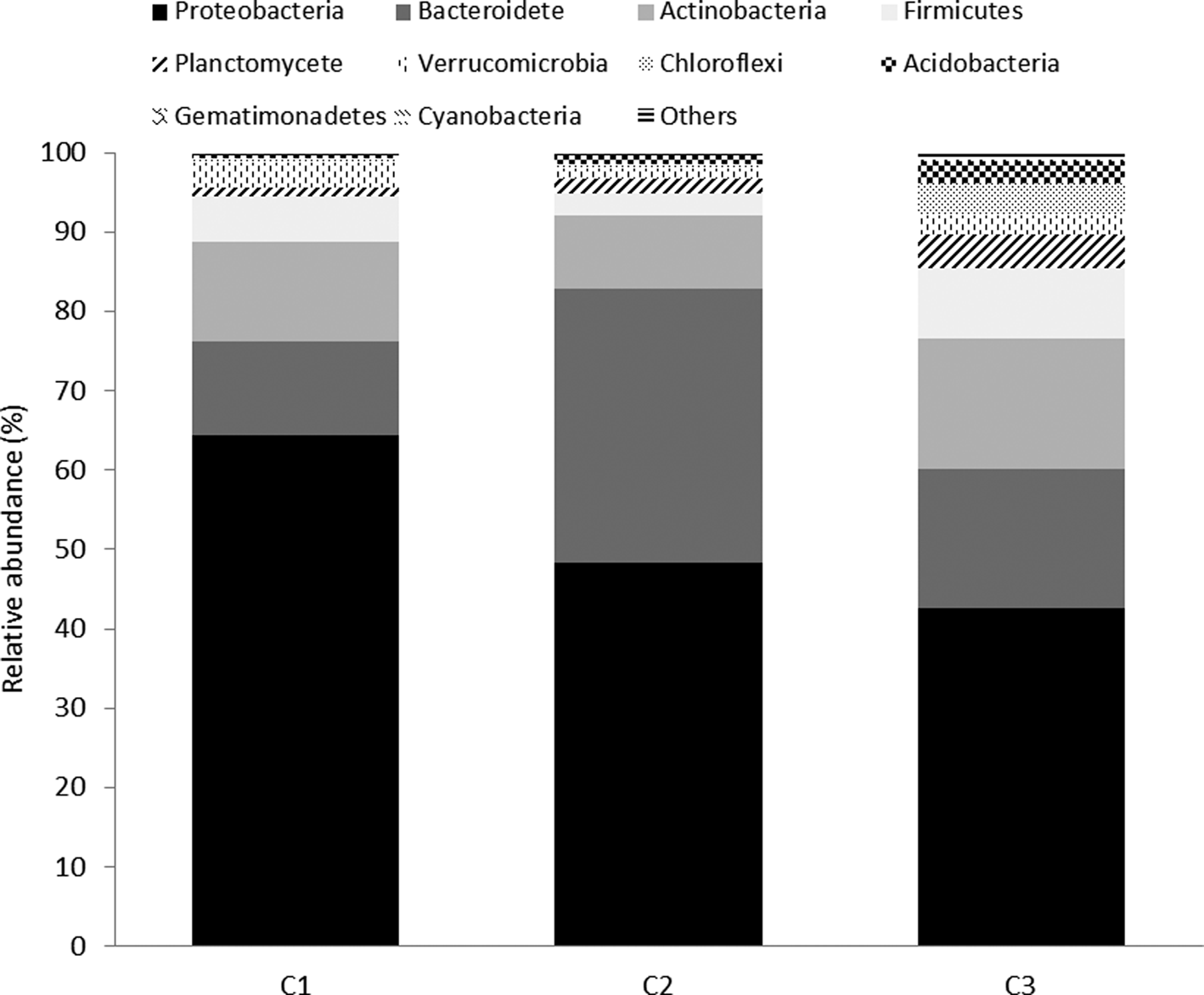
Sequencing analysis of the 16S rRNA gene showed 4147 OTUs after clustering at 97% similarity. In the three composts, Proteobacteria (52%), Bacteroidetes (21%) and Actinobacteria (13%) were the dominant microbial populations (Fig. 2).
Relative abundance of different bacterial phyla (16S rRNA) within different composts (relative abundance >0.5%).

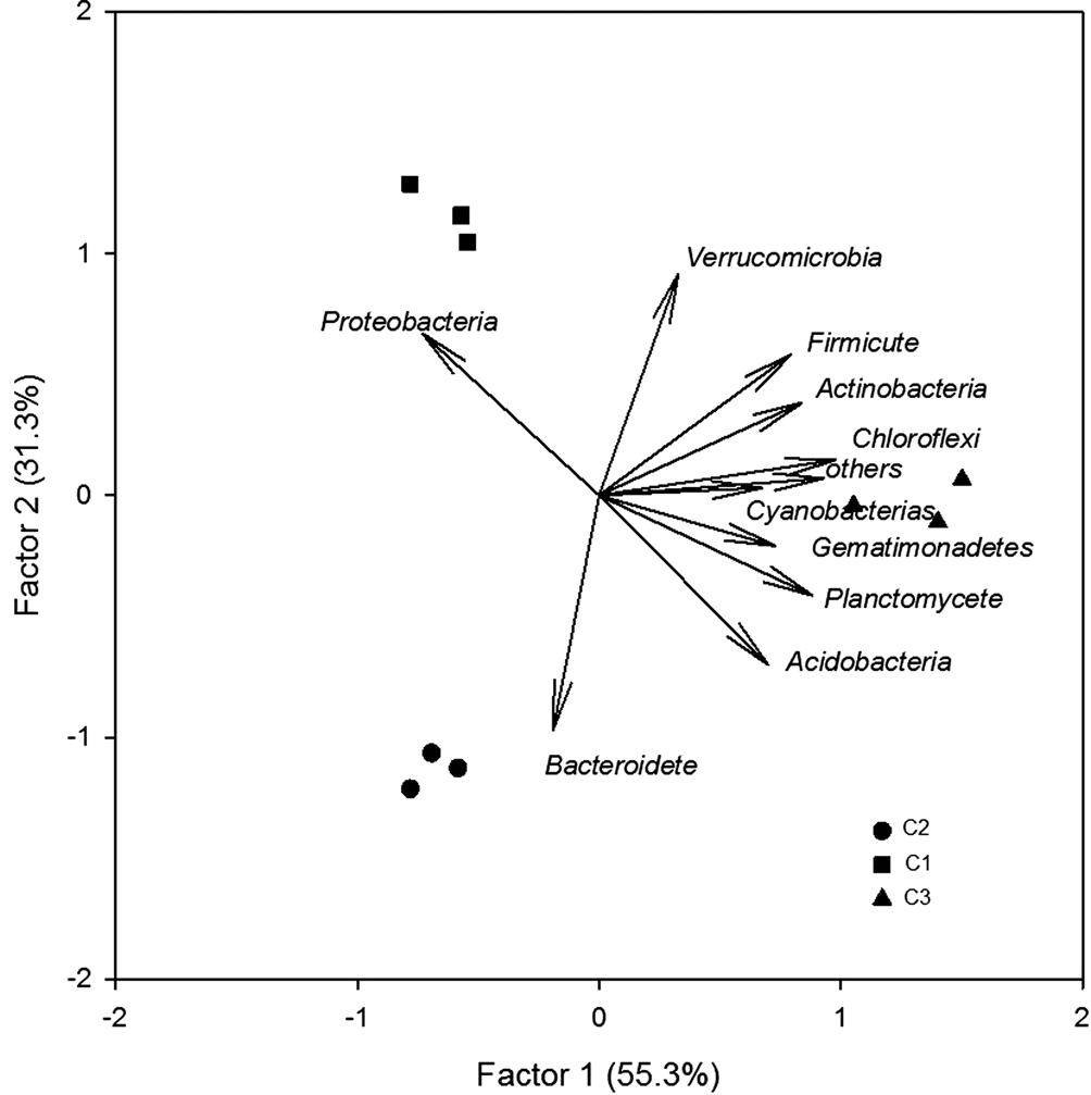
PCA of 16S rRNA sequences showed that, according to factor 1 (55% of the total system), the composts separated significantly (P < 0.05) into two groups: one group containing C1 and C2 and the other group containing C3 alone (Fig. 3). C1 and C2 showed a significantly (P < 0.05) higher relative abundance of Proteobacteria than C3 (Fig. 2).
PCA of bacteria phyla based on relative abundance of the phyla.

Within the dominant phylum, Proteobacteria, the most abundant subgroups were Gammaproteobacteria and Alphaproteobacteria, which made up almost 85% of the Proteobacteria. Furthermore, while Alphaproteobacteria, Sphingomonadales and Rhodobacteriales increased, Rhodospirillales decreased significantly in C1 and C2 (Table S1). Within the Gammaproteobacteria, the relative abundance of Xanthomonodales and Cellvibrionales was higher in C1 and C2 than in C3, while that of Nevskiales was significantly lower (P < 0.05) in C1 and C2 (Table S1). Within the Bacteroidetes, the orders Cytophagales and Chitinophagales showed significantly (P < 0.05) lower relative abundance in C1 and C2 than in C3, while Flavobacteriales and Sphingobacteriales showed a higher relative abundance in C1 and C2 (Table S1). The relative abundance of some orders belonging to Actinobacteria such as Micrococcales increased in C1 and C2, while others like Acidimicrobiales decreased significantly (P > 0.05) (Table S1).
The sequencing of the ITS2 region indicated the dominance of Ascomycota (95%) followed by Basidiomycota (3%) and Mucoromycota (2%). Within Ascomycota, Microascales and Sordariales were the dominant orders. Composts C1 and C2 showed a significantly lower (P < 0.05) relative abundance of Sordariales than compost C3. Furthermore, compost C1 and C2 showed a significantly higher relative abundance of Mucoromycota (Morteriella) than compost C3.
Composition of the bacterial community determined through metaproteomics
Metaproteomics made it possible for us to identify 367 proteins in the composts. These proteins were identified (99.6–100%) as bacterial proteins. According to phylogenetic analysis of the proteins, the bacterial composition of compost was dominated by Proteobacteria (61%), Actinobacteria (20%) and Thermotogae (11%) (Fig. 4). C1 and C2 showed a higher relative abundance of Proteobacteria and Deinococcus than C3 (Fig. 4).
Phylogenetic assignment of bacterial proteins in composts (relative abundance >0.5%).

Within the dominant phylum (Proteobacteria), the most abundant subgroups were Gammaproteobacteria and Alphaproteobacteria, which accounted for almost 71% of this phylum. Within Gammaproteobacteria, the most abundant bacterial proteins belonged to the orders Xanthomonadales, Enterobacteriales and Pseudomonodales. The Xanthomonadales were higher (P < 0.05) in C1 and C2 than in C3, while the Enterobacteriales and Pseudomonodales were lower (P < 0.05) in C1 and C2 than in C3 (Table S2). Within Alphaproteobacteria, the most abundant order was Rhizobiales, which had a greater abundance in C1 and C2 than in C3. In Betaproteobacteria, the most abundant order was Burkholderiales, and the levels were higher in C1 and C2 than in C3. The order Kosmotogales (Thermotogae), on the other hand, was significantly greater in C3 (P < 0.05). Within Actinobacteria, the order Micromonosporales showed higher relative abundance in C1 and C2 than in C3, while the relative abundance of Chloroflexi (Chloroflexales) was lower in the first two composts (P < 0.05) (Table S2).
Microbial functionality
The relative abundance of proteins involved in different functions like cell wall structure (M), carbohydrate transportation and metabolism (G) and inorganic ion transport and metabolism (P) was significantly higher (P < 0.05) in C1 and C2 than in C3. On the other hand, functions like energy production and conversion (C) and lipid transport and metabolism (I) were significantly lower (P < 0.05) in C1 and C2 than in C3 (Fig. 5). PCA indicated that the functional structures of C1 and C2 were similar to each other yet clearly different from the functional structure of C3 (Fig. 6).
Functional classification of microbial proteins in composts (% relative COGs >2%). Abbreviations: M, secondary metabolites biosynthesis, transport and catabolism; O, transcription; V, defense mechanism; J, nucleotide transport and metabolism; K, posttranslational modification, protein turnover, chaperons; C, cell cycle control, cell division, chromosome partitioning; ET, amino acid transport and metabolism-signal transduction mechanisms; G, energy production and conversion; P, translation, ribosomal structure and biogenesis; I, inorganic ion transport and metabolism.

PCAs of microbial protein-functional groups (COGs) of composts.

Correlation of sequencing and metaproteomics data to P. nicotianae disease incidence
A main objective of this analysis of composts was to correlate taxonomic and functional data with P. nicotianae disease incidence in composts that could be used as bioindicators.
We found a significant correlation between P. nicotianae disease incidence and PCA1 at the phylogenetic level (F = 0.917; P < 0.05) and at the functional level (F = 0.864; P < 0.01). At both levels, the relative abundance of Proteobacteria showed a significant negative correlation with disease incidence [(F = −0.700; P < 0.05) and (F = −0.762; P < 0.05), respectively]. The PCA1 of the COGs showed a significant correlation (F = 0.917; P < 0.01) with disease incidence. Between the different functions, V, C and I showed a significant positive correlation with disease incidence (F = 0.819; P < 0.01; F = 0.859; P < 0.01; F = 0.935; P < 0.01, respectively). On the other hand, M, G and P showed a significant negative correlation with disease incidence (F = −0.789; P < 0.05; F = −0.894; P < 0.01; F = −0.902; P < 0.01, respectively).
Discussion
It is clear that compost suppressiveness is pathogen-specific and related to the mechanism(s) of disease suppression attributed to either the chemical or biological factors of the composts (Hadar and Papadopoulou, Reference Hadar and Papadopoulou2012). Two of the three composts used in this experiment (C1 and C2) were found to be suppressive against P. nicotianae in pepper, with a disease incidence of <50%. The third compost, C3, however, was found to be non-suppressive against or even conductive to P. nicotianae in pepper, with a disease incidence >50% (Blaya et al., Reference Blaya, Lloret, Ros and Pascual2015). Among different physico-chemical characteristics, the effect of high EC levels has also been used in controlling root pathogens belonging to Phytophthora spp. on Gerbera (Thinggaard and Anderssen, Reference Thinggaard and Andersen1995). Accordingly, we found a negative correlation between EC and P. nicotianae disease incidence, indicating that composts with high levels of EC like C1 (3.35 mS cm−1) and C2 (2.26 mS cm−1) can help control P. nicotianae without interfering in seedling growth (Lemaire et al., Reference Lemaire, Dartigues and Rivière1985).
Special attention should be paid to the starting material used for the composting process (Castaño et al., Reference Castaño, Borreo and Aviles2011). The composts utilized in this study had different compositions of raw materials and microorganisms (Hoitink and Boehm, Reference Hoitink and Boehm1999; Ishii and Takki, Reference Ishii and Takii2003), some of which could contribute to suppressiveness. Garlic residue, for instance, is a substance that shows a broad-spectrum of antimicrobial activity against many genera of bacteria and fungi (Adetumbi et al., Reference Adetumbi and Lau1983). Compost C1 included garlic residue (25%), which would support the abovementioned hypothesis concerning pathogen suppression. Compost C3, however, was not found to be suppressive, and its composition also included garlic, but in a lower percentage (3%). This could indicate that not only the type of raw materials, but also the proportion and mixture, could influence the composts’ suppressive activity against P. nicotianae.
A positive correlation found between compost microbial activity and Phytophthora spp. and Pythium spp. suppression (Ntougias et al., Reference Ntougias, Papadopoulou and Zervakis2008; Blaya et al., Reference Blaya, Lloret, Ros and Pascual2015) suggests that microbial activity could be used as potential indicator of the suppressive capacity of a particular compost. However, this fact is not consistent with the results obtained in our study. Dehydrogenase activity, a widely recognized indicator of potential microbial activity (Garcia et al., Reference Garcia, Hernandez, Costa, Cecanti, Masciandaro and Gallardp-Lancho1993), did not show any correlation with disease incidence. For this reason, exploring the composition and functionality of microorganisms from suppressive/non-suppressive composts is important for making the best compost choices.
The use of high-throughput sequencing technologies has proven to be a powerful tool for identifying the variability in the bacterial and fungal communities in compost (de Gannes et al., Reference de Gannes, Eudoxie and Hickey2013; Neher et al., Reference Neher, Weicht, Bates, Leff and Fierer2013) to better manage soil-borne pathogens (Blaya et al., Reference Blaya, Marhuenda, Pascual and Ros2016; Mengesha et al., Reference Mengesha, Gill, Powell, Evans and Bary2017). Moreover, according to Bastida et al. (Reference Bastida, Torres, Moreno, Baldrian, Ondoño, Ruiz-Navarro, Hernandez, Richnow, Starke, García and Jehmlich2016), functional and phylogenetic analyses of proteins in soils complement DNA-based community analyses and make it possible to distinguish lifestyles, which could not be achieved solely by the amplification of taxonomical genes.
In our study, we found differences in the abundance of microbial populations in the different composts both through amplicon sequencing and metaproteomics. As described above, 16S rDNA amplicon sequencing indicated that Proteobacteria (alpha- and gamma-), Bacteroidete and Actinobacteria are the dominant phyla, which is consistent with previous compost sequencing studies (Danon et al., Reference Danon, Franke-Whittle, Insam, Chen and Hadar2008; Partanen et al., Reference Partanen, Hultman, Paulin, Auvinen and Romantschuk2010; de Gannes et al., Reference de Gannes, Eudoxie and Hickey2013). Using metaproteomic analysis, the dominant phyla were Proteobacteria (alpha- and gamma-), Actinobacteria, and Thermotogae, which was also consistent with protein-based compost analysis (Liu et al., Reference Liu, Li and XI2015). Both 16S rDNA gene sequencing and the functional analysis of proteins made it possible to decipher the key microbial players that are potentially involved in suppressive composts.
The Proteobacteria encompass an enormous level of morphological, physiological and metabolic diversity and are of great importance to global carbon, nitrogen and sulfur cycling (Kersters et al., Reference Kersters, De Vos, Gillis, Swings, Vandamme, Stackebrandt, Dwarkin, Falkow, Rosenberg, Schleifer and Stackebrandt2006). Proteobacteria species have shown significant negative correlation with disease incidence, indicating their dominance and a link between taxonomy and functionality in suppressive composts (Blaya et al., Reference Blaya, Marhuenda, Pascual and Ros2016; Mehta et al., Reference Mehta, Yu, Srivastava, Sinkkonen, Kurola, Gupta, Jääskeläinen, Palni, Sharma and Romantschuk2016). We therefore consider that this phylum could be an indicator of suppressive composts. Several orders of Proteobacteria have been identified by sequencing and protein analysis that could be involved in suppressive activities toward soil pathogens. The abundance of Xanthomonadales and Burkholderiales, for instance, was found to be higher in suppressive composts, which harbor genera and species that act against plant pathogenic fungi (Postma et al., Reference Postma, Sheper and Schilder2010). Pseudomonadales have also been identified as antagonists of soil-borne pathogens (Gonzalez-Sanchez et al., Reference Gonzalez-Sanchez, Perez-Jimenez, Pliego, Ramos, de Vicente and Cazorla2010) and inducers of systemic resistance in plants (Tian et al., Reference Tian, Yang and Zhang2007). Furthermore, proteins matched to Rhizobiales have also been found to be higher in suppressive composts. In fact, several rhizobial strains have been reported to induce plant growth and exhibit biocontrol properties and a resistance spectrum available against various abiotic stresses in a variety of agricultural crops (Gopalakrishnan et al., Reference Gopalakrishnan, Srinivas, Alekhya, Prakash, Kudapa, Rathore and Varshney2015).
The Actinobacteria harbor populations with antifungal activity against plant pathogen fungi (Postma et al., Reference Postma, Sheper and Schilder2010). The proteins of Micromonosporales, an Actinobacterial order, are found in greater abundance in suppressive than in non-suppressive composts. Researchers have also shown that non-streptomycete Actinobacteria are a useful source of novel bioactive compounds (Stackebrandt and Ebers, Reference Stackebrandt and Ebers2006; Kurtboke, Reference Kurtboke2012). The AntiBiotic Literature (ABL) database demonstrates that members of the family Micromonosporaceae account for the highest proportion (38%) of bioactive metabolite-producing strains among non-streptomycete families.
In the current study, proteins associated with carbohydrate processes, cell wall structure and inorganic ion transport and metabolism were more deeply involved in the suppressive composts than the non-suppressive composts. Data suggest that these COGs could be indicators of suppressive composts.
In our study, we found a significantly higher amount of bacterial proteins than fungal proteins, which were nearly non-existent. This is probably related to the poor coverage of fungal genomes in the general metagenome databases used for protein searches (Liu et al., Reference Liu, Li and XI2015).
Conclusions
Our study concludes that there are differences between suppressive and non-suppressive composts at the phylogenetic level (sequencing) and at the functional level (analysis of COGs). It is also important to highlight the differences between the DNA and protein analyses. Proteobacteria could be a good indicator of compost suppressiveness due to their high correlation with P. nicotianae disease incidence. The presence of certain microorganisms can also contribute to the suppressive ability of compost, although they are not determinants of it. Other types of physico-chemical factors such as EC may also drive the suppressiveness of composts. In the future, the development of metagenome databases with a higher number of fungal genomes would further extend our knowledge of the biological mechanisms of suppressiveness mediated by fungi.
Supplementary material
The supplementary material for this article can be found at https://doi.org/10.1017/S1742170518000467.
Acknowledgements
The authors would like to thank the Spanish Ministry for the project AGL2014-52732-C2-1-R and the Fundacion Seneca of the Region of Murcia for its financial support within the Research Groups of Excellence programme of the Region of Murcia (19896/GERM/15). The authors are grateful for use of the analytical facilities of the Centre for Chemical Microscopy (ProVIS) at the Helmholtz-Centre for Environmental Research, which is supported by European Regional Development Funds (EFRE – Europe funds Saxony) and the Helmholtz-Association.