The phylum Nematoda constitutes one of the largest and most abundant groups of animals. Although most species of these worms are free-living, this phylum also contains parasites of virtually every multicellular organism on the planet (Ref. Reference Schmidt and Roberts1). This, allied to the fact that in many cases parasitism has severe and even fatal consequences for the host, dictates that nematodes are of substantial medical, veterinary and economic importance. For this reason, parasitic nematodes have been the subject of much recent study.
Nematodes are generally small, but the existence of larger parasitic species (e.g. Ascaris lumbricoides, which may grow to over 40 cm in length) has been known of for thousands of years (Ref. Reference Schmidt and Roberts1). Typically, the worms are slender, bilaterally symmetrical organisms that are encased in a noncellular cuticle. The cuticle is shed four times during the course of a nematode's life and the five stages are often referred to as L1–L5, with L5 being the adult stage (Ref. Reference Schmidt and Roberts1).
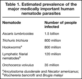
Adult parasitic nematodes can inhabit two locations within humans: the gastrointestinal tract or the tissues. The most common gastrointestinal nematodes of humans are A. lumbricoides, Trichuris trichiura, Necator americanus and Ancylostoma duodenale (the latter two species are ‘hookworms’) (Table 1). An infected individual may harbour more than one type of these parasites, and collectively, these four species of worm may infect more than one third of the world's population. The major tissue-dwelling nematodes of humans are the filarial nematodes, the adult worms of which may live in the lymphatic system (e.g. Wuchereria bancrofti, Brugia malayi) or the subcutaneous tissues (Onchocerca volvulus) (Ref. Reference Schmidt and Roberts2). Infection with gastrointestinal nematodes may arise through ingestion of eggs [A. lumbricoides (Ref. Reference Schmidt and Roberts3), T. trichiura (Ref. Reference Schmidt and Roberts4)] or penetration of the skin by L3 larvae (hookworms) (Ref. Reference Schmidt and Roberts5). Infection with filarial nematodes occurs when L3 larvae are transferred to humans by blood-feeding insects that act as vectors (Ref. Reference Schmidt and Roberts2). The majority of people infected with nematodes tend to be asymptomatic, but some species can cause very severe problems such as elephantiasis (e.g. W. bancrofti, B. malayi) and blindness (O. volvulus) (Ref. Reference Schmidt and Roberts2).
Estimated prevalence of the major medically important human nematode parasites

a Ancylostoma duodenale and Necator americanus;
b Wuchereria bancrofti and Brugia malayi
Parasitic nematodes can protect humans from allergy and autoimmunity
One surprising feature of nematodes is their longevity. It is reported, for example, that W. bancrofti can live in excess of a decade (Ref. Reference Subramanian6) and, although a number of factors may well be involved, it is assumed that a major factor contributing to long-term infection is suppression of the host immune system. In relation to this, analysis of the immune response during human infection generally indicates that people having patent infection – that is infection with larval stages (microfilariae) in their bloodstreams – demonstrate reduced lymphocyte proliferative responses and decreased production of the cytokine interferon-γ (IFN-γ), but increased production of the cytokines interleukin- (IL) 4 and IL-10 and the antibody type IgG4 (Ref. Reference Nutman and Kumaraswami7). As IL-4 is a cytokine that is classically associated with helper T-cells of the type 2 category (Ref. Reference Nakamura8) and IL-10 is an anti-inflammatory cytokine (Ref. Reference Moore9), such an immune response may be characterised as being of a type-2 T-helper cell (Th2), anti-inflammatory phenotype. There is probably a benefit for humans in the generation of such an immunological phenotype because, as alluded to earlier, although filarial nematode infection can result in severe pathology, such as elephantiasis, the majority of infected individuals exhibit little evidence of an inflammatory response or overt tissue destruction or disruption (Ref. Reference Rajan10). Similar conclusions are now being made with respect to gastrointestinal nematodes (Ref. Reference Bradley and Jackson11) and indeed, it is now accepted that a Th2-like, somewhat anti-inflammatory immunological phenotype is a feature of nematode infection. An interesting characteristic of this phenotype is that it has been reported in some cases to extend to subsequent responses to non-nematode antigens–for example to antigens in vaccines targeting bacterial infections that rely on Th1 responses for efficacy (Ref. Reference van Riet, Hartgers and Yazdanbakhsh12). One immediate thought, therefore, is whether such a change in immune-responsiveness could lead to changes in susceptibility to diseases associated with aberrant Th1-associated inflammatory responses, such as the autoimmune diseases. In relation to this, there is currently great interest in the idea that the recent alarming increase in autoimmune and allergic diseases in the industrialised world, but not developing countries, reflects increased hygiene, causing a reduction in infection with pathogens such as parasitic worms, an idea known as the ‘hygiene hypothesis’ (Ref. Reference Wills-Karp, Santeliz and Karp13). Consistent with this, it has been suspected for many years that allergic and autoimmune diseases may be less prevalent in areas of nematode transmission (see Ref. Reference Elliott, Summers and Weinstock14). Interestingly, results from human and animal studies find evidence for infection associated not only with reduced autoimmunity, but also with decreased allergy, an initially surprising observation given the Th2-associated status of this type of disease (Ref. Reference Busse and Lemanske15).
Evidence from human studies
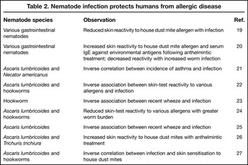
Although there are cases in which no association, or indeed increased susceptibility, has been observed, the majority of studies support the idea that nematode infection can protect against allergy, as measured by factors such as skin reactivity to allergens or wheezing, and more consistently, by comparing symptoms before and after anthelmintic treatment (for reviews, see Refs Reference van Riet, Hartgers and Yazdanbakhsh12, Reference Elliott, Summers and Weinstock14, Reference Cooper16, Reference Falcone and Pritchard17, Reference Maizels18) (Table 2). Autoimmune diseases have not been as well studied as allergic disease, but again there is evidence supporting an inverse relationship between nematode infection and conditions such as inflammatory bowel disease, type-1 diabetes, rheumatoid arthritis and multiple sclerosis (for reviews, see Refs Reference Elliott, Summers and Weinstock14, Reference Hunter and McKay28, Reference Zaccone29, Reference Reddy and Fried30). Furthermore, an inverse relationship between infection with Strongyloides stercoralis and autoimmune liver disease has recently been reported (Ref. Reference Aoyama31). Thus, overall, the idea that helminths can protect against human allergic and autoimmune inflammatory disease is compelling.
Nematode infection protects humans from allergic disease

Evidence from animal studies
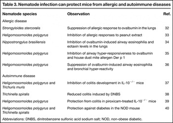
At the outset, it has to be appreciated that using animal models to investigate whether nematodes are likely to protect humans against allergy and autoimmunity has the limitation of differences in the pattern of exposure. Models invariably employ one or two exposures to a significant number of nematodes, whereas humans will generally acquire their nematodes one or a few at a time, over a period of many years. There are a number of recent mouse model studies that show nematode infection can protect against allergic disease, in particular lung-associated disease (Table 3). This type of approach has also provided evidence that nematodes can protect against autoimmune diseases, such as colitis and diabetes. Hence, the use of mouse models in this area has produced a series of convincing findings supporting the hypothesis that nematodes may protect humans from allergy and autoimmunity.
Nematode infection can protect mice from allergic and autoimmune diseases

Abbreviations: DNBS, dinitrobenzene sulfonic acid sodium salt; NOD, non-obese diabetic.
Mechanism of action
The role of Th2 polarisation and regulatory T cells
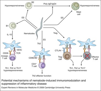
As mentioned above, nematode infections are chronic and have been classically characterised by the induction of a Th2 immune response (Ref. Reference Finkelman41). However, more recently they have also been associated with regulatory T-cell (Treg) responses (Ref. Reference Diaz and Allen42). It is convenient to simply view these two types of immune response in terms of the cytokines generally associated with them, for example IL-4, IL-5 and IL-13 for Th2 responses, and IL-10 and transforming growth factor-β (TGF-β) for (certain) regulatory responses (Fig. 1). Although the host may have evolved the latter to deal with any persistent significant antigen challenge, it is clear that nematodes are particularly adept at polarising immune responses towards Th2, in particular by their effects on dendritic cells, the sentinels of the immune system that sample and process foreign molecules for presentation to T cells. Presentation of peptides derived from antigens is accompanied by signals generated by dendritic cell surface costimulatory molecules and secreted cytokines interacting with receptors on T cells, and this combination impacts on T-cell polarisation (Ref. Reference Banchereau43). Thus, nematode antigens must contain information that facilitates dendritic cell-driven Th2 polarisation. Interestingly, compared with antigens that induce Th1 responses, nematode-derived molecules have generally been shown to have little or only a partial effect on dendritic cell activation as measured by upregulation of markers such as costimulatory molecules (Refs Reference Whelan44, Reference Balic45). The induction of Th2 responses may reflect particular characteristics of nematode molecules (see Ref. Reference Harnett and Harnett46). However, the advantage of this to the worms is uncertain, because there is evidence from rodent models of gastrointestinal nematode infection that Th2 immune responses are protective (Ref. Reference Gause, Urban and Stadecker47). This is also supported by some human epidemiological studies (Ref. Reference Bradley and Jackson11). Moreover, Th2 responses are associated with inflammatory pathology during gastrointestinal nematode infection (Ref. Reference Scales48). Nevertheless, it is possible to perceive that the induction of an immunological environment polarised towards Th2 may provide a situation in which the autoimmune diseases generally considered to be Th1 dependent (Ref. Reference Liblau, Singer and McDevitt49) would find it difficult to flourish. Consistent with this, a reduction in inflammation is observed in IL-10−/− mice with colitis upon infection with Heligomosomoides polygyrus, which reflects an inhibition of mucosal production of IFN-γ and IL-12 p40, and the increased production of IL-13 (Ref. Reference Elliott39).
Potential mechanisms of nematode-induced immunomodulation and suppression of inflammatory disease. Infection with nematodes can induce T-cell hyporesponsiveness by directly or indirectly (through antigen-presenting cells such as dendritic cells) suppressing T-cell activation. For example, infection can result in the induction of T regulatory (Treg) cells that are refractory to antigen and can prevent maximal T-effector responses of Th1-, Th2- or Th17-based inflammation. Moreover, worm products such as ES-62 can induce B1 cells to produce IL-10, which acts to suppress dendritic cell maturation and hence results in T-cell anergy, preventing Th1-Th2- or Th17-mediated inflammation. Alternatively, the worms can modulate dendritic cell maturation to polarise T-cell responses towards a Th2 phenotype and hence counteract Th1-based inflammation. By contrast, it has been proposed that nematodes can antagonise Th2-driven inflammation by producing polyspecific IgE or IgG4 to block allergen-induced mast-cell degranulation. Moreover, certain excretory-secretory (ES) products have been shown to directly induce mast-cell responsiveness by subverting FcɛRI signalling (see Fig. 2). Abbreviations: CTLA, cytotoxic T-lymphocyte antigen; MHC, major histocompatibility complex; TCR, T-cell receptor.

Considering regulatory responses, these may be generated in response to any chronic inflammatory insult in an attempt to prevent it getting out of control, but with respect to nematodes, recent evidence intriguingly suggests that they may contribute to worm persistence during infection via inhibition of protective immunity (Ref. Reference Taylor50). Of particular relevance to this review however, is the fact that regulatory responses do not only inhibit inflammatory responses to nematodes, but also to heterologous antigens. This means that responses to other infections (Ref. Reference van Riet, Hartgers and Yazdanbakhsh12) or even vaccines (Ref. Reference van Riet, Hartgers and Yazdanbakhsh12) may be affected, and in addition, susceptibility to diseases associated with aberrant inflammatory responses altered. The Th2 dominance associated with nematode infection provides an attractive argument to explain the ability of the helminths to inhibit autoimmune diseases, but it obviously does not hold true with respect to allergic diseases, which are themselves Th2 dependent (Ref. Reference Busse and Lemanske15). However, both Th1 and Th2 responses may be dampened down by anti-inflammatory regulatory responses (Ref. Reference Diaz and Allen42) and hence the ability of nematodes to protect against allergy may be explained in this way. Consistent with the latter, the ability of H. polygyrus to inhibit allergic airway hyper-responsiveness in the mouse has been shown by the use of adoptive transfer experiments to be dependent on CD4+ CD25+ Tregs (Ref. Reference Wilson35). In terms of cytokines, C57BL/6 strain mice showed increased IL-10 production in response to infection, whereas BALB/c mice demonstrated augmented TGF-β. The latter result may explain why the use of BALB/c-knockout mice showed no role for the anti-inflammatory molecule IL-10 in generating the protective response (Ref. Reference Wilson35) – a result not observed with C57BL/6 animals (Ref. Reference Kitagaki36). These results are also consistent with the ability of T-cell lines expressing IL-10 or TGF-β to protect against allergy in the ovalbumin-sensitisation model (Ref. Reference Carvalho, Bastos and Araujo51).
It has already been mentioned that IL-10−/− mice with colitis show a reduction in inflammation when infected with H. polygyrus (Ref. Reference Elliott39). There could be a regulatory component operating in this system, because interestingly, the protective effect can be transferred by mesenteric lymph node T cells from infected animals, and these cells were shown to have increased Foxp3 mRNA expression – a key marker of Treg function. It was recently demonstrated that H. polygyrus infection is associated with the generation of a regulatory CD8+ T-cell population in the lamina propria (Ref. Reference Metwali52). Interestingly, H. polygyrus also causes increased expression of TLR4 on mucosal T cells, and interaction of TLR4 with its ligand LPS resulted, not in release of the expected IL-12 or TNF-α, but rather of the cytokine associated with regulatory responses TGF-β (Ref. Reference Ince53).
Role of nonspecific IgE production in allergy prevention
The idea of regulatory responses preventing the development of allergic responses during nematode infection is clearly supported by experimental evidence; however, other possible explanations have been tested. Nematodes have a tendency to induce considerable nonspecific IgE secretion during infection (which may prevent specific mast cell- and/or basophil-mediated attack) and it has been suggested that by saturating FcɛRI sites, these antibodies may block mast-cell degranulation that would be mediated by allergens (reviewed in Ref. Reference Erb54). Certainly, an association between high levels of circulating nonspecific IgE and protection against allergy has been shown in a number of studies (see Ref. Reference Erb54). Moreover, increased cutaneous allergic responses following anthelmintic treatment are associated with a corresponding decrease in nonspecific IgE (Ref. Reference Lynch20). However, it has been found that Nippostrongylus brasiliensis infection does not reduce mast-cell degranulation in response to ovalbumin in the skin of immunised mice, in spite of the infection generating a massive increase in nonspecific IgE production (Ref. Reference Wohlleben34). It has also been reported, again using N. brasiliensis, that a small amount of IgE specific for the parasite antigen Nb-Ag1, is still able to facilitate mast-cell degranulation in the presence of much greater levels of nonspecific IgE (Ref. Reference Pochanke55). Finally, studies on humans infected with a variety of filarial nematodes indicate that the relative levels of polyclonal IgE and filaria-specific IgE do not tend to reach that required for inhibition of filaria-induced mast-cell or basophil degranulation (Ref. Reference Mitre, Norwood and Nutman56). The same outcome was observed when examining dust-mite-specific IgE. Thus overall, it appears that saturation of FcɛRI by polyspecific IgE antibodies during nematode infection cannot alone explain protection from allergy (Fig. 1).
Interestingly, although nematode infection increases polyspecific IgE production and as mentioned above, does not appear to inhibit the IgE response to allergens, there have been reports where the opposite is the case. Thus for example, S. stercoralis infection of mice results in reduced anti-ovalbumin IgE levels in the lungs (Ref. Reference Wang32). Likewise, H. polygyrus infection results in decreased circulating IgE levels in mice in response to peanut allergen (Ref. Reference Bashir33). H. polygyrus infection has also been shown to increase ovalbumin-specific IgE as well as total IgE in a mouse model of atopic asthma (Ref. Reference Kitagaki36).
Nematode infections can also be associated with the production of high levels of IgG4, an antibody that competes with IgE for allergen binding but does not promote mast-cell or basophil degranulation (Ref. Reference Hussain, Poindexter and Ottesen57) (Fig. 1). However, studies investigating whether high levels of IgG4 can protect against allergic reactions in humans have produced conflicting results (Ref. Reference Carvalho, Bastos and Araujo51).
IL-10, Tregs and mast-cell function
As alluded to earlier (e.g. Ref. Reference Wilson35) a feature of some nematode infections is the production of IL-10 and/or Tregs, and some recent papers suggest that both of these could have an impact on mast-cell function. Thus, it was shown that IL-10 inhibited the release of TNF-α, IL-8 and histamine by human mast cells activated via FcɛRI (Ref. Reference Royer58). Furthermore, the use of IL-10 transgenic mice demonstrated that overexpression of the cytokine reduces passive cutaneous anaphylaxis responses (Ref. Reference Kennedy Norton59). Also, staying with mice, studies on mast cells have revealed that FcɛRI expression and IgE-mediated release of leukotriene C4 can both be inhibited by Tregs (Ref. Reference Kashyap60). Thus, although these observations have not been related to nematode infections as yet, it is possible that nematode induction of IL-10 and/or Tregs could interfere with mast-cell function and hence allergic responses.
Molecules involved in immunomodulation in animal models
Excretory-secretory products of Nippostrongylus brasiliensis
An excretory-secretory (ES) extract, containing as-yet-undefined components of adult stage N. brasiliensis, inhibits the development of asthma (ovalbumin-hypersensitivity model) in the lungs of mice in a similar manner to infection with the parasite itself (Ref. Reference Trujillo-Vargas61). Administration of adult ES during the sensitisation period entirely blocked eosinophilia and mucus production in the lungs and also significantly inhibited airway hyper-reactivity development. Ovalbumin-specific IgG1 and IgE levels in the bloodstream and the amount of both IL-4 and IL-5 in the lungs were all reduced by exposure to the ES. No role was found for Toll-like receptor (TLR)2, TLR4, IFN-γ or IL-10. Moreover, such ES-induced parasite-specific Th2 responses develop as normal. The active agent appears unlikely to be a protein, because it is resistant to both heat and proteinase K. Similarly, intratracheal installation of ES derived from the N. brasiliensis L3 stage reduces neutrophil influx in response to LPS in the lungs of Wistar rats (Ref. Reference Keir62).
Ascaris suum PAS1
It was reported in 2002 that an A. suum extract could inhibit lung inflammation and hyper-responsiveness and suppress IL-4, IL-5 and eotaxin in response to hen's egg white (Ref. Reference Lima63). More recently, it was discovered that A. suum contains a protein, APAS3, which, upon immunisation and challenge is able to induce Th2-dependent, eosinophilic airway hyperreactivity in BALB/c mice (Ref. Reference Itami64). Interestingly, another A. suum product, PAS1, inhibits this response (Ref. Reference Itami64). Inhibition is associated with reduced Th2 cytokines (IL-4, IL-5) and chemokines [eotaxin, RANTES (regulated upon activation, normal T-cell expressed and secreted)], and considerably elevated IL-10 levels in bronchoalveloar lavage fluid. Measurement of serum levels also showed that production of IgG1 and IgE was blocked. PAS1 is most active when administered prophylactically at both immunisation and challenge stages; administration at immunisation only reveals a partial effect, but PAS1 is completely ineffective when given only at challenge. PAS1 also induces IL-10 and TGF-β production in macrophages and this is associated with loss of proinflammatory cytokine production (Ref. Reference Oshiro, Macedo and Macedo-Soares65).
Dirofilaria immitis-derived antigen (DiAg)
A purified 15 kDa protein from the canine filarial nematode, Dirofilaria immitis known as DiAg (Dirofilaria immitis-derived antigen), causes polyclonal proliferation of B cells and nonspecific IgE production (Ref. Reference Imai and Fujita66). Interestingly DiAg prevents the spontaneous generation of IgG anti-insulin antibodies seen in the NOD mouse, and this is associated with a lack of development of Th1-dependent autoimmune diabetes. Consistent with this, DiAg increases levels of IL-4 and IL-10 when injected into BALB/c mice. DiAg can also inhibit passive cutaneous anaphylaxis (PCA) reactions in rats (Ref. Reference Imai and Fujita66). This was found not to be due to an effect on the number or viability of mast cells or to differences in the amount of histamine released, but interestingly, given the relative dismissal of this mode of action in the human situation, appears to be due to nonspecific saturation of FcɛRI.
ES-62
ES-62 is a phosphorylcholine (PC)-containing glycoprotein secreted by filarial nematodes, which has been shown to have a wide range of immunomodulatory properties that are largely anti-inflammatory (Ref. Reference Harnett and Harnett67). Consequently, the molecule has been tested in mouse models of autoimmune and allergic disease and has been found to be active against collagen-induced arthritis (CIA) (Ref. Reference McInnes68) and type-I hypersensitivity in the skin and lungs (Ref. Reference Melendez69).
With respect to CIA, ES-62 was effective as a prophylactic treatment and also therapeutically, and in both cases, ex vivo analyses revealed that the effects were associated with inhibition of collagen-specific proinflammatory (TNF-α, IL-6) and Th1 (IFN-γ) cytokines and (IgG2a) antibody production (Ref. Reference McInnes68). In the prophylactic experiment, increased production of collagen-specific IL-10 was also observed. More recent work on the CIA model, involving a comparison of ES-62, PC-free ES-62 and PC conjugated with an irrelevant protein (ovalbumin), has revealed that the antiarthritis effect of ES-62 is dependent on its PC moiety, although interestingly, not on a reduction in collagen-specific IgG2a production (Ref. Reference Harnett70). ES-62 was also found to suppress LPS-induced proinflammatory cytokine production by cells obtained from human synovial fluid and membranes (Ref. Reference McInnes68), and human cells are also susceptible to the anti-inflammatory action of PC conjugated to ovalbumin.
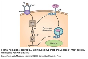
Turning to allergy, ES-62 was found to be active in two mouse models – ovalbumin-induced airway hypersensitivity and immediate-type hypersensitivity to oxazolone in the skin (Ref. Reference Melendez69). In the former, ES-62 was found to reduce peribronchial inflammation and mucosal hyperplasia, inhibit eosinophila and prevent release of IL-4, the signature cytokine required for airway inflammation development. In the latter model, a reduction in ear swelling indicated that ES-62 targeted inflammation. Subsequent in vitro analysis revealed that ES-62 directly prevented mast cell degranulation and release of mediators of allergy induced via ligation of FcɛRI. The mechanism of action involved inhibition of key signal transduction events involved in mast cell activation, including phospholipase-D-coupled, sphingosine-kinase-mediated calcium mobilisation and NF-κB activation. It was found that ES-62 mediates these effects by forming a complex with TLR4, which results in the sequestration and perinuclear degradation of protein kinase C (PKC)-α, a molecule found to be critical for mast-cell activation (Ref. Reference Oshiro, Macedo and Macedo-Soares65), by a caveolae or lipid raft proteosomal-independent mechanism (Fig. 2).
Filarial nematode-derived ES-62 induces hyporesponsiveness of mast cells by disrupting FcɛRI signalling. Antigen (Ag)-driven ligation of IgE bound to FcɛRI induces mast cell degranulation and generation of pro-inflammatory mediators and cytokines by triggering a signalling cascade involving protein kinase C-α (PKC-α) complexing with phospholipase D (PLD) to stimulate PLD-coupled, sphingosine kinase (SK)-mediated, calcium mobilisation. ES-62 directly blocks mast cell activation by subverting TLR4 signalling such that it results in sequestration and trafficking of PKC-α for perinuclear degradation by a caveolae/lipid raft, proteosomal-independent mechanism. Such sequestration of PKC-α uncouples FcɛRI-mediated mast cell responses. Abbreviations: ES, excretory-secretory product; PA, phosphatidic acid; S-1-P, sphingosine-1-phosphate; TLR, toll-like receptor.

The finding that ES-62 can inhibit components of a Th2 response, such as IL-4 production (Ref. Reference Melendez69), indicates that the original hypothesis where ES-62 was solely a Th2-polarising agent (Ref. Reference Whelan44) was too simplistic. It is possible in fact that a target of ES-62 may perhaps be a cytokine, such as IL-17, which appears to act as a master regulator of both Th1- and Th2-type inflammation (Ref. Reference Kollis and Linden71) and indeed plays a critical role in CIA (Ref. Reference Nakae, Nambu, Sudo and Iwakura72), the first model of inflammation in which ES-62 was found to be protective.
Currently, work is underway to develop molecules that mimic the activity of ES-62 for use as drugs for treating allergic and autoimmune diseases. Unlike the parent molecule, these will be too small to be immunogenic and given its importance in promoting ES-62 activity, will be based around PC, but possibly with slight structural modifications to avoid interaction with natural anti-PC antibodies.
Clinical implications/applications
The weight of evidence in support of parasitic nematodes preventing allergy and autoimmunity not surprisingly led scientists to consider their therapeutic potential. Within the past few years therefore, parasitic nematodes have been used in clinical trials aimed at ameliorating inflammatory diseases. It has been possible to carry out these trials because of the known or predicted lack of pathogenicity of certain species.
An example of a ‘safe’ species is Trichuris suis, which is similar to the human whipworm T. trichiura, but can persist for only a limited period in the human gastrointestinal tract following infection. These factors have dictated that it has been selected for testing in human trials as a treatment for inflammatory bowel disease. Encouragingly, in a series of trials, the majority of people suffering from inflammatory bowel disease have shown improvement in symptoms following ingestion of embryonated ova of this worm (reviewed in Refs Reference Elliott, Summers and Weinstock14, Reference Reddy and Fried30).
N. americanus has also been employed in trials, although because this human helminth has pathogenic potential, low numbers must be used to treat people. When infecting patients suffering from Crohn's disease with 50 larvae, those with moderately active (but not inactive or mild) disease showed some improvement (Ref. Reference Croese73). A trial determining the effect of hookworms on asthma has also recently started (Ref. Reference Falcone and Pritchard17).
The ultimate aim in this area of research is to use defined nematode molecules (or derivatives thereof) in treating human disease. As proof that this can work, one need look no further than the recombinant nematode anticoagulant protein c2 (rNAPc2), a serine protease inhibitor derived from the original hookworm (Ancylostoma caninum) protein (Ref. Reference Stassens74). The anticoagulant activity of this protein has been found to inhibit thrombin generation in patients undergoing elective percutaneous coronary intervention (Ref. Reference Moons75), to reduce the likelihood of deep venous thrombosis occurring during total knee arthroplasty (Ref. Reference Lee76) and to reduce new thrombin generation and ischaemia in patients with non-ST-segment elevation acute coronary syndrome (Ref. Reference Giugliano77). The anticoagulant activity of the protein is due to inhibition of the catalytic complex of subendothelial tissue factor/activated factor VII by a unique mechanism (see Ref. Reference Giugliano77). It is to be hoped that nematode-derived molecules and/or their derivatives will soon also be used in the treatment of human allergic and autoimmune diseases.
Acknowledgements and funding
The authors would like to thank the Wellcome Trust, the Medical Research Council and the Biotechnology and Biological Sciences Research Council for funding their work cited in this review.