In recent years, considerable progress has been made in the field of non-invasive cardiac imaging, with evolution from the so-called unidimensional mode towards three-dimensional representations. In this review, we concentrate on two of the innovative technologies used for three-dimensional imaging, namely three-dimensional echocardiography and cardiac computed tomographic angiography.
Appreciation of complex intracardiac anatomy and spatial relationships is inherent to the diagnosis and treatment of patients with congenitally malformed hearts. Beginning over 30 years ago, and until recently, our ability clinically to image the heart non-invasively was limited to cross-sectional echocardiography.Reference Tajik, Seward, Hagler, Mair and Lie1 This technique has fundamental limitations. The very nature of obtaining a two-dimensional echocardiographic slice, which has no thickness, necessitates the use of multiple orthogonal sweeps to provide an impression of overall cardiac structure. This requires the imager mentally to reconstruct the global anatomy, and then use the structure of his or her report to express this mentally reconstructed vision. This means that the only three-dimensional image of the heart is the virtual representation that exists in the mind of the imager, who then translates this vision into words. It is not easy for an untrained, albeit interested, observer to understand the images obtained in the course of a sweep. On the contrary, expert interpretation is required. Importantly, since myocardial motion occurs in three dimensions, cross-sectional techniques inherently do not lend themselves to accurate quantitation. Recognition of the limitations of cross-sectional imaging led to burgeoning research and clinical interest in three-dimensional imaging.
Three-Dimensional Echocardiography
Early techniques providing three-dimensional echocardiographic images used reconstructive approaches, which were based on acquisition of cross-sectional images that were subsequently stacked and aligned based on phases of the cardiac cycle in order to recreate a three-dimensional dataset.Reference Matsumoto, Matsuo and Kitabatake2 These approaches proved to be accurate, but the time needed for analysis, and the equipment required for offline processing, imposed fundamental limitations on their clinical applicability. By 1990, nonetheless, results were published using a matrix-array transducer that provided real-time images of the heart in three dimensions.Reference von Ramm and Smith3 Although this was an important breakthrough, the transducer used to obtain the images could not be steered in the third dimension, namely elevation. Since then, dramatic technological advances have facilitated the ability to perform live three-dimensional echocardiographic scanning, including the ability to steer the beam in three dimensions, and to render images in real time.Reference Shirali4
As we write, three-dimensional echocardiography has three broad areas of clinical application among patients with congenitally malformed hearts, first visualization of morphology, second, assessment of the sizes of chambers and ventricular function, and third, use for transoesophageal interrogation and image-guided interventions.
Visualization of morphology
From early in development of three-dimensional echocardiography, the structural complexity inherent to the congenitally malformed heart has been identified as a fertile substrate for exploration. We illustrate the utility of this technology in a series of clinical situations.
Atrioventricular valvar disease
Three-dimensional echocardiography is invaluable in delineating the morphology of the atrioventricular valves. Its role in delineating congenital abnormalities of the mitral valve has been well described,Reference Espinola-Zavaleta, Vargas-Barron and Keirns5 while others have demonstrated the additive value of epicardial interrogation.Reference Rawlins, Austin and Simpson6 Specific findings demonstrated in children include the morphology of the leaflets, their cordal attachments, the subcordal apparatus, the mechanism and origin of regurgitation, and the geometry of the regurgitant volume. The utility of the technique in in patients with Ebstein’s malformation of the tricuspid valve has also been emphasised.Reference Vettukattil, Bharucha and Anderson7 It was shown that three-dimensional echocardiography provided clear visualization of the morphology of the abnormal valvar leaflets, including the extent of their formation, the level of their attachment, and their degree of coaptation. They were also able to visualize the mechanism of regurgitation or stenosis.
Atrioventricular septal defect
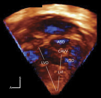
Our group studied patients with atrioventricular septal defects and common atrioventricular junction, showing that gated three-dimensional echocardiographic views could be cropped to obtain “en face” views of the atrial and ventricular septal structures enclosing the septal defect (Fig. 1).Reference Hlavacek, Crawford, Chessa and Shirali8 This perspective is useful in determining the precise location of the interventricular communication relative to the bridging leaflets, and in demonstrating how these relationships determine the level of shunting, be it at atrial or ventricular level, or both levels. Useful additional information obtained by the technique included precise characterization of valvar regurgitation, unique views of surgically significant features such as zone of apposition between the left ventricular components of the bridging leaflets, the substrate for subaortic stenosis, and the presence and location of additional septal defects.
Side view of the ventricular septum in a patient with atrioventricular septal defect and common atrioventricular junction, looking from left to right. The free walls of the left atrium and left ventricle have been cut away. This view demonstrates the disproportion of the inlet and outlet dimensions of the septum characterizing the lesion. Note the crescentic inferior margin of the atrial septum, and the scooped-out edge of the ventricular septum. This view provides excellent anatomic detail regarding the relationships between the bridging leaflets of the common atrioventricular valve (CAVV) and the septal structures. Abbreviations: ASD – interatrial component; LVI – left ventricular inflow tract; LVO – left ventricular outflow tract; VSD – interventricular component; A – anterior; S – superior.

Atrial and ventricular septal defects
Many groups have now studied interatrial and interventricular communications, demonstrating novel views of the holes which permit improved accuracy in quantifying their size when compared with cross-sectional interrogation.Reference Cheng, Xie, Wang, Wang and Lu9 In our practice, we have found three-dimensional echocardiography to be of great value in evaluating the side view of the ventricular septum, and in assessing malformations of the outflow tracts which involve malalignment of the outlet septum.
Aortic arch, pulmonary arteries, and aortopulmonary shunts
Our group used three-dimensional echocardiographic colour flow Doppler to provide echocardiographic angiograms of the patterns of flow in the aortic arch, the right and left pulmonary arteries, and across Blalock-Taussig shunts.Reference Hlavacek, Lucas, Baker, Chessa and Shirali10 Three-dimensional echocardiography provided additional diagnostic information to the standard cross-sectional echocardiogram. In over half of the patients, the diagnosis as obtained using the three-dimensional technique was validated via surgery, cardiac catheterization, magnetic resonance imaging, or computed tomographic angiography.
Characterization of left ventricular noncompaction
We also used three-dimensional echocardiography to evaluate patients with left ventricular noncompaction.Reference Baker, Pereira, Hlavacek, Chessa and Shirali11 We found that the three-dimensional modality enhanced diagnosis, providing detailed characterization of the affected myocardium, including easy visualization of trabecular projections, intertrabecular recesses, endocardial borders, and abnormalities of mural motion. The technique also provided easy differentiation between the compact and trabecular layers of the myocardium.
Assessment of sizes of chambers and ventricular function
Some of the most exciting applications of three-dimensional echocardiography have focused on the ability of the new technique directly, and accurately, to capture the volume of chambers throughout the cardiac cycle. Once acquired, a wide variety of analyses can be performed with various software tools.
Software programs for volumetric analysis utilize a semi-automated algorithm to track the endocardium throughout the cardiac cycle. Through volume rendering, this information is used to create a cast of the ventricular chamber. Measurement of left ventricular mass and volume in adults and children using these tools has shown excellent correlation and agreement with magnetic resonance imaging, and superior accuracy to cross-sectional echocardiography.Reference Caiani, Corsi and Zamorano12, Reference Poutanen and Jokinen13 In addition, our group recently demonstrated that quantification of left ventricular volumes can be performed rapidly, with excellent inter- and intra-observer reproducibility.Reference Baker, Flack and Hlavacek14 More recently, quantification of right ventricular volumes has been validated in normal hearts, as well as in the setting of congenital malformations, including hypoplastic left heart syndrome and tetralogy of Fallot.Reference Niemann, Pinho and Balbach15 This technology promises to provide great insight into the serial evaluation of ventricular function in patients who have undergone palliation or repair of congenital cardiac disease. Segmental analysis provides additional insights into regional ventricular function. For example, segmental ejection fractions can be calculated and compared, and regional differences in mural deformation can be examined. The temporal dispersion in the timing of change in regional volumes provides a measure of dyssynchrony.
Evaluation of dyssynchrony is assuming an expanding role in the evaluation of children with congenitally malformed hearts. In a recent multi-centric study, it was shown that patients with congenitally malformed hearts who undergo resynchronization exhibited a significant increase in mean ejection fraction.Reference Dubin, Janousek and Rhee16 Measurement of dyssynchrony by three-dimensional echocardiography involves calculating the time from end-diastole to the minimal systolic volume for each ventricular sub-volume. The dispersion of these times can be used to create several indexes of dyssynchrony. Our group recently demonstrated that children with left ventricular dysfunction exhibit increased intra-ventricular dyssynchrony in a pattern that is negatively correlated with left ventricular systolic function.Reference Baker, Hlavacek, Chessa, Fleming and Shirali17 This method of evaluation of dyssynchrony has correlated well with tissue-Doppler based indexes.Reference Vieira, Cury and Naccarato18 Three-dimensional echocardiography has also demonstrated left ventricular dyssynchrony in patients who had undergone repair of lesions involving the right heart, including tetralogy of Fallot.Reference Liang, Cheung, Wong and Cheung19, Reference Raedle-Hurst, Mueller, Rentzsch, Schaefers, Herrmann and Abdul-Khaliq20
To date, ventricular analysis by three-dimensional echocardiography has progressed from global analysis of the left and right ventricles, through segmental analysis of the left ventricular wall, to evaluation of dyssynchrony. A potential next step in this evolution is the development of a clinically practical and accurate method of acquiring pressure volume loops.
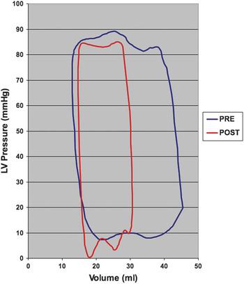
Research using such analysis has played a fundamental role in developing current concepts of cardiac pathophysiology and performance.Reference Burkhoff, Mirsky and Suga21, Reference Grossman, Braunwald, Mann, McLaurin and Green22 It provides vital information regarding systolic and diastolic cardiac performance, myocardial energetics, and ventriculovascular interactions.Reference Sugawa23 Such analysis, however, has failed to make the transition from application in research to widespread clinical use. One of the major reasons for this failure is the difficulty of acquiring data under varying conditions of ventricular loading. While accurate and effective, these methods are cumbersome and expensive. Recently, innovative techniques have been developed to derive indexes of ventricular pressure related to volume without altering the load, but using a single cardiac cycle.Reference Karunanithi and Feneley24 Our laboratory is currently working on methodology that utilizes three-dimensional echocardiography to capture left ventricular volume and mass, paired with simultaneously collected data related to pressure, and using this to provide loops during a diagnostic catheterization (Fig. 2). If the application is successful, it would transform this highly robust measurement of ventricular function into a clinically-practical modality.
Example of a three-dimensional echocardiographic pressure volume loop of the left ventricle in a patient before and after device closure of a patent arterial duct. Abbreviations: LV – left ventricular; mmHg – millimeters of mercury; ml – milliliters.

Three-dimensional transoesophageal echocardiography and image-guided interventions
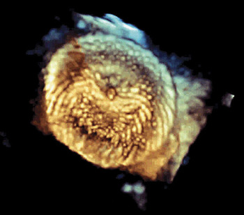
Three-dimensional echocardiography using the transoesophageal portal is a new application, albeit one with great potential in the care of patients with congenitally malformed hearts.Reference Baker, Shirali, Ringewald, Hsia and Bandisode25 We found this modality to be useful in routine diagnostic studies, as well as the perioperative setting, and also to guide trans-catheter interventions, particularly for guiding percutaneous interventions involving the atrial septum (Fig. 3). Thus far, we have been able to use the available probe in patients who weigh over 22 kg. The development of a miniaturized probe should greatly increase the practicality of this modality in the setting of congenital cardiac disease.
Three-dimensional transesophageal echocardiographic image of a device that has been placed within the oval fossa to close a large atrial septal defect. The marker band in the center of the left atrial disc can readily be identified. The image can be cropped and rotated to visualize how well the device is seated on the rims of the fossa prior to release of the device in the catheterization laboratory.

This is something to anticipate, since use of the three-dimensional approach holds great promise for guidance of cardiac interventions. Our group described its value in guiding endocardial biopsy in children.Reference Scheurer, Bandisode, Ruff, Atz and Shirali26 Another group has used it to provide intracardiac navigation during closure of atrial septal defects in a swine model. The goal of this latter approach is to permit cardiac surgery without the use of cardiopulmonary bypass.Reference Suematsu, Martinez and Wolf27
Cardiac Computed Tomographic Angiography
The commercial introduction of single slice helical scanners in 1997 initiated the current age of cardiac computed tomographic angiography. Prior to that time, specialized applications of computed tomography had provided limited advances in cardiac imaging, but had never achieved widespread acceptance. The introduction of single slice helical scanners was followed by multislice scanners, which allowed computed tomographic angiography to develop into a clinically practical imaging modality. The ensuing generations of scanners brought marked improvements in the time required for scanning, from 50 seconds to less than one second, in temporal resolution, from 0.5 seconds to 0.083 seconds, and in the thickness of the collimator slices, from 10 millimeters to 0.5 millimeters.Reference Hurlock, Higashino and Mochizuki28 As the technology improved, so did computed tomographic angiography gain considerable popularity within the field of cardiology. In adult patients, it has been shown to be an accurate and predictive imaging modality for the detection of coronary arterial disease.Reference Budoff, Dowe and Jollis29, Reference Hadamitzky, Freissmuth and Meyer30 As the technology advances, its accuracy in detecting coronary arterial lesions has continued to increase.Reference Hurlock, Higashino and Mochizuki28
While the evaluation of patients with acquired coronary arterial disease has been the major driving force behind many of the innovations, this modality has also proven to be very useful for patients with congenitally malformed hearts. Although echocardiography remains the primary modality for imaging these patients, computed tomographic angiography serves as a useful adjunct to image structures which might be poorly defined. The high spatial resolution, and isotropic nature, of the resulting datasets allows viewing in multiple cross-sectional or curvilinear planes, along with the ability for excellent three-dimensional reconstructions. This inherent three-dimensional property of computed tomographic angiography makes it useful for imaging tortuous vessels, and in defining complex anatomical relationships. With a scan time of only 1 to 5 seconds, it is possible to image most children without sedation.
Until recently, magnetic resonance imaging was the only multiplanar imaging tool available to evaluate patients with congenitally malformed hearts. While it remains an important imaging modality, it has some disadvantages in comparison to computed tomography. These include limited availability and limited technical expertise, poorer spatial resolution, a strong susceptibility to metallic artifact, contraindications in patients with pacemakers, less ability simultaneously to image airway and vascular structures, and increased need for general anaesthesia in children.Reference Garg, Powell, Sena, Marshall and Geva31, Reference Taylor32 In addition, magnetic resonance requires significantly longer periods of scanning, making it less practical in critically ill patients, infants with poor thermoregulation, or poorly cooperative children. For these reasons, multiple authors have suggested a complimentary role for cardiac computed tomography and magnetic resonance imaging in patients that require advanced cardiac imaging.Reference Taylor32, Reference Boxt33
The indications for computed tomographic angiography in patients with congenitally malformed hearts are evolving. To date, its use has been documented for a variety of cardiac lesions. We have selected several clinical situations as illustrative of its distinct benefits in children, namely vascular rings, anomalies resulting in small and tortuous vessels, such as pulmonary venous anomalies and systemic-to-pulmonary collateral arteries, congenital and acquired anomalies of the coronary arteries, and patients with metallic implants.
Vascular rings
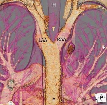
The ability of computed tomographic angiography simultaneously to image vascular structures and the airways makes it an ideal modality with which to diagnose and characterize vascular rings and pulmonary slings prior to surgical intervention.Reference Taylor32 Using this technology, in addition to detailing the precise anatomy, and providing measurements of vascular structures, the imager is able to describe and quantify involvement of the trachea or bronchuses. Commercially available software allows for automatic three-dimensional reconstructions of the airway, thus providing virtual bronchoscopy. This information allows the surgeon to plan the approach for surgical division of the ring (Fig. 4), and helps determine whether tracheal intervention will also be necessary.
Computed tomographic angiogram of a double aortic arch, viewed from behind. The spine and posterior rib cage have been cut away. This image demonstrates how both aortic arches encircle the trachea. This angiogram reveals that the left aortic arch is larger than the right, directing the surgical approach via a right thoracotomy. Abbreviations: T – trachea; LAA – left aortic arch; RAA – right aortic arch; H – head; L – left; R – right; F – foot.

Pulmonary venous anomalies and systemic-to-pulmonary collateral arteries
Congenital anomalies associated with small and tortuous vessels can be challenging to describe using echocardiography. There are at least 2 situations in which such vessels are commonly present, namely anomalous pulmonary venous connections and tetralogy of Fallot with pulmonary atresia and multiple collateral arrteries. The high resolution, large-field, three-dimensional nature of computed tomographic angiography in these settings permits characterization of the course and connections of these vessels in a much more straightforward manner. Limited spatial resolution, and a poor ratio of signal to noise in infants, makes magnetic resonance imaging problematic in these settings.Reference Taylor32 Experience with computed tomographic angiography has been well documented in both situations.Reference Ucar, Fitoz, Tutar, Atalay and Uysalel34, Reference Hayabuchi, Inoue and Watanabe35 In addition, the ability precisely to characterize the course of these vessels, and their relationship to other vascular structures and the airways, has clear potential in pre-operative planning.
Coronary arterial anomalies
Computed tomographic angiography is particularly robust for evaluating coronary arterial disease. Its use is well documented in patients with congenital anomalies, such as anomalous origin of a coronary artery, and coronary arterial fistulas.Reference Goo, Seo and Yun36, Reference Lerner, Frush and Boll37 Its use is also well documented in patients with Kawasaki disease.Reference Kwon, Oh and Han38 Quantification of myocardial perfusion has previously been a distinct advantage of magnetic resonance imaging over computed tomographic angiography. Recent investigations using newer technology, however, have validated the ability of cardiac computed tomography to characterize myocardial perfusion, possibly with greater accuracy than magnetic resonance imaging.Reference Schuleri, Centola and George39
Metallic implants
Many patients with congenitally malformed hearts require placement of metallic devices, such as coils and stents. Echocardiographic analysis of such stented vessels is challenging at best. On the other hand, computed tomographic angiography allows excellent visualization of metallic objects. Evaluation of vascular stents is highly accurate when compared to traditional angiography.Reference Eichhorn, Jourdan, Hill, Raman, Cheatham and Long40 The ability to image metallic objects is also useful when evaluating patients with vascular structures in close proximity to sternal wires who present for repeat sternotomy.
Exposure to radiation
An obvious concern when considering the use of computed tomographic angiography is the exposure to ionizing radiation. This concern is particularly relevant in children, who have a greater sensitivity to radiation.Reference Brenner, Elliston, Hall and Berdon41 While the quantification of risk resulting from diagnostic radiation exposure is controversial,Reference Alkadhi42 most experts agree that every effort should be made to minimize exposure whenever possible. The potential risks of malignant changes should be thoughtfully weighed against the inherent risks of other imaging modalities, including inadequate or delayed diagnosis, complications of invasive angiography, or increased need for sedation.
Given the increasing utility of cardiac computed tomographic angiography, a considerable amount of research has been directed towards strategies to minimize exposure to radiation without sacrificing diagnostic quality. At our institution, over nine-tenths of computed tomographic angiograms performed on children are done without cardiac gating. This results in a substantial drop in exposure to radiation. Cardiac gating is only used in patients who require detailed evaluation of coronary arterial or intracardiac anatomy. For those who require cardiac gating, newer prospective protocols, and high-pitch acquisitions, have resulted in impressive reductions in doses of radiation.Reference Achenbach, Marwan and Schepis43, Reference Blankstein, Shah and Pale44 Additional savings in dosage can be obtained by using lower power for the tube, or the settings for its current, automated algorithms to reduce dosage, and detectors of greater efficiency.Reference Budoff45, Reference Hausleiter and Meyer46 As the technology advances, and the newer strategies introduced for reducing dosage become standard, it likely that the amount of radiation needed will continue to decrease.Reference Alkadhi42
We have published our own initial strategies for reducing the dosage of radiation in children, with those imaged using our protocol of 80 kilovolts exposed to an average dose of only 1 millisievert.Reference Herzog, Mulvihill and Nguyen47 In comparison, the average person in the United States of America, is exposed to approximately 3 millisieverts of radiation each year through natural sources. Using anthropomorphic phantoms, we also compared the exposure from computed tomographic angiography to that produced when performing diagnostic catheterizations in chidlren.Reference Hlavacek, Mulvihill, Gonzalez, Shirali and Frey48 Unlike findings reported for adults, we found that the exposure to radiation from computed tomographic angiography in our patients was considerably lower than that resulting from diagnostic catheterization. In the last 3 years, we have further refined our strategies, and have expanded our protocol using low doses to all patients weighing less than 45 kilograms. As a result, most of our studies are performed with an exposure of less 0.5 millisieverts.
Conclusion
Recent innovations in the fields of three-dimensional echocardiography and cardiac computed tomographic angiography have allowed these modalities to offer clinically useful and complimentary information in the evaluation of patients with congenitally malformed hearts. Three-dimensional echocardiography offers excellent anatomical imaging and functional analysis in a manner that is easily obtained and clinically practical. Cardiac computed tomographic angiography offers robust, high-resolution, multiplanar data in patients who require additional imaging. Technological advances continue to minimize the risk due to radiation from this modality. Sustained progress in the development of both modalities will continue to improve our capabilities to diagnose and manage patients with congenitally malformed hearts.
Financial Support:
Dr Hlavacek was supported by a T32 Training grant from the National Institutes of Health (HL007710-11A1) for research related to this article.
Dr Baker was supported by a grant from the American Society of Echocardiography for research related to this article.
Dr Shirali is a consultant and member of the Advisory Board at Philips Medical Systems.