Introduction
Hippocampal activity is abnormal in schizophrenia and has been proposed as a key driver of illness pathophysiology (Grace, Reference Grace2016; Lieberman et al., Reference Lieberman, Girgis, Brucato, Moore, Provenzano, Kegeles and Small2018). Measures of basal hippocampal activity, including cerebral blood flow (CBF) and cerebral blood volume, have shown that the hippocampus is hyperactive in individuals at high risk for psychosis (Allen et al., Reference Allen, Chaddock, Egerton, Howes, Bonoldi, Zelaya and McGuire2016; Provenzano et al., Reference Provenzano, Guo, Wall, Feng, Sigmon, Brucato and Small2020; Schobel et al., Reference Schobel, Chaudhury, Khan, Paniagua, Styner, Asllani and Small2013), in the early stage of psychosis (McHugo et al., Reference McHugo, Talati, Armstrong, Vandekar, Blackford, Woodward and Heckers2019), and in chronic schizophrenia (Kawasaki et al., Reference Kawasaki, Suzuki, Maeda, Urata, Yamaguchi, Matsuda and Takashima1992; Malaspina et al., Reference Malaspina, Harkavy-Friedman, Corcoran, Mujica-Parodi, Printz, Gorman and Van Heertum2004, Reference Malaspina, Storer, Furman, Esser, Printz, Berman and Van Heertum1999; Scheef et al., Reference Scheef, Manka, Daamen, Kühn, Maier, Schild and Jessen2010; Schobel et al., Reference Schobel, Lewandowski, Corcoran, Moore, Brown, Malaspina and Small2009; Talati et al., Reference Talati, Rane, Kose, Blackford, Gore, Donahue and Heckers2014; Talati, Rane, Skinner, Gore, & Heckers, Reference Talati, Rane, Skinner, Gore and Heckers2015). Hippocampal dysfunction in schizophrenia has also been observed using task-related functional magnetic resonance imaging (fMRI). Collectively, fMRI studies suggest that hippocampal activation is decreased in schizophrenia during tasks that typically recruit the hippocampus in healthy individuals (Achim et al., Reference Achim, Bertrand, Sutton, Montoya, Czechowska, Malla and Lepage2007; Francis et al., Reference Francis, Hummer, Vohs, Yung, Liffick, Mehdiyoun and Breier2016; Ongür et al., Reference Ongür, Cullen, Wolf, Rohan, Barreira, Zalesak and Heckers2006; Ragland et al., Reference Ragland, Layher, Hannula, Niendam, Lesh, Solomon and Ranganath2017; Tamminga et al., Reference Tamminga, Thomas, Chin, Mihalakos, Youens, Wagner and Preston2012). Underlying hippocampal hyperactivity may create an environment in which neuronal resources are unavailable for task conditions where the hippocampus is engaged, resulting in decreased task activation observed with fMRI (Heckers et al., Reference Heckers, Rauch, Goff, Savage, Schacter, Fischman and Alpert1998).
Current models of psychosis pathophysiology have proposed that hippocampal dysfunction progressively worsens with illness stage (Heckers & Konradi, Reference Heckers and Konradi2015; Lieberman et al., Reference Lieberman, Girgis, Brucato, Moore, Provenzano, Kegeles and Small2018). While there is a growing body of work examining longitudinal changes in hippocampal structure over the course of psychotic illness (Haukvik et al., Reference Haukvik, Hartberg, Nerland, Jørgensen, Lange, Simonsen and Agartz2016; Ho et al., Reference Ho, Holt, Cheung, Iglesias, Goh, Wang and Zhou2017a, Reference Ho, Iglesias, Sum, Kuswanto, Sitoh, De Souza and Holt2017b; Makowski et al., Reference Makowski, Bodnar, Shenker, Malla, Joober, Chakravarty and Lepage2017; Mamah et al., Reference Mamah, Harms, Barch, Styner, Lieberman and Wang2012; Olabi et al., Reference Olabi, Ellison-Wright, McIntosh, Wood, Bullmore and Lawrie2011), there are limited longitudinal data available on hippocampal function (reviewed in González-Vivas et al., Reference González-Vivas, Soldevila-Matías, Sparano, García-Martí, Martí-Bonmatí, Crespo-Facorro and Sanjuan2019). Individuals at risk for psychosis show persistently increased hippocampal activity across 1–2-year follow-up periods (Allen et al., Reference Allen, Chaddock, Egerton, Howes, Bonoldi, Zelaya and McGuire2016, p. 201; Schobel et al., Reference Schobel, Chaudhury, Khan, Paniagua, Styner, Asllani and Small2013). Extant data in clinical psychosis come primarily from short-term studies examining the effect of antipsychotic treatment on hippocampal function. Broadly, these studies have shown that the hyperactivity observed at baseline in schizophrenia is attenuated at follow-up (Bolding et al., Reference Bolding, White, Hadley, Weiler, Holcomb and Lahti2012; Lahti, Holcomb, Weiler, Medoff, & Tamminga, Reference Lahti, Holcomb, Weiler, Medoff and Tamminga2003; Lahti, Weiler, Holcomb, Tamminga, & Cropsey, Reference Lahti, Weiler, Holcomb, Tamminga and Cropsey2009; Liddle, Lane, & Ngan, Reference Liddle, Lane and Ngan2000). Longitudinal task-related fMRI studies have consistently found reduced activation of the hippocampus in schizophrenia at the baseline assessment and at follow-up, with inter-scan intervals ranging from 6 weeks to 6 months (Bergé et al., Reference Bergé, Carmona, Salgado, Rovira, Bulbena and Vilarroya2014; Cadena et al., Reference Cadena, White, Kraguljac, Reid, Maximo, Nelson and Lahti2018; Reske et al., Reference Reske, Kellermann, Habel, Jon Shah, Backes, von Wilmsdorff and Schneider2007); but see Gurler et al. (Reference Gurler, White, Kraguljac, Ver Hoef, Martin, Tennant and Lahti2020) for an exception. To our knowledge, there have been no studies examining how hippocampal activation changes over longer time periods in individuals with a psychotic disorder diagnosis. Consequently, there is little data to support or refute the hypothesis that hippocampal dysfunction progresses over stages of psychosis.
In this study, we examined hippocampal activation during a task as a proxy measure for hippocampal hyperactivity over a 2-year period in individuals in the early stage of psychosis and healthy volunteers. The hippocampus is integrally involved in learning and memory, and hippocampal-dependent memory deficits have been well-studied in schizophrenia (Achim & Lepage, Reference Achim and Lepage2005; Aleman, Hijman, de Haan, & Kahn, Reference Aleman, Hijman, de Haan and Kahn1999). However, there are distinct functional properties found along the hippocampal anterior–posterior axis that extend beyond a role in memory encoding and retrieval. The anterior hippocampus is involved in emotion, stress, and processing the global or wholistic aspects of a stimulus, while the posterior hippocampus is preferentially responsible for spatial reasoning and processing of fine details of a stimulus (Fanselow & Dong, Reference Fanselow and Dong2010; Poppenk, Evensmoen, Moscovitch, & Nadel, Reference Poppenk, Evensmoen, Moscovitch and Nadel2013; Strange, Witter, Lein, & Moser, Reference Strange, Witter, Lein and Moser2014). The distinction between anterior and posterior hippocampal functions is particularly important in light of the growing evidence for differential impairment of these regions in schizophrenia. Hyperactivity has been primarily observed in the anterior hippocampus in clinical high-risk individuals (Provenzano et al., Reference Provenzano, Guo, Wall, Feng, Sigmon, Brucato and Small2020; Schobel et al., Reference Schobel, Chaudhury, Khan, Paniagua, Styner, Asllani and Small2013) and in early and chronic stages of psychosis (McHugo et al., Reference McHugo, Talati, Armstrong, Vandekar, Blackford, Woodward and Heckers2019; Ragland et al., Reference Ragland, Layher, Hannula, Niendam, Lesh, Solomon and Ranganath2017; Talati et al., Reference Talati, Rane, Kose, Blackford, Gore, Donahue and Heckers2014).
Recent work has identified a specific role for the anterior hippocampus in the perception and construction of scenes (Hodgetts, Shine, Lawrence, Downing, & Graham, Reference Hodgetts, Shine, Lawrence, Downing and Graham2016; Zeidman & Maguire, Reference Zeidman and Maguire2016). Individuals with schizophrenia show reduced activation of the anterior hippocampus during scene encoding (Francis et al., Reference Francis, Hummer, Vohs, Yung, Liffick, Mehdiyoun and Breier2016). Our group has recently shown that a deficit in hippocampal recruitment during scene processing in early psychosis is linked to hippocampal hyperactivity (McHugo et al., Reference McHugo, Talati, Armstrong, Vandekar, Blackford, Woodward and Heckers2019). Here, we test the hypothesis that (A) hippocampal function is impaired at illness onset and (B) declines over the course of the first 2 years of illness.
Methods
Participants
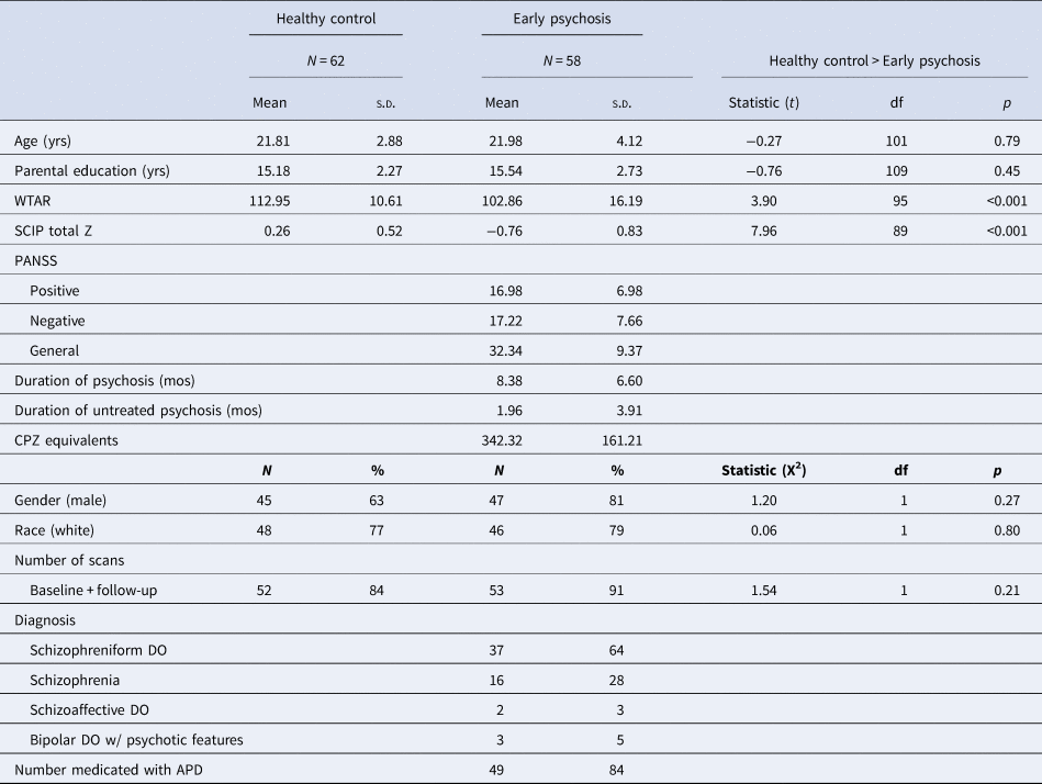
Participants (N = 147) were 76 individuals in the early stage of a psychotic disorder (EP) and 71 healthy control individuals (HC) recruited between May 2013 and February 2018 for a prospective 2-year longitudinal study on hippocampal structure and function in the early stages of psychosis (Table 1). To specifically target early pathology (Newton et al., Reference Newton, Rouleau, Nylander, Loze, Resemann, Steeves and Crespo-Facorro2018), the majority of early psychosis participants were recruited during the initial months of illness (i.e. the average duration of psychosis was approximately 8 months). Early psychosis participants were recruited from the inpatient and outpatient clinics of the Vanderbilt University Medical Center Psychiatric Hospital and healthy controls were recruited from the surrounding community through advertisements. Groups were recruited to be matched for mean age, gender, race, and parental education. Data from participants in this cohort have been included in previous reports (Armstrong, Avery, Blackford, Woodward, & Heckers, Reference Armstrong, Avery, Blackford, Woodward and Heckers2018; Avery et al., Reference Avery, McHugo, Armstrong, Blackford, Woodward and Heckers2019, Reference Avery, Armstrong, McHugo, Vandekar, Blackford, Woodward and Heckers2021, Reference Avery, McHugo, Armstrong, Blackford, Woodward and Heckers2021; McHugo et al., Reference McHugo, Talati, Woodward, Armstrong, Blackford and Heckers2018, Reference McHugo, Talati, Armstrong, Vandekar, Blackford, Woodward and Heckers2019), but the longitudinal fMRI data and analyses presented here are novel. All participants provided written informed consent and received monetary compensation for their time. The Vanderbilt University Institutional Review Board approved the study.
Participant baseline demographics and clinical characteristics

HC, healthy control; EP, early psychosis; yrs, years; mos, months; WTAR, Wechsler Test of Adult Reading; SCIP, Screen for Cognitive Impairment in Psychiatry; PANSS, Positive And Negative Symptom Scale; CPZ, chlorpromazine; APD, antipsychotic drug.
Parental education unavailable for one EP; WTAR unavailable for two EP, six HC; SCIP unavailable for two EP.
Inclusion criteria for patients were a diagnosis of schizophreniform disorder, schizophrenia, or schizoaffective disorder, with a duration of psychosis less than 2 years. Exclusion criteria for all participants included the presence of significant head injury, major medical illnesses, pregnancy, mental, claustrophobia, and current substance abuse or dependence within the past month at the time of study enrollment. Participants were excluded for data quality, including low task performance, motion, or fMRI coverage. Baseline MRI scans that passed quality control were available on 58 early psychosis and 62 control participants. Fifty early psychosis (86%) and 47 healthy control individuals (76%) completed the study. Details regarding participant attrition are included in online Supplementary Fig. S1. Early psychosis participants who completed the study did not differ from those who did not complete the study on demographic or clinical characteristics (all p's > 0.07).
Clinical and cognitive characterization
We collected clinical data during in-person interviews at baseline and at the end of the study. Psychiatric diagnoses were assessed with the Structured Clinical Interview for DSM-IV, TR [SCID (First, Spitzer, Miriam, & Williams, Reference First, Spitzer, Miriam and Williams2002)]. All data gathered during the in-person interviews were augmented by an extensive review of all available medical records. Taking into account all available information, diagnostic consensus meetings were held and final diagnoses were made by psychiatrist SH. Clinical symptoms at the time of scanning were characterized using the Positive and Negative Symptom Scale [PANSS (Kay, Fiszbein, & Opler, Reference Kay, Fiszbein and Opler1987)]. The onset of psychosis was determined through the Symptom Onset in Schizophrenia Inventory [SOS (Perkins et al., Reference Perkins, Leserman, Jarskog, Graham, Kazmer and Lieberman2000)], a standardized measure for rating prodromal v. psychotic symptoms. The duration of psychosis was calculated as the amount of time between the date of onset of psychosis (determined with the SOS) and study enrollment. The duration of untreated psychosis was calculated as the time between the date of onset of psychosis (determined with the SOS) and the date of first antipsychotic treatment. Chlorpromazine equivalents were calculated using published formulas (Gardner, Murphy, O'Donnell, Centorrino, & Baldessarini, Reference Gardner, Murphy, O'Donnell, Centorrino and Baldessarini2010; Leucht et al., Reference Leucht, Samara, Heres, Patel, Woods and Davis2014). Premorbid IQ was estimated using the Wechsler Test of Adult Reading [WTAR (Wechsler, Reference Wechsler2001)]. Cognitive function was assessed at baseline and 2-year follow-up using the Screen for Cognitive Impairment in Psychiatry [SCIP (Purdon, Reference Purdon2005)]. Clinical and cognitive characteristics of the sample are described in Table 1 and online Supplementary Table S1.
Data acquisition
Imaging data were collected at baseline and after 2 years (median time to follow-up in months: 24). We acquired a 3D T1-weighted image on one of two identical 3 T Philips Intera Achieva scanners with a 32-channel head coil (Philips Healthcare, Inc., Best, The Netherlands) at the Vanderbilt University Institute of Imaging Science (voxel size = 1 mm3; field of view = 256 mm2; number of slices = 170; gap = 0 mm; TE = 3.7 ms; TR = 8.0 ms). Each structural image was visually inspected for motion or other artifacts prior to inclusion (no images were removed). We collected 111 volumes of whole-brain fMRI data during the task with an echo planar imaging sequence (38 ascending slices, oriented at −15° relative to the intercommissural plane; voxel size = 3.0 × 3.0 × 3.2 mm; TR = 2 s; TE = 28.0 ms; flip angle = 90°). This acquisition protocol and sequence parameters were designed to maximize signal in the hippocampus and ventral brain regions.
Task fMRI
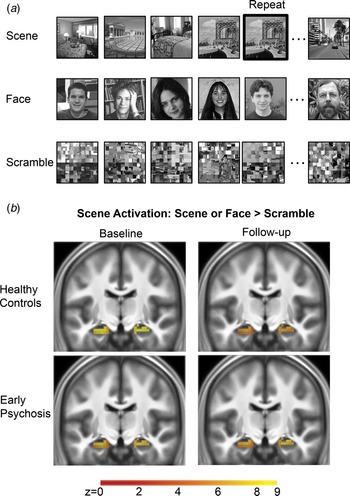
Participants completed a single run of the scene processing task during fMRI scanning at baseline and again after 2 years (Fig. 1a). The run was a block design 1-back task, composed of nine blocks of 16 scene, face, or scrambled images, separated by fixation periods. Block order was consistent across participants and at baseline and follow-up. Each image was presented for 750 ms with a 250 ms interstimulus interval. Participants were instructed to respond by buttonpress if the current image was a repeat of the immediately preceding image (0–3 target matches per block). Stimuli were black and white images that consisted of indoor or outdoor scenes, scenes featuring male or female faces, and scrambled versions of scene images. Different stimulus sets matched for the presence of indoor/outdoor scenes and male/female faces were used at baseline and follow-up. Task performance was measured using mean hit rate, correct rejection rate, and reaction time (Supplementary Methods and Results). Participants with a low hit rate or correct rejection rate (<50% in any condition) were excluded (online Supplementary Fig. S1; baseline excluded: two early psychosis and three healthy control participants; follow-up excluded: one early psychosis and two healthy control participants).
(a) Scene processing task. The same task was presented at baseline and follow-up, with unique stimuli presented at each timepoint. Participants viewed nine 16s blocks of scene, face, or scrambled images. Each block contained 16 images presented for 750 ms each, followed by a 250 ms fixation period. Participants were instructed to respond by buttonpress when an image was repeated (example indicated by bold outline). (b) Hippocampal activation in response to scenes is present in the healthy control and early psychosis groups at baseline and follow-up.

fMRI data processing and analysis
We analyzed structural and functional data with SPM12 (http://www.fil.ion.ucl.ac.uk/spm) in Matlab 2018a (Mathworks, Natick, Massachusetts, Inc.) using standard parameters. Functional images were realigned to the mean image. The structural image was then coregistered to the mean functional image, segmented, and normalized to MNI space. The realigned functional images were normalized by applying the deformation fields derived from structural image processing, then spatially smoothed with a 6 mm full-width at half-maximum Gaussian kernel. Framewise displacement of functional data for each participant was calculated using FSL's fsl_motion_outliers (http://fsl.fmrib.ox.ac.uk/fsl). Participants were excluded for gross motion (>6 mm or 4 degrees of motion) or incomplete coverage of the hippocampus based on visual inspection of the first-level masks. Data from included participants of both groups had similar levels of motion at baseline (mean framewise displacement: early psychosis = 0.15, healthy control = 0.13; t 102 = −1.21, p = 0.23) and follow-up (mean framewise displacement: early psychosis = 0.15, healthy control = 0.13; t 87 = −1.73, p = 0.09).
The first-level fMRI analysis included separate regressors for the scene, face, and scramble conditions, modeling the onset of each image in each condition with a stimulus duration = 750 ms, convolved with the canonical hemodynamic response. A high-pass filter with a cutoff of 128 s was applied. Face images used in the task were composed of people presented in the context of a background scene (Fig. 1a). As a result, we measured activation during scenes using a first-level contrast calculated as the difference in the average response to scene and face images compared to scrambled images (hereafter referred to as ‘scene’). A secondary region of interest analysis confirmed that the main findings were present in the anterior hippocampus and were observed when including only the indoor/outdoor scene condition compared to scrambled images (methods detailed in Supplementary Methods and Results).
Statistical analysis
We conducted voxelwise, region-of-interest group-level statistical analyses using the Sandwich Estimator Toolbox for SPM (SwE: http://www.nisox.org/Software/SwE/). This toolbox was designed for flexible analysis of longitudinal neuroimaging data and has been shown to provide robust control of false-positive results in unbalanced data (Guillaume et al., Reference Guillaume, Hua, Thompson, Waldorp, Nichols and Alzheimer's Disease Neuroimaging Initiative2014). We fit a model with first-level scene contrast images as the dependent variable, and dummy-coded variables representing the intercept for each group at each time point (healthy control: baseline; healthy control: follow-up; early psychosis: baseline; early psychosis: follow-up). This framework allowed us to test both aspects of our main hypothesis within a single model using all available data. Mean framewise displacement was included as a covariate to adjust for differences in motion. We conducted hypothesis tests using linear contrasts on the fitted model (described below). Analyses were carried out within a mask of the bilateral hippocampus generated in a previous study (McHugo et al., Reference McHugo, Talati, Armstrong, Vandekar, Blackford, Woodward and Heckers2019) and corrected for multiple comparisons using a voxelwise threshold of p < 0.01 and cluster corrected for p FWE = 0.05 with a non-parametric wild bootstrap method (Guillaume, Nichols, & ADNI, Reference Guillaume, Nichols and ADNI2015). This approach is not dependent on random field theory assumptions and the potential reduced control of false-positive rates (e.g. Eklund, Nichols, & Knutsson, Reference Eklund, Nichols and Knutsson2016).
To confirm hippocampal activation in response to scene processing at baseline and follow-up, we conducted separate voxelwise, one-sample t tests in SwE using the first-level scene contrast images in each group at each time point. Next, we tested whether the hippocampal function is impaired at illness onset using a two-sample t test comparing the average response at baseline to scenes in the early psychosis group to the healthy control group. We have previously shown lower hippocampal activation in early psychosis using a subset of the longitudinal cohort reported here (McHugo et al., Reference McHugo, Talati, Armstrong, Vandekar, Blackford, Woodward and Heckers2019). Consequently, this test represents a confirmation of our previous finding in the full longitudinal cohort. Finally, we tested whether the hippocampal function in early psychosis over the first 2 years of illness differs from controls using a group by time interaction t test. Follow-up tests were used to determine within-group changes in activation over time. Additional analyses of the reliability of task activation over time are reported in the supplement (Supplementary Methods and Results: Scene processing task activation reliability). We conducted exploratory analyses to examine whether scene activation was associated with clinical symptoms assessed by the PANSS, duration of psychosis, and medication load (CPZ equivalents, medication status). We used Spearman correlations to test for an association between continuous variables and percent signal change during scenes (described in Supplementary Methods) and t tests to examine differences related to medication status (yes/no).
Results
Scene processing
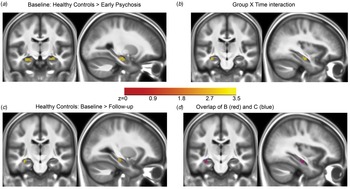
We identified robust bilateral activation of the hippocampus to scenes in both healthy control and early psychosis participants at baseline and follow-up (Fig. 1b). Location and statistics of significant clusters are reported in supplementary materials (online Supplementary Table S2). Consistent with our hypothesis, hippocampal activation during scene processing was lower in the early psychosis group at baseline (Fig. 2a). We observed a significant group by time interaction in a cluster of voxels centered in the anterior hippocampus (Fig. 2b). However, follow-up tests showed that this interaction was driven by a decrease in activation in the healthy control group from baseline to follow-up (Fig. 2c, d). No significant clusters were observed in a between-group comparison of scene activation at follow-up.
Voxelwise analyses of scene activation in a hippocampal region of interest. (a) At baseline, a between-group comparison confirms lower activation in the anterior hippocampus in early psychosis. (b) A group-by-time interaction shows that scene activation over time differs between groups. (c) Contrary to our hypothesis, the healthy control group had reduced activation at follow-up compared to baseline. (d) Simultaneous display of the thresholded maps from (b) and (c) indicates that reduced activation in healthy controls is driving the group by time interaction in the anterior hippocampus.

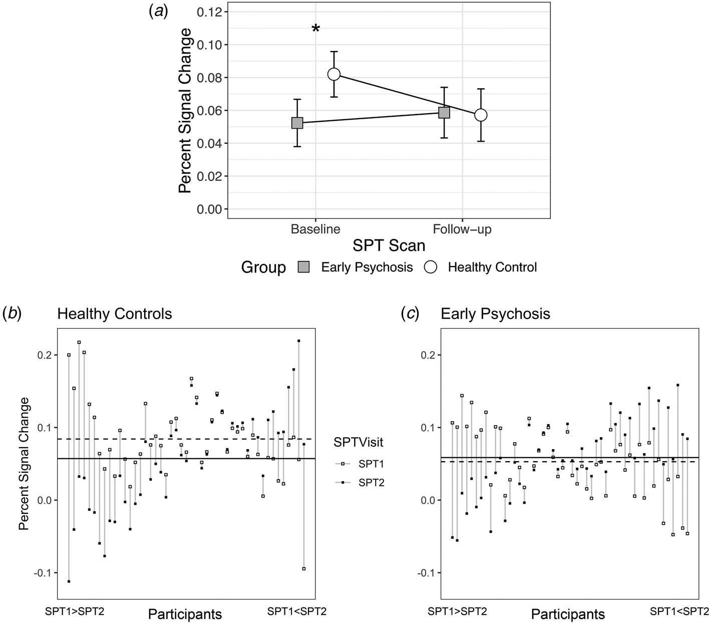
A region of interest analysis of percent signal change data from the anterior hippocampus showed a similar pattern of results (Fig. 3). We observed a group × time interaction (F 1112 = 4.24, p = 0.04) due to reduced activation in the early psychosis group only at baseline (t 212 = −2.92, p = 0.008), not at 2-year follow-up (t 212 = 0.13, p = 1.0). Contrary to our hypothesis, hippocampal activation did not decline further in the early psychosis group (t 212 = 0.59, p = 1.0), but decreased slightly over time in the healthy control group (t 212 = −2.32, p = 0.04). Our primary results did not change when using the average response to the scene-only condition, rather than the average of scene and face conditions (group × time interaction: F 1,112 = 6.02, p = 0.02).
(a) We observed lower anterior hippocampal scene activation in early psychosis participants relative to healthy individuals only at baseline, not at follow-up. Unexpectedly, this resulted from a decrease in activation over 2 years in the healthy control group rather than a change in the early psychosis group. Asterisk denotes a significant between-group post-hoc test at corrected p < 0.05. Error bars indicate the 95% confidence interval of the estimated marginal mean. Patterns of anterior hippocampal scene activation across time vary across individual participants in the healthy control (b) and early psychosis groups (c). Dashed horizontal lines indicate group mean activation at baseline (SPT1); solid horizontal lines indicate group mean activation at follow-up (SPT2). Each vertical line represents the change in activation of an individual participant from SPT1 (open square) to SPT2 (filled square). Within each group, there are a subset of individuals showing a decrease in activation from baseline to follow-up, others showing relatively stable activation between visits, and a third subset showing increased activation from baseline to follow-up.

Association of anterior hippocampal activation and clinical factors in early psychosis
We explored whether the anterior hippocampal response to scenes was associated with clinical characteristics in the early psychosis group. At baseline, anterior hippocampal scene activation was not associated with positive symptoms (r = 0.05, p = 0.73), negative symptoms (r = −0.04, p = 0.76), general psychopathology (r = −0.09, p = 0.48), duration of psychosis (r = 0.07, p = 0.58), or duration of untreated psychosis (r = 0.11, p = 0.41). Antipsychotic dosage (measured by chlorpromazine equivalents) was not related to the anterior hippocampal activation to scenes (r = −0.02, p = 0.88), and anterior hippocampal activation did not differ between medicated and unmedicated patients at baseline (t = −0.77, p = 0.52). We did not observe any relationship between clinical characteristics and anterior hippocampal activation at follow-up (all p's > 0.1; detailed statistics are provided in online Supplementary Table S3).
Discussion
Current models of psychosis propose that hippocampal function deteriorates over time. In the present study, we found that hippocampal activation during the visual processing of scenes is impaired in the early stage of a non-affective psychotic disorder and that hippocampal activation was unchanged 2 years later, while it declined in healthy control participants.
Consistent with previous studies, we found reduced recruitment of the hippocampus in individuals with psychosis at baseline (Achim et al., Reference Achim, Bertrand, Sutton, Montoya, Czechowska, Malla and Lepage2007; Francis et al., Reference Francis, Hummer, Vohs, Yung, Liffick, Mehdiyoun and Breier2016; Heckers et al., Reference Heckers, Rauch, Goff, Savage, Schacter, Fischman and Alpert1998; Ongür et al., Reference Ongür, Cullen, Wolf, Rohan, Barreira, Zalesak and Heckers2006; Ragland et al., Reference Ragland, Layher, Hannula, Niendam, Lesh, Solomon and Ranganath2017; Tamminga et al., Reference Tamminga, Thomas, Chin, Mihalakos, Youens, Wagner and Preston2012; Weiss et al., Reference Weiss, Schacter, Goff, Rauch, Alpert, Fischman and Heckers2003). The present finding replicates, in a larger sample, our previous cross-sectional result that included a subset of the individuals in the current study. This hippocampal hypoactivation is thought to represent a ceiling effect, driven by underlying hyperactivity, so that individuals with psychosis lack the available resources to recruit the hippocampus when necessary (Heckers et al., Reference Heckers, Rauch, Goff, Savage, Schacter, Fischman and Alpert1998; McHugo et al., Reference McHugo, Talati, Armstrong, Vandekar, Blackford, Woodward and Heckers2019; Weiss et al., Reference Weiss, Schacter, Goff, Rauch, Alpert, Fischman and Heckers2003). Hippocampal hyperactivity has also been reported in individuals at high risk for psychosis (Allen et al., Reference Allen, Chaddock, Egerton, Howes, Bonoldi, Zelaya and McGuire2016; Schobel et al., Reference Schobel, Chaudhury, Khan, Paniagua, Styner, Asllani and Small2013), but does not predict conversion to clinical psychosis (Provenzano et al., Reference Provenzano, Guo, Wall, Feng, Sigmon, Brucato and Small2020). Collectively, these results suggest that hyperactivity may be a significant risk factor for developing a psychotic disorder and longer-term studies are needed to understand how hippocampal dysfunction changes with progression to chronic stages of schizophrenia.
We found a significant group by time interaction in hippocampal activation, but the pattern we observed precludes a simple interpretation. In a longitudinal case–control study, the control group is included to establish the normative pattern against which cases may be compared. In the present study, activation in the healthy control group declined slightly from baseline to follow-up. In contrast, activation in the early psychosis group did not change over time. Here we discuss two potential explanations: (1) hippocampal activation habituates over time in healthy individuals but not in early psychosis and (2) hippocampal activation during scene processing does not change in the first 2 years of a non-affective psychotic disorder.
First, repeated exposure to a stimulus or task is associated with an altered response upon repetition that may manifest as habituation, sometimes termed repetition suppression, or behavioral priming. Reduced activation with task repetition (i.e. habituation) is a common finding in healthy individuals (Kim, Reference Kim2017). Although habituation is often described as a short-term form of implicit learning, long-term habituation effects are well known (Rankin et al., Reference Rankin, Abrams, Barry, Bhatnagar, Clayton, Colombo and Thompson2009). Additionally, priming effects have been observed over years (Cave, Reference Cave1997) and both perceptual and conceptual forms of priming have been described (Meister, Buelte, Sparing, & Boroojerdi, Reference Meister, Buelte, Sparing and Boroojerdi2007). Recent work has shown that habituation induced by long-term conceptual familiarity is similar in nature to the shorter-term habituation observed over minutes or days with fMRI (Poppenk, McIntosh, & Moscovitch, Reference Poppenk, McIntosh and Moscovitch2016). For the healthy control group in our study, it is likely that the reduced activation observed at follow-up reflects these types of perceptual and behavioral facilitation effects due to prior exposure to the scanner environment, task, and perceptually similar stimuli.
With the current data, we cannot disentangle the extent to which patients and controls differ in terms of habituation. However, impaired habituation has been observed in schizophrenia (Avery et al., Reference Avery, McHugo, Armstrong, Blackford, Woodward and Heckers2019; Holt et al., Reference Holt, Weiss, Rauch, Wright, Zalesak, Goff and Heckers2005; Lee et al., Reference Lee, Reavis, Engel, Altshuler, Cohen, Glahn and Green2019; Williams, Blackford, Luksik, Gauthier, & Heckers, Reference Williams, Blackford, Luksik, Gauthier and Heckers2013) and a genetic disorder associated with increased risk for psychosis (Larsen et al., Reference Larsen, Mørup, Birknow, Fischer, Olsen, Didriksen and Siebner2019), but see Kovács, Grotheer, Münke, Kéri, and Nenadić (Reference Kovács, Grotheer, Münke, Kéri and Nenadić2019) for an exception. We have previously found that greater short-term habituation in healthy controls is associated with better subsequent memory performance (Avery et al., Reference Avery, McHugo, Armstrong, Blackford, Vandekar, Woodward and Heckers2020b). In contrast, patients with schizophrenia who showed reduced habituation (i.e. a sustained response) had better subsequent memory. Recently, we have shown habituation deficits that persist over 2 years using a different task in a largely overlapping cohort (Avery et al., Reference Avery, McHugo, Armstrong, Blackford, Woodward and Heckers2021). Short- and long-term habituation are likely to rely on distinct neuronal mechanisms. Short-term habituation is thought to result from reduced excitatory neurotransmission (McDiarmid, Bernardos, & Rankin, Reference McDiarmid, Bernardos and Rankin2017) whereas long-term habituation may depend on changes in inhibitory tone within neuronal networks (Ramaswami, Reference Ramaswami2014). Current models of hippocampal hyperactivity in schizophrenia suggest the presence of altered excitatory and inhibitory signaling (Heckers & Konradi, Reference Heckers and Konradi2015; Lieberman et al., Reference Lieberman, Girgis, Brucato, Moore, Provenzano, Kegeles and Small2018; Tamminga, Southcott, Sacco, Wagner, & Ghose, Reference Tamminga, Southcott, Sacco, Wagner and Ghose2012). Collectively, these findings support the hypothesis that patients do not show the same pattern of reduced response to repeated stimulus and task exposure as healthy controls because of impairments in habituation that stem from underlying hyperactivity (Holt, Reference Holt2019). Future studies incorporating this task should include a subsequent memory test or a modified behavioral task to probe the extent to which group differences in memory impact the findings observed with longer-term task repetition.
Second, the stable pattern of hippocampal activation in the patient group is consistent with other reports that brain activation patterns are not showing progressive deterioration in the early stages of psychosis (Bergé et al., Reference Bergé, Carmona, Salgado, Rovira, Bulbena and Vilarroya2014; Cadena et al., Reference Cadena, White, Kraguljac, Reid, Maximo, Nelson and Lahti2018; Niendam et al., Reference Niendam, Ray, Iosif, Lesh, Ashby, Patel and Carter2018; Reske et al., Reference Reske, Kellermann, Habel, Jon Shah, Backes, von Wilmsdorff and Schneider2007; Smucny et al., Reference Smucny, Lesh, Zarubin, Niendam, Ragland, Tully and Carter2020). In contrast to studies of persistent deficits in patients, we observed a stable pattern in the context of declining hippocampal activation in our healthy participants. At baseline, the early psychosis group showed reduced activation compared to the healthy control group. At follow-up, the decline in activation from baseline in the healthy control group was not observed in the early psychosis group. Indeed, there was no between-group difference in activation at follow-up. In the early psychosis group, hippocampal recruitment may have improved over time, resulting in no apparent change in the longitudinal pattern of activation. Because baseline recruitment in the early psychosis group was reduced, improved hippocampal recruitment would manifest as either an increase or no change from baseline (as we observed). This interpretation is supported by the observation that clinical features (positive, negative, and general symptom scores on the PANSS, see online Supplementary Table S1) and overall cognition (SCIP scores, see online Supplementary Table S1) significantly improved in our patient cohort. Consequently, the hippocampal recruitment deficit we observed at baseline but not at follow-up may be related to the relatively greater burden of psychosis at baseline. Although we did not observe a correlation between task activation and clinical features, our ability to observe such a relationship is limited in part by the low reliability of the anterior hippocampal activation pattern.
The primary limitation of our study is that the group average trajectories of hippocampal activation do not capture the trajectories of individuals. Examination of task activation patterns over time at the individual participant level revealed substantial heterogeneity (Fig. 3b, c). In both groups, there appeared to be three patterns of activation trajectories over time: strong habituation (i.e. a decrease from baseline to follow-up), relatively stable activation from baseline to follow-up, and sensitization (i.e. an increase from baseline to follow-up). Over 2 years, the reliability of task activation was extremely low (Supplementary Methods and Results, online Supplementary Table S5; ICCs ~0). In contrast, the reliability of the task within a session was in the range commonly reported for fMRI tasks (online Supplementary Table S6; ICCs = 0.21–0.51) (Elliott et al., Reference Elliott, Knodt, Ireland, Morris, Poulton, Ramrakha and Hariri2020). Multiple factors may contribute to low fMRI reliability, even in healthy individuals, including longer length of follow-up (Bennett & Miller, Reference Bennett and Miller2010); between-subject variability in tasks that robustly activate a region (Hedge, Powell, & Sumner, Reference Hedge, Powell and Sumner2018); and low temporal SNR (Raemaekers et al., Reference Raemaekers, Vink, Zandbelt, van Wezel, Kahn and Ramsey2007).
The poor test-retest reliability of anterior hippocampal task activation over 2 years in healthy individuals raises the possibility that the observed group × time interaction reflects state-dependent effects or noise, rather than a true group effect. We explored whether low reliability was selective to the anterior hippocampus by examining the reliability and activation patterns in the posterior hippocampus and retrosplenial cortex, regions connected to the anterior hippocampus and involved in scene processing, respectively. Reliability was higher in both regions, but still low (range: 0.01–0.3; online Supplementary Table S7; Fig. S3; Supplementary Material: Scene processing task activation reliability). We observed a group × time interaction in response to scene processing in the retrosplenial cortex but not the posterior hippocampus, suggesting that the group effect we observed in the anterior hippocampus is not due to noise alone. However, an important direction for this line of research is to improve the reliability of the task by increasing the number of samples acquired in each condition and task modifications that facilitate the assessment of behavior in order to confirm our findings. Future studies are needed using state of the art multivariate fMRI methods (Kragel, Han, Kraynak, Gianaros, & Wager, Reference Kragel, Han, Kraynak, Gianaros and Wager2020), possibly in combination with resting-state fMRI data (Elliott et al., Reference Elliott, Knodt, Cooke, Kim, Melzer, Keenan and Hariri2019), CBF measures (Khalili-Mahani et al., Reference Khalili-Mahani, Rombouts, van Osch, Duff, Carbonell, Nickerson and van Gerven2017), or calibrated fMRI (Blockley, Griffeth, Simon, & Buxton, Reference Blockley, Griffeth, Simon and Buxton2013) to more fully characterize individual differences in hippocampal dysfunction in the context of an evolving psychotic disorder.
The main strengths of our study include a focus on the early stage of psychosis and high retention of individuals in both groups over the 2-year study period (>75%). There are several limitations to the present work. Our early psychosis sample was predominantly medicated at the time of baseline assessment, and we had a majority of male and white individuals. We also did not examine whether the anterior hippocampal dysfunction observed at baseline is present across psychosis spectrum disorders, including affective psychosis, or is limited to non-affective psychosis. An important task for future studies is to examine hippocampal function across a broader psychosis population. Hippocampal function is impacted by cognitive factors including attention (Aly & Turk-Browne, Reference Aly, Turk-Browne, Hannula and Duff2017) and cognitive control (Anderson, Bunce, & Barbas, Reference Anderson, Bunce and Barbas2016). Although overall accuracy was high (>93%), it is possible that attentional differences between groups may have influenced the activation observed here. Moreover, individuals in the early psychosis group had lower current cognition and lower premorbid IQ than healthy individuals. Future studies are needed to clarify the role of cognitive and prefrontal deficits on hippocampal function in psychosis. Finally, we have focused on the hippocampus because of its hypothesized role in psychosis pathophysiology. While prior evidence points to primary dysfunction within the anterior hippocampus in early psychosis (Avery et al., Reference Avery, McHugo, Armstrong, Blackford, Woodward and Heckers2019; Blessing et al., Reference Blessing, Murty, Zeng, Wang, Davachi and Goff2020; Lieberman et al., Reference Lieberman, Girgis, Brucato, Moore, Provenzano, Kegeles and Small2018; McHugo et al., Reference McHugo, Talati, Woodward, Armstrong, Blackford and Heckers2018, Reference McHugo, Talati, Armstrong, Vandekar, Blackford, Woodward and Heckers2019), additional work using tasks that are dependent on posterior hippocampal function are needed to confirm the specificity of our finding.
Our study provides novel evidence that deficits in hippocampal recruitment are already apparent in the early stage of psychosis and do not show evidence of a further decline in the first 2 years of illness. Longitudinal follow-up extending beyond the first 2 years of illness is needed to better characterize the long-term trajectories of hippocampal dysfunction in psychosis and how this might relate to outcomes.
Supplementary material
The supplementary material for this article can be found at https://doi.org/10.1017/S0033291721001318.
Acknowledgements
This work was supported by the Charlotte and Donald Test Fund, NIMH grant R01-MH70560 (Heckers), Jack Martin MD Research Professor in Psychopharmacology (Blackford), the Vanderbilt Psychiatric Genotype/Phenotype Project, the Vanderbilt Institute for Clinical and Translational Research (through grant 1-UL-1-TR000445 from the National Center for Research Resources/NIH) and the Advanced Computing Center for Research and Education at Vanderbilt University, Nashville, TN. The authors would like to thank the participants for their involvement and Xinyu Liu, Rachel McKinney, Margo Menkes, Margaret Quinn, Caitlin Ridgewell, and Katherine Seldin for their assistance in data collection.
Financial support
This work was supported by the Charlotte and Donald Test Fund, NIMH grant R01-MH70560 (Heckers), Jack Martin MD Research Professor in Psychopharmacology (Blackford), the Vanderbilt Psychiatric Genotype/Phenotype Project, the Vanderbilt Institute for Clinical and Translational Research (through grant 1-UL-1-TR000445 from the National Center for Research Resources/NIH) and the Advanced Computing Center for Research and Education at Vanderbilt University, Nashville, TN.
Conflict of interest
None.