In mammalian cells, reactive oxygen species (ROS) and reactive nitrogen species (RNS) are continuously generated as a consequence of normal cell metabolism. The superoxide radical is formed by processes mediated by enzymes such as NAD(P)H oxidases and xanthine oxidase or nonenzymatically, by redox-reactive compounds such as the semiubiquinone compound of the mitochondrial electron transport chain. Superoxide dismutases (SODs) convert superoxide into hydrogen peroxide and oxygen. In the presence of reduced transition metals (e.g. ferrous or cuprous ions), hydrogen peroxide can be converted into the highly reactive hydroxyl radical, which is capable of causing several oxidation-mediated modifications in biomolecules (Ref. Reference Chance, Sies and Boveris1). The nitric oxide radical is produced in higher eukaryotes by the oxidation of L-arginine in a reaction catalysed by nitric oxide synthases (Ref. Reference Palmer, Rees, Ashton and Moncada2). Nitric oxide itself is not particularly toxic in vivo, but it can react with superoxide to form the powerful oxidant peroxynitrite (Ref. Reference Pacher, Beckman and Liaudet3). Oxidative stress arises when the rate of ROS and RNS production exceeds their rate of clearance by antioxidant compounds and enzymes (Ref. Reference Gutteridge and Halliwell4). Owing to their high oxygen consumption and high lipid content, neural tissues are particularly sensitive to oxidative stress. It is therefore perhaps unsurprising that oxidative stress has been implicated in the pathogenesis of several neurodegenerative diseases, such as amyotrophic lateral sclerosis (ALS), Alzheimer disease (AD), Parkinson disease (PD) and Huntington disease (HD) (Refs Reference Goodall and Morrison5, Reference Halliwell6, Reference Lin and Beal7, Reference Browne and Beal8).
Phase II detoxifying and antioxidant enzymes are the primary means by which neural cells protect themselves from toxic ROS and RNS, and electrophiles (compounds with an electron-deficient center). The induction of such enzymes depends almost exclusively on de novo gene transcription, which is governed by the transcription factor nuclear factor erythroid-2-related factor 2 (Nrf2, encoded by NFE2L2) (Refs Reference Moi, Chan and Asunis9, Reference Itoh, Igarashi and Hayashi10), which acts on the electrophile response element or antioxidant response element (ARE; Ref. Reference Rushmore and Pickett11). Nrf2 activation protects neurons and astrocytes from oxidative damage in many different acute paradigms, and it is a promising therapeutic target in chronic neurodegeneration.
Nrf2 activation and regulation
Nrf2 is a redox-sensitive transcription factor and a member of a protein family characterised by the presence of a conserved basic region leucine zipper (bZip) dimerisation domain, which was first identified in the Drosophila transcription factor Cap'n'Collar [CNC (Ref. Reference Andrews, Erdjument-Bromage and Davidson12)]. The CNC–bZip protein family includes the p45 subunit of the nuclear factor erythroid 2 (NF-E2), which was originally identified as an erythroid-restricted DNA-binding protein that recognises regulatory elements in the globin gene (Refs Reference Ney, Sorrentino, Lowrey and Nienhuis13, Reference Strauss and Orkin14). Other family members are Nrf1 (NFE2L1), Nrf3 (NFE2L3), BACH1 and BACH2. CNC–bZip transcription factors must heterodimerise with members of the Maf proto-oncogene family in order to bind to regulatory elements in the DNA (Refs Reference Itoh, Chiba and Takahashi15, Reference Motohashi, O'Connor and Katsuoka16).
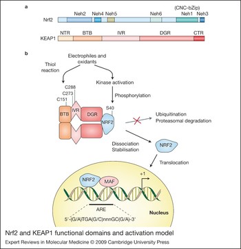
Comparison of human Nrf2 (Ref. Reference Moi, Chan and Asunis9) and the chicken homologue ECH (erythroid-derived CNC homology protein) (Ref. Reference Itoh, Igarashi and Hayashi10), led to the identification of Nrf2–ECH homology domains (Neh; Fig. 1). One of these domains (Neh2) was found to mediate the association of Nrf2 with the cytoplasmic protein KEAP1 [kelch-like ECH-associated protein 1 (Ref. Reference Itoh, Wakabayashi and Katoh17)]. When the murine Keap1 gene was disrupted, constitutive activation of Nrf2 and its target genes caused juvenile lethality as a result of hyperkeratotic lesions in the esophagus and forestomach. In vivo evidence of an interaction between Nrf2 and KEAP1 was obtained when the lethality of the Keap1-null mutation was reversed by the simultaneous knockout of Nrf2 (Ref. Reference Wakabayashi, Itoh and Wakabayashi18). The mechanism of this interaction and that of Nrf2 transactivation remains a topic of active research. It has been postulated that under basal conditions Nrf2 remains in the cytoplasm associated with the actin cytoskeleton through KEAP1 (Ref. Reference Itoh, Wakabayashi and Katoh17). In unstressed conditions, Nrf2 is rapidly degraded and displays a half-life of between 10 and 40 min (Refs Reference Itoh, Wakabayashi and Katoh17, Reference Stewart, Killeen and Naquin19, Reference Alam, Killeen and Gong20, Reference McMahon, Thomas and Itoh21). KEAP1 has been shown to function as an adaptor for the Cullin-3-based E3 ligase in the cytoplasm, where it targets Nrf2 for ubiquitin-mediated proteasomal degradation (Refs Reference Cullinan, Gordan and Jin22-Reference McMahon, Thomas and Itoh24). In the presence of ARE inducers, the Nrf2–KEAP1 interaction is suppressed, and Nrf2 translocates to the nucleus, where it dimerises with small Maf proteins to increase the transcription rate of ARE-driven genes (Fig. 1).
Nrf2 and KEAP1 functional domains and activation model. (a) Diagram representing the functional domains of Nrf2 and KEAP1. Nrf2 has six highly conserved regions Neh1–Neh6 (Nrf2-ECH homology). Neh1 contains the CNC homology region and basic leucine zipper domain (CNC-bZip). The N-terminus (Neh2) and C-terminus (Neh3) of the proteins are also highly conserved. KEAP1 binds Nrf2 at Neh2 and the serine at residue 40 (S40) is located in this domain. Additionally, there are two conserved acidic domains (Neh4 and Neh5) as well as a serine-rich conserved region (Neh6). KEAP1 presents two characteristic domains, the bricabrac, tramtrack and broad complex (BTB) domain and the double glycine repeat (DGR) domain. KEAP1 bridges the Cullin-3-based E3 ligase and Nrf2 using its BTB and the central intervening region (IVR) to bind Cul3 and its DGR to bind the Neh2 domain of Nrf2. Two additional regions are present in Nrf2: the N-terminal region (NTR) and the C-terminal region (CTR). (b) A model for KEAP1–Nrf2 interaction and activation. The BTB domain participates in KEAP1 dimerisation. Under basal conditions, Nrf2 is continuously degraded. Electrophiles and oxidants directly modify reactive cysteine residues in KEAP1, disrupting its dimerisation or KEAP1–Cul3 interaction, and ubiquitination of Nrf2 is interrupted. Alternative activated kinases can phosphorylate Nrf2 at S40 and disrupt KEAP1–Nrf2 interaction. Nrf2 then translocates to the nucleus and increases ARE-driven transcription.

Since several electrophilic ARE inducers are able to modify sulfhydryl groups by alkylation or oxidation, and because KEAP1 contains 25 cysteines that are conserved in human, mouse and rat, it was proposed that disruption of the Nrf2–KEAP1 complex might result from the direct modification of cysteine thiols in KEAP1 (Ref. Reference Dinkova-Kostova, Holtzclaw and Cole25). These cysteine residues are highly reactive to electrophiles and, at least in cell culture, Cys151, Cys273 and Cys288 are essential for proper KEAP1 function. Upon modification, an increase in Nrf2 activity is observed (Refs Reference Zhang and Hannink26, Reference Levonen, Landar and Ramachandran27, Reference Wakabayashi, Dinkova-Kostova and Holtzclaw28, Reference Yamamoto, Suzuki and Kobayashi29). In addition to direct modification of thiol groups in KEAP1, activation of several kinases in response to electrophilic or oxidative stress causes translocation of Nrf2 to the nucleus. Phosphorylation of Nrf2 at serine and threonine residues by protein kinase C, phosphatidyl-inositol 3-kinase, PKR-like endoplasmic reticulum kinase, JNK, ERK and p38 MAPK (MK14) has been reported to facilitate the release of Nrf2 from KEAP1 (Ref. Reference Kobayashi and Yamamoto30).
Interestingly, it has also been proposed that KEAP1 shuttles between the nucleus and the cytoplasm, thus limiting Nrf2 activity by escorting the transcription factor out of the nucleus (Refs Reference Sun, Zhang, Chan and Zhang31, Reference Nguyen, Sherratt and Nioi32, Reference Velichkova and Hasson33). However, the subcellular distribution of KEAP1 remains under investigation (Ref. Reference Watai, Kobayashi and Nagase34). In general, it has become clear that the major regulatory steps in Nrf2 activation are increased protein stability (by dissociating from KEAP1) and its translocation to the nucleus, prompted by post-translational modifications of Nrf2 or KEAP1. Hence, most chemical inducers increase Nrf2 protein without affecting Nrf2 mRNA levels. An exception to the above mechanism is observed with some members of the fibroblast growth factor family (FGF-1, FGF-7, FGF-10), which increase Nrf2 protein levels, at least in part, by increasing gene transcription (Refs Reference Braun, Hanselmann and Gassmann35, Reference Vargas, Pehar and Cassina36), and therefore might contribute to the activation of ARE-driven genes without any additional post-translational modification of Nrf2 or KEAP1.
Genes regulated by the Nrf2–ARE pathway
Nrf2 activation can prevent or reduce cellular damage associated with several types of injury in many different tissues and organs (Ref. Reference Lee, Li and Johnson37). Nrf2-mediated protection depends on the expression of a battery of genes with detoxification, antioxidant and anti-inflammatory capacity. Although it is clear that the coordinated expression of this group of genes has an important cytoprotective role, two components are particularly important for the prevention of oxidative-mediated damage in nervous cells: the haem oxygenase (HO) system and a group of enzymes involved in glutathione synthesis and utilisation.
Haem oxygenase
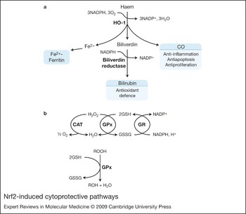
Haem oxygenase is a microsomal enzyme that, in concert with the NADPH cytochrome P450 reductase, catalyses the degradation of the haem group to yield biliverdin, iron and carbon monoxide (Refs Reference Tenhunen, Marver and Schmid38, Reference Maines39). Biliverdin is subsequently converted to bilirubin by biliverdin reductase (Fig. 2A). Two isoforms of haem oxygenase have been characterised: a constitutive isoform, HO-2 (HMOX2) and an inducible enzyme, HO-1 (HMOX1; Refs Reference Shibahara, Müller, Taguchi and Yoshida40, Reference Maines, Trakshel and Kutty41). HO-2 protein is widely expressed throughout the rodent nervous system, whereas basal HO-1 expression in the normal brain is confined to small groups of scattered neurons and glial cells (Ref. Reference Barañano and Snyder42). The induction of HO-1 is considered to counteract oxidative damage and confer cytoprotection, as suggested by studies with either deficient or increased HO-1 expression (Refs Reference Poss and Tonegawa43, Reference Yachie, Niida and Wada44, Reference Chen, Gunter and Maines45). In the nervous system, HO-1 can be highly induced in glia by its substrate haem, Nrf2 activation, and by a variety of pro-oxidant and inflammatory stimuli (Refs Reference Dwyer, Nishimura, De Vellis and Yoshida46, Reference Dwyer, Nishimura and Lu47, Reference Kitamura, Furukawa and Matsuoka48, Reference Schipper, Bernier, Mehindate and Frankel49, Reference Regan, Guo and Kumar50, Reference Mehindate, Sahlas and Frankel51, Reference Fauconneau, Petegnief and Sanfeliu52).
Nrf2-induced cytoprotective pathways. (a) Haem degradation pathway. Haem oxygenase-1 (HO-1) catalyses the degradation of the haem group to iron [Fe(II)], biliverdin and carbon monoxide (CO). Iron is sequestered by ferritin and biliverdin is subsequently converted to bilirubin by biliverdin reductase. Biliverdin and bilirubin are potent antioxidants. (b) Disposal of hydrogen peroxide (H202) and organic peroxides (ROOH) by glutathione (GSH)-utilising enzymes. In the reaction catalysed by the GSH peroxidase (GPx), GSH serves as an electron donor in the reduction of hydrogen peroxide to water, and in the reduction of organic hydroperoxides to the corresponding alcohol. The oxidised glutathione disulfide (GSSG) is then recycled by the glutathione reductase (GR) in an NADPH-consuming reaction. The peroxisomal catalase (CAT) also participates in the disposal of H202 to water and oxygen.

The products of haem degradation – biliverdin, iron and carbon monoxide – are all biologically active molecules. Induction of HO-1 might protect cells against oxidative damage by augmenting the breakdown of the pro-oxidant haem group to the radical-scavenging bile pigments, biliverdin and bilirubin (Refs Reference Stocker, Yamamoto and McDonagh53, Reference Doré and Snyder54). Furthermore, both these pigments, as well as carbon monoxide, have been shown to have potent immunomodulatory properties. Experimental observations indicate that the extent of HO-1 induction may be crucial because excessive haem degradation could result in toxic levels of carbon monoxide, bilirubin and, more importantly, iron. However, cytoprotection by HO-1 is attributable to its augmentation of iron efflux from the cell (Ref. Reference Ferris, Jaffrey and Sawa55). Moreover, in many tissues, coinduction of ferritin provides a ‘sink’ for the excess of intracellular iron. In some situations, haem-derived iron and carbon monoxide might exacerbate intracellular oxidative stress and cellular injury by promoting free radical generation within the mitochondrial compartment (Refs Reference Song, Su, Song, Paudel and Schipper56, Reference Schipper57). Increased HO-1 expression has been reported in neurodegenerative diseases involving glial activation, such as AD, PD, multiple sclerosis (Refs Reference Takeda, Itoyama and Kimpara58, Reference Schipper59, Reference van Horssen, Schreibelt and Drexhage60) and ALS (Refs Reference Vargas, Pehar and Cassina36, Reference Ferrante, Shinobu and Schulz61). Although the cytoprotective role of HO-1 is not yet completely understood, it is likely that the elevated HO-1 levels represent an attempt to restore the redox homeostasis and attenuate inflammation (Ref. Reference Cuadrado and Rojo62).
Glutathione
SOD activity is a major defence mechanism that combats oxygen toxicity by converting superoxide to hydrogen peroxide. The efficacy of SOD as an antioxidant relies on the decomposition of hydrogen peroxide by catalase (CAT) and glutathione peroxidase (GPx) to prevent its conversion to the hydroxyl radical. The peroxisomal CAT quickly decomposes hydrogen peroxide to water and molecular oxygen, but specific activity of CAT in the brain is much lower than in peripheral tissues (Refs Reference Ho, Magnenat and Bronson63, Reference Ho, Xiong, Ma, Spector and Ho64). GPx (a selenium-dependent enzyme) probably has a major role in disposing of hydrogen peroxide and organic hydroperoxides in neural tissue. Since there is no CAT in mitochondria, glutathione is particularly important for peroxide detoxification in this organelle during normal or pathological conditions. Although mitochondrial glutathione comprises only about 10% of the total glutathione in the cell, it represents a distinctly regulated pool with major implications for antioxidant protection (Ref. Reference Lash65).
In the reaction catalysed by GPx, the tripeptide glutathione [γ-l-glutamyl-l-cysteinylglycine (GSH)] serves as an electron donor in the reduction of hydrogen peroxide to water and organic hydroperoxides to the corresponding alcohol (Ref. Reference Dringen, Pawlowski and Hirrlinger66). The oxidised GSH – glutathione disulfide (GSSG) – is then recycled by glutathione reductase in an NADPH-consuming reaction (Fig. 2B). In addition to its role in peroxide detoxification, GSH functions as the main cysteine storage in the cell, maintains the cellular redox homeostasis (sustaining the thiol status of proteins) and is a key component in the detoxification of xenobiotics and their metabolites through GSH conjugation. The glutathione S-transferases (GSTs) form a group of multigene isoenzymes involved in the cellular detoxification of both xenobiotic and endobiotic compounds (Ref. Reference Hayes, Flanagan and Jowsey67). Catalysis occurs by conjugation with GSH and the less toxic and more hydrophilic conjugated products are then eliminated from the cell by the transmembrane multidrug resistance-associated proteins (MRPs). In particular, MRP1 and MRP2 can export GSH conjugates to the extracellular compartment (Ref. Reference Cole and Deeley68).
GSH is synthesised by the consecutive action of the enzymes glutamate cysteine ligase (GCL) and glutathione synthase. GCL is the rate-limiting enzyme in GSH synthesis and it is feedback inhibited by GSH (Ref. Reference Meister69). Nrf2 regulates the basal and inducible levels of GCL-modifier subunit (GCLM), GCL-catalytic subunit (GCLC), glutathione synthase and glutathione reductase (Refs Reference Moinova and Mulcahy70, Reference Wild, Moinova and Mulcahy71, Reference Chan and Kwong72, Reference Harvey, Thimmulappa and Singh73); hence Nrf2 activation might regulate GSH homeostasis by affecting de novo synthesis and/or GSH redox state. Nrf2 also regulates the expression of several GSTs (Ref. Reference Chanas, Jiang and McMahon74), MRP1 (Ref. Reference Hayashi, Suzuki, Itoh, Yamamoto and Sugiyama75) and the cysteine–glutamate exchange transporter that maintains intracellular GSH levels by regulating cysteine influx (Refs Reference Sasaki, Sato and Kuriyama-Matsumura76, Reference Shih, Erb and Sun77). Following Nrf2 activation, the cell will experience an increase in the enzymes involved in GSH synthesis and utilisation, which will have a major role in resistance to oxidative stress. Remarkably, even in the absence of a net increase in the amount of GSH, in a cell with enough redox potential (a supply of NADPH), the increase in glutathione reductase and GSH-utilising enzymes induced by Nrf2 activation should still provide increased antioxidant defences.
Nrf2 in cell survival
The cytoprotective effect of Nrf2 expression has been studied in many different cell models. Nrf2-deficient cells are more sensitive to peroxides, nitric oxide, mitochondrial toxins, endoplasmic reticulum stress and glucose deprivation (Ref. Reference Lee, Li and Johnson37). Although Nrf2-knockout mice develop normally, aged females develop a systemic autoimmune disease (Refs Reference Yoh, Itoh and Enomoto78, Reference Li, Stein and Johnson79), and it is possible that Nrf2 has an important role in modulating the innate immunological response (Ref. Reference Thimmulappa, Lee and Rangasamy80). In vivo, electron paramagnetic resonance analyses of the disappearance of the spin probe Carbamoyl-PROXYL in Nrf2-knockout mice demonstrated a primary decrease of tissue-reducing activity and thus an impaired capacity to deal with electrophiles and ROS detoxification (Ref. Reference Hirayama, Yoh and Nagase81). Consequently, Nrf2-deficient mice are more sensitive to pulmonary inflammatory diseases, chemical hepatotoxicity and carcinogenesis, and chemopreventive agents have no or reduced efficacy (Ref. Reference Kensler, Wakabayashi and Biswal82). It has also been proposed that the decline in the cellular defences against oxidative, toxicological and pathological insults associated with ageing is due to a decrease in Nrf2-mediated GCL expression (Ref. Reference Suh, Shenvi and Dixon83). However, this observation was restricted to changes in hepatic GSH levels and it remains to be determined whether this phenomenon is relevant to other tissues.
Nrf2-dependent GSH levels regulate the sensitivity of the cells to Fas and TNF-α-induced apoptosis (Refs Reference Kotlo, Yehiely and Efimova84, Reference Morito, Yoh and Itoh85). There is an inverse correlation between the intracellular levels of glutathione and the ability of the receptor to initiate apoptotic downstream signaling pathways, suggesting that GSH levels regulate the sensitivity of the cell to death-receptor signals. In motor neurons, apoptosis induced by p75NTR (another member of the death receptor family, also known as TNFRSF16) or mediated by Fas, involves increased production of ROS and RNS (Refs Reference Raoul, Estévez and Nishimune86, Reference Pehar, Cassina and Vargas87). Accordingly, increased GSH levels mediated by Nrf2 activation completely prevent p75NTR- and Fas-mediated motor neuron apoptosis (Ref. Reference Pehar, Vargas and Robinson88).
Role of the astrocyte–neuron interaction in neuroprotection
In the presence of astrocytes, neurons are more resistant to the oxidative stress induced by several compounds (Refs Reference Bronstein, Perez-Otano and Sun89, Reference Desagher, Glowinski and Premont90, Reference Tanaka, Toku and Zhang91). GSH levels are key in this observation, because GSH depletion in cocultures caused the loss of astrocyte-mediated neuroprotection (Refs Reference Drukarch, Schepens and Jongenelen92, Reference Gegg, Clark and Heales93). Astrocytes have more efficient GSH synthesis systems and higher GSH content than neurons (Refs Reference Sagara, Miura and Bannai94, Reference Sagara, Makino and Bannai95, Reference Dringen96, Reference Sun, Shih and Johannssen97). One of the reasons for this difference resides in the fact that Nrf2-driven genes are preferentially activated in astrocytes over neurons (Refs Reference Lee, Calkins and Chan98, Reference Shih, Johnson, Wong, Kraft, Jiang, Erb, Johnson and Murphy99, Reference Kraft, Johnson and Johnson100). Consequently, astrocytes have more enzymes involved in GSH synthesis (GCL and GS), and can better increase its production and release to the extracellular medium upon stimulation. The coordinated increase in GSH biosynthesis and release is a fundamental component in the neuronal protection conferred by Nrf2 activation in astrocytes (Refs Reference Shih, Johnson, Wong, Kraft, Jiang, Erb, Johnson and Murphy99, Reference Kraft, Johnson and Johnson100, Reference Vargas, Pehar and Cassina101).
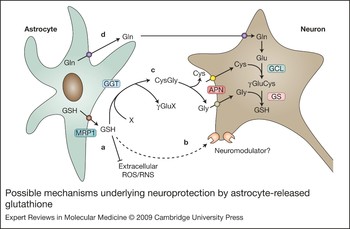
Neurons cannot take up GSH, so how can an Nrf2-mediated increase in GSH release from astrocytes protect neighbouring neurons? GSH will react nonenzymatically with a variety of radicals, and therefore might function as a first line of defence against ROS and RNS in the extracellular space (Ref. Reference Drukarch, Schepens and Stoof102). Because GSH possesses unique and specific binding sites in membranes of both neurons and astrocytes, it has also been proposed that GSH might be a glia-derived neuromodulator and/or neurotransmitter (Refs Reference Guo and Shaw103, Reference Janáky, Ogita and Pasqualotto104, Reference Hermann, Varga and Oja105). However, the best understood protective mechanism is that the increased secretion of GSH by astrocytes boosts the content of GSH in the cocultured neurons (Refs Reference Bolaños, Heales and Peuchen106, Reference Dringen, Pfeiffer and Hamprecht107).
Cysteine is extremely unstable extracellularly and rapidly auto-oxidises to cystine (Ref. Reference Meister108). Because neurons cannot use cystine, they must rely on the presence of cysteine as a GSH precursor (Ref. Reference Dringen, Pfeiffer and Hamprecht107). Extracellular GSH is the most important source of cysteine for neurons. Most cells readily take up cysteine, and the availability of cysteine determines the level of neuronal GSH (Ref. Reference Dringen, Pfeiffer and Hamprecht107). GSH and GSH-conjugates are exported from astrocytes by MRP1 (Refs Reference Hirrlinger, König and Keppler109, Reference Hirrlinger and Dringen110, Reference Minich, Riemer and Schulz111) and the release of GSH from astrocytes is probably the process that consumes most of the intracellular GSH. Glutamate and cysteine in GSH are linked through the γ-carboxyl group of glutamate rather than the α-carboxyl group. This unconventional link is subject to hydrolysis by γ-glutamyltranspeptidase (GGT), which is present on the external surfaces of astrocytes (Refs Reference Dringen, Pfeiffer and Hamprecht107, Reference Dringen, Kranich and Hamprecht112). GGT produces the dipeptide cysteinyl-glycine, which can then be hydrolysed by aminopeptidase N (Ref. Reference Dringen, Gutterer, Gros and Hirrlinger113) to release cysteine and glycine. Astrocytes also release glutamine, and hence make available all the necessary precursors for neuronal GSH synthesis (Fig. 3). Although all the data supporting this working model come from cell culture systems, we have recently provided in vivo evidence that modification of the astrocytic glutathione system might help surrounding neurons to cope with noxious stimuli (Ref. Reference Vargas, Johnson and Sirkis114). Transgenic overexpression of Nrf2 in astrocytes resulted in a ~25% increase in glutathione content in the spinal cord and protected motor neurons from the toxicity of ALS-linked mutant SOD1 (see below).
Possible mechanisms underlying neuroprotection by astrocyte-released glutathione. At least three different mechanisms could account for the neuroprotective effect of increased glutathione (GSH) release from astrocytes. (a) First, GSH reacts nonenzymatically with a variety of radicals and might function as a first line of defence against ROS or RNS in the extracellular space. (b) Second, it has been proposed that GSH might be a glia-derived neuromodulator and/or neurotransmitter. (c) Third, released GSH is hydrolysed by the γ-glutamyltranspeptidase (GGT) present on the external surfaces of astrocytes. The GGT produces the dipeptide cysteinyl-glycine (CysGly), which then can be hydrolysed by the aminopeptidase N (APN) to release cysteine and glycine. (d) Astrocytes also release glutamine and hence make available all the necessary precursors for neuronal GSH synthesis. X, any acceptor of the γ-glutamyl moiety; MRP1, multidrug-resistance-associated protein 1; GCL, glutamate cysteine ligase; GS, glutathione synthase.

Nrf2-mediated neuroprotection
Although the role of oxidative stress in the etiology of neurodegenerative diseases remains controversial, it is clear that oxidative stress markers (e.g., protein oxidation and nitration, lipid peroxidation and nucleic acid oxidation) are found in many neurodegenerating tissues (Refs Reference Browne and Beal8, Reference Ischiropoulos and Beckman115, Reference Nunomura, Moreira and Lee116). Oxidative stress might occur as the primary pathological mechanism or as a secondary event, originating from insults such as mitochondrial dysfunction and excitoxicity. In addition, antioxidant defences decrease with ageing, which is the greatest risk factor for neurodegenerative diseases. In consequence, much attention has been given to Nrf2 as a promising therapeutic target to combat oxidative stress in neurodegeneration (Refs Reference van Muiswinkel and Kuiperij117, Reference Calkins, Johnson and Townsend118, Reference de Vries, Witte and Hondius119, Reference Johnson, Johnson and Kraft120).
Amyotrophic lateral sclerosis
ALS is the most common adult-onset motor neuron disease, caused by the progressive degeneration of motor neurons in the spinal cord, brainstem and motor cortex (Ref. Reference Rowland and Shneider121). The aetiology of most ALS cases remains unknown (sporadic ALS); however, ~10% are inherited in a dominant manner (familial ALS). Both forms of ALS share the same pathological features, including progressive muscle weakness, atrophy and spasticity. ALS is usually fatal within 3–5 years of onset because of denervation of the respiratory muscles and diaphragm. Approximately 10–20% of familial ALS is caused by a toxic gain-of-function induced by mutations in the Cu/Zn-superoxide dismutase (SOD1) (Ref. Reference Rosen, Siddique and Patterson122). Rodents overexpressing mutated forms of human SOD1 generally develop an ALS-like phenotype (Refs Reference Gurney, Pu and Chiu123, Reference Howland, Liu and She124). Several hypotheses, including oxidative stress, glutamate excitotoxicity, formation of high molecular weight aggregates, defective axonal transport, decreased trophic support and mitochondrial dysfunction have all been proposed to explain the toxic effect of mutated human SOD1 (Refs Reference Beckman, Estévez, Crow and Barbeito125, Reference Cleveland and Rothstein126, Reference Bruijn, Miller and Cleveland127, Reference Manfredi and Xu128).
Although the molecular mechanism underlying the selective death of motor neurons remains unknown, toxicity requires the expression of mutant human SOD1 in non-neuronal cells, as well as in motor neurons. The proposed non-cell-autonomous mechanism of the disease suggests that, although the expression of the mutant enzyme in motor neurons affects disease onset (Ref. Reference Boillée, Yamanaka and Lobsiger129), expression within the glial compartment influences disease progression (Refs Reference Boillée, Yamanaka and Lobsiger129, Reference Beers, Henkel and Xiao130, Reference Yamanaka, Chun and Boillee131). Whether it is a primary etiological component or a secondary event, it is clear that oxidative stress occurs in ALS, and probably affects the course of the disease (Refs Reference Pacher, Beckman and Liaudet3, Reference Goodall and Morrison5, Reference Ischiropoulos and Beckman115). Both nitric oxide and superoxide are produced by motor neurons in response to apoptotic stimuli, such as trophic factor deprivation and the activation of Fas and p75NTR pathways (Refs Reference Raoul, Estévez and Nishimune86, Reference Pehar, Cassina and Vargas87, Reference Estévez, Spear and Manuel132). Additionally, in ALS patients and rodent models, a strong glial reaction typically surrounds degenerating motor neurons and oxidative markers are present in these cells, as well as in motor neurons (Ref. Reference Barbeito, Pehar and Cassina133).
Recently Sarlette and co-workers (Ref. Reference Sarlette, Krampfl and Grothe134) reported a reduction of expression of Nrf2 mRNA and protein in neurons from primary motor cortex and spinal cord from ALS postmortem tissue samples. A similar decrease in mRNA encoding Nrf2 was observed in embryonic motor neurons isolated from ALS model rats (Ref. Reference Pehar, Vargas and Robinson88). However, no altered expression of Nrf2 or ARE-driven genes was found in laser-capture microdissected motor neurons from ALS model mice (Ref. Reference Ferraiuolo, Heath and Holden135). Despite these apparent discrepancies, it is clear that there is an increase in ARE-driven genes in the spinal cord of animal models for ALS upon disease onset. A strong increase in HO-1 was found at onset in model rats (Ref. Reference Vargas, Pehar and Cassina36), whereas a direct assessment of ARE activation was obtained by mating the model mice with mice transgenic for ARE-driven human placental alkaline phosphatase activity (Ref. Reference Kraft, Resch, Johnson and Johnson136). This protective response might prevent neuronal degeneration in early stages of the disease, but it is overwhelmed by other mechanisms that drive apoptosis.
Transgenic mice expressing mutant human SOD1 exclusively in astrocytes displayed astrocytosis but failed to develop the disease (Ref. Reference Gong, Parsadanian and Andreeva137). However, astrocytes isolated from rats (Ref. Reference Vargas, Pehar and Cassina101) or mice (Refs Reference Vargas, Johnson and Sirkis114, Reference Nagai, Re and Nagata138) expressing mutated human SOD1G93A are toxic to cocultured wild-type motor neurons. Overexpression of Nrf2 completely reverses the toxicity of ALS astrocytes to motor neurons in vitro. Moreover, overexpression of Nrf2 under the control of the astrocyte-specific promoter for the glial fibrillary acidic protein delays onset and increases the lifespan in ALS mouse models (Ref. Reference Vargas, Johnson and Sirkis114). These results suggest that the astrocytic Nrf2-mediated increase in GSH content and secretion does not directly modify the toxic component of astrocytes but instead improves the ability of motor neurons to cope with them. Increased glutathione secretion from astrocytes might also alter the way in which they interact with other cells and contribute to the decreased microglial response observed (Ref. Reference Vargas, Johnson and Sirkis114). This was the first in vivo evidence that Nrf2 activation in astrocytes can be beneficial to protect neurons in chronic neurodegeneration and a proof-of-principle demonstrating that Nrf2 activation should be a suitable therapeutic target in ALS. In addition, since Nrf2 activation in motor neurons critically modulates apoptotic pathways (Ref. Reference Pehar, Vargas and Robinson88), the direct activation of Nrf2 in neurons deserves further investigation in the context of ALS.
Parkinson disease
PD is associated with progressive loss of dopaminergic neurons in the substantia nigra pars compacta, which project to the striatum, as well as with more widespread neuronal changes that cause complex and variable motor and nonmotor symptoms (Ref. Reference Thomas and Beal139). Decreased activity of mitochondrial complex I, as well as increased lipid, protein and DNA oxidation, are found in individuals affected with PD (Refs Reference Alam, Daniel and Lees140, Reference Alam, Jenner and Daniel141, Reference Dexter, Holley and Flitter142). Studies of familial PD have revealed a prominent role for mitochondrial dysfunction in the pathogenesis of the disease, and impaired mitochondrial function increases oxidative stress (Refs Reference Valente, Abou-Sleiman and Caputo143, Reference Clements, McNally and Conti144). In many cases, Nrf2 status has been inferred from ARE-driven gene expression and distribution. The enzyme NAD(P)H:quinone oxidodreductase (NQO1) catalyses the two-electron reduction of quinones, preventing their participation in redox cycling and subsequent generation of ROS. NQO1 is a prototypical ARE-driven gene, which, along with HO-1, is strongly upregulated in glial cells in postmortem PD brain (Refs Reference van Muiswinkel, de Vos and Bol145, Reference Schipper146). Nrf2 distribution is mainly nuclear in parkinsonian nigral neurons – a pattern that is consistent with a neuronal response to increased oxidative stress (Ref. Reference Ramsey, Glass and Montgomery147).
Nrf2 activation protects against the neurotoxic dompamine analogue 6-hydroxydopamine and 1-methyl-4-phenyl-1,2,3,6-tetrahydropyridine (MPTP) in vitro (Refs Reference Yamamoto, Sawada and Izumi148, Reference Jakel, Townsend, Kraft and Johnson149, Reference Wruck, Claussen and Fuhrmann150). Moreover, MPTP administration resulted in a greater loss of dopamine transporter levels in the striatum of Nrf2-knockout mice (Ref. Reference Burton, Kensler and Guilarte151), whereas intrastriatal transplantation of Nrf2-overexpressing astrocytes protects against 6-hydroxydopamine in mice (Ref. Reference Jakel, Townsend, Kraft and Johnson149). Nrf2 deficiency also increases MPTP sensitivity. Furthermore, Nrf2 overexpression restricted to astrocytes in a Nrf2-knockout background is sufficient to completely protect against MPTP toxicity (Ref. Reference Chen, Vargas and Pani152), indicating that modulation of the Nrf2–ARE pathway in astrocytes represents a suitable therapeutic approach to reduce or prevent neuronal degeneration in PD.
Alzheimer disease
AD is a complex and genetically heterogeneous disease, characterised by progressive memory deficits, cognitive impairment and personality changes, which are accompanied by specific structural abnormalities in the brain. Loss of neurons and synapses in the neocortex, hippocampus and other subcortical regions of the brain are common features of AD (Ref. Reference Bertram and Tanzi153). The main histological features of AD are extracellular protein deposits termed β-amyloid (or senile) plaques, β-amyloid deposits in blood vessels and intraneuronal neurofibrillary tangles. The β-amyloid peptide is organised in fibrils intermixed with non-fibrillar forms of the peptide and forms the major component of the amyloid deposits (Ref. Reference Bertram and Tanzi153). Altered metal biology, mitochondrial dysfunction and increased ROS and RNS production by reactive glial cells are believed to contribute to the oxidative damage in lipids, proteins and DNA observed in AD (Refs Reference Marshak, Pesce, Stanley and Griffin154, Reference Smith, Harris, Sayre and Perry155, Reference Schipper, Bennett and Liberman156, Reference Wang, Su and Perry157, Reference Shaftel, Griffin and O'Banion158). In postmortem temporal cortex and hippocampus of patients with AD, the percentage of astrocytes expressing HO-1 is significantly greater than in non-demented individuals (Ref. Reference Schipper, Bennett and Liberman156). Moreover, NQO1 activity and expression is increased in neurons and astrocytes in AD (Refs Reference Raina, Templeton and Deak159, Reference Wang, Santa-Cruz, DeCarli and Johnson160, Reference SantaCruz, Yazlovitskaya and Collins161). However, Nrf2 immunostaining was found to be predominantly cytoplasmic in hippocampal neurons from individuals with AD (Ref. Reference Ramsey, Glass and Montgomery147), unlike the nuclear distribution in dopaminergic neurons in PD. This differential distribution of Nrf2 in AD and PD, might reflect a difference in the way that affected neurons in these diseases respond to oxidative stress.
In a mouse model of AD (APP/PS1 mice) the decrease in the expression of mRNA encoding Nrf2, NQO1, GCLC and GCLM correlates with an increased accumulation of β-amyloid deposits (Ref. Reference Kanninen, Malm and Jyrkkänen162). Recently, the GSH:GSSG ratio was found to be reduced in brain homogenates in a different mouse model of AD (3 × Tg-AD; Ref. Reference Resende, Moreira and Proença163). In addition, Nrf2 activation protects neural cells against β-peptide-induced neurotoxicity in vitro (Refs Reference Kanninen, Malm and Jyrkkänen162, Reference Wruck, Götz and Herdegen164), suggesting that activation of the Nrf2–ARE pathway might be beneficial in the context of AD pathology.
Huntington disease
HD is an autosomal dominant neurodegenerative disorder that results from a polyglutamine repeat expansion in the first exon of the huntingtin gene (HTT). HD is characterised by the progressive development of involuntary choreiform movements, cognitive impairment, neuropsychiatric symptoms and premature death (Ref. Reference Browne and Beal8). Neuronal degeneration is mainly observed in the striatum and cerebral cortex, and it is believed to be due to the effect of mutant huntingtin on mitochondrial function and energy metabolism, but the exact mechanism remains unknown. Severe deficits in mitochondrial complex II, III and IV activity were observed in the striatum of postmortem HD brains (Refs Reference Brennan, Bird and Aprille165, Reference Gu, Gash and Mann166), and reduced complex IV activity, together with elevated nitric oxide and superoxide production, has been observed in one genetic mouse model of HD (Refs Reference Tabrizi, Workman and Hart167, Reference Bogdanov and Andreassen168). Although, there is no available information on the status of the Nrf2–ARE pathway in HD, the protective effect of Nrf2 activation has been demonstrated in models of mitochondrial complex II toxicity, where ROS production increases upon disruption of the electron transport chain. Mitochondrial complex II inhibition with 3-nitropropionic acid (3NP) or malonate produces characteristic striatal degeneration, which is similar to that observed in HD (Refs Reference Schapira169, Reference Brouillet, Condé, Beal and Hantraye170). Nrf2-knockout mice are more sensitive to the striatal lesions caused by administration of these inhibitors (Refs Reference Shih, Imbeault and Barakauskas171, Reference Calkins, Jakel and Johnson172). Chemical activation or viral overexpression of Nrf2 significantly reduces lesion size caused by 3NP (Ref. Reference Shih, Imbeault and Barakauskas171) and intrastriatal transplantation of Nrf2-overexpressing astrocytes has a remarkable neuroprotective effect against malonate toxicity in vivo (Ref. Reference Calkins, Jakel and Johnson172).
In addition to its role in neurodegenerative diseases, Nrf2 activation protects neurons from ischaemia and intracerebral haemorrhage (Refs Reference Shih, Li and Murphy173, Reference Zhao, Kobori, Aronowski and Dash174, Reference Zhao, Sun and Zhang175, Reference Wang, Fields and Zhao176). Interestingly, in the case of ischaemia or reperfusion injury, activation of Nrf2 and increased expression of HO-1, mainly in neurons and not astrocytes, seems to be central to the observed protection (Refs Reference Satoh, Okamoto and Cui177, Reference Satoh, Kosaka and Itoh178). Moreover, Nrf2 activation attenuates the disruption in the blood–brain barrier observed in traumatic brain injury, by preventing the loss of tight junction proteins and endothelial cell death (Refs Reference Zhao, Moore, Redell and Dash179, Reference Zhao, Moore, Clifton and Dash180). Since neuronal function is intimately linked to vascular function, it might be necessary to activate Nrf2 in endothelial, glial and neuronal cells, to effectively protect the neurovascular unit and achieve significant protection against trauma and neurodegenerative diseases.
Nrf2 activation through an increase in GSH also helps cells to cope with the oxidative stress associated with viral infection of the central nervous system (CNS) (Refs Reference Qiang, Kuang and Liu181, Reference Jiang, Scofield and Yan182). Although microglial production of intracellular and extracellular ROS is intrinsic to the role of microglia in the CNS, persistent ROS and cytokine release will result in chronic neuroinflammation and neuronal damage. Neuroinflammation is a hallmark of viral infection, ischaemia and trauma, and the role of Nrf2 – in particular the role of the astrocytic Nrf2–ARE pathway – in modulating the magnitude and duration of the microglial response is a very interesting possibility (Refs Reference Min, Yang and Kim183, Reference Innamorato, Rojo and García-Yagüe184).
Conclusions and outstanding questions
It is clear that Nrf2 activation represents an exceptional defence mechanism for many cell types. Particularly in the CNS, astrocytic Nrf2 activation has a major role in protecting neurons from noxious stimuli. Many laboratories, including ours, have generated a considerable amount of data firmly indicating the potential therapeutic value of Nrf2 activation in acute lesions of the CNS. It is likely that more evidence of the beneficial role of Nrf2 activation in chronic neurodegeneration will soon be available. Although potent activators of Nrf2 are available, the therapeutic challenge resides in identifying chemicals that, in addition to their role in activation of Nrf2, can efficiently penetrate the blood–brain barrier to reach astrocytes. In cell-replacement therapies, the intrinsic complications of replacing highly specialised cells such as neurons, make astrocytes a much more appealing target. Glial-restricted precursors (GRPs) are an exciting candidate for this purpose. However, in the context of a chronic neurodegenerative disease, transplantation of GRPs that go on to produce astrocytes might not be enough; Nrf2-overexpressing GRPs might offer a much better solution. Because of the extraordinary capacity of astrocytes to activate the Nrf2–ARE pathway, most of the research so far has concentrated on this particular aspect of Nrf2 protection. Although the exact mechanism governing astrocytic Nrf2-mediated neuroprotection is still under investigation, enough data exists to support the proof-of-principle for this concept and the challenge ahead is to determine the role of neuronal Nrf2 activation in neurodegeneration.
Acknowledgements and Funding
We thank the reviewers for their comments and suggestions. This work was supported by grants from the ALS Association and the Robert Packard Center for ALS Research at Johns Hopkins, ES08089 and ES10042 from the National Institute of Environmental Health Sciences (NIEHS) to J.A.J. M.R.V. is a recipient of the Milton Safenowitz postdoctoral fellowship for ALS research.