INTRODUCTION
Among the species of the order Dendrochirotida Grube, Reference Grube1840, members of the family Psolidae Burmeister, Reference Burmeister1837 are characterized by the presence of large overlapping scales which cover dorsally and laterally their body; ventral surface is modified into a sole; dorsal papillae are absent; mouth and anus are dorsal; and presence of 10 oral tentacles (O'Loughlin & Whitfield, Reference O'Loughlin and Whitfield2010; Smirnov, Reference Smirnov2012). These holothurians are mainly epibenthic suspension feeders (Smith, Reference Smith1962; Gutt, Reference Gutt1991).
To date, seven species of the genus Psolus Oken, Reference Oken1815 have been reported for the North-east Atlantic Ocean. Previous studies from different oceanographic cruises carried out in the Bay of Biscay, reported holothurian data from the continental shelf and slope, i.e. ‘Caudan’, ‘Thalassa’, ‘BIOGAS’, ‘Hespérides 76’, ‘Cantábrico 83’, ‘CAP-89’, ‘COCACE’ or ‘INDEMARES’ (Koehler, Reference Koehler1896; Cherbonnier, Reference Cherbonnier1969; Sibuet, Reference Sibuet1977; Monteiro, Reference Monteiro1980; López-Ibor, Reference López-Ibor1987; de la Hoz & García, Reference de la Hoz and García1991; Louzao et al., Reference Louzao, Anadón, Arrontes, Álvarez-Claudio, Fuente, Ocharan, Anadón and Acuña2010; IEO, 2014). During these cruises only two species of the genus Psolus (P. phantapus and P. tessellatus) were found (Koehler, Reference Koehler1896; Louzao et al., Reference Louzao, Anadón, Arrontes, Álvarez-Claudio, Fuente, Ocharan, Anadón and Acuña2010). Aditionally, Psolus tessellatus was collected in the Bay of Biscay during trawling operations during the ‘VITAL’ campaign (Massin, Reference Massin2013).
The examination of the holothurian material collected in 2012 during the BIOCANT oceanographic cruise in the central Cantabrian Sea and the Avilés Canyon System, revealed the presence of an undescribed species of the genus Psolus.
The aim of the present paper is to describe and illustrate the new species, to characterize the mitochondrial cytochrome c oxidase subunit I (COI) gene and to compare the new species with other members of the genus Psolus.
MATERIALS AND METHODS
Collection of the specimens and morphological examination
The studied specimens were collected during the first BIOCANT cruise (carried out in March 2012 aboard RV ‘Sarmiento de Gamboa’), with an Agassiz dredge (Romero-Romero et al., Reference Romero-Romero, Molina-Ramírez, Höfer and Acuña2016) in sandy bottoms at 1500 m depth in the boundaries of La Gaviera and El Corbiro canyons (from the Avilés Canyon System, ACS). Freshly collected holothurians were placed on a Kaiser rePRO 5602 motorized reprography column (Kaiser Fototechnik GmbH & Company KG, Buchen, Germany) and photographed with a Canon EOS Mark III DSLR camera (Canon, Tokyo, Japan) onboard. All the specimens were fixed in 10% neutral buffered formalin and later transferred to 70% ethanol. Fragments of one specimen were fixed directly in 70% ethanol for molecular analysis. The studied specimens were deposited in the Museo Nacional de Ciencias Naturales of Madrid (MNCN-CSIC), under the accession numbers MNCN 29.04/148 (holotype), MNCN 29.04/149 to MNCN 29.04/156 (paratypes) and MNCN 29.04/157 (non-type material).
Fragments of tentacles, dorsal and ventral tegument were dissected from selected specimens, cleaned in a 3% sodium hypochlorite solution for 10–30 min to remove the attached organic matter; washed four to five times in distilled water, transferred to slides with absolute ethanol and finally mounted as permanent slides for posterior examination. Photomicrographs were taken with a Nikon Digital Sight DS-L1 camera mounted on a Nikon Eclipse E600 microscope. Selected ossicles for scanning electron microscopy (SEM) were mounted on aluminium stubs, gold coated and imaged with a JEOL 6610 LV scanning electron microscope.
Two voucher specimens of Psolus squamatus (MNCN/29.04/32 and MNCN/29.04/37) stored in the Museo Nacional de Ciencias Naturales of Madrid (MNCN-CSIC) were used as comparative material. Systematics and nomenclature follow the World Register of Marine Species (WoRMS, 2017).
DNA extraction, PCR amplification and sequencing
DNA was extracted from 10–50 mg of ethanol-preserved tissues from the dorsal side and the tentacles of one of the studied specimens, using E.Z.N.A Mollusc DNA Kit (Omega Bio-tek, Omega Bio-Tek, Norcross, GA, USA) following the manufacturer's protocol. A horizontal electrophoresis (1% agarose gel) was done to check the success of the extraction, and DNA samples were stored at −20°C. Mitochondrial cytochrome c oxidase subunit I (COI) gene was amplified by polymerase chain reaction (PCR) in a total volume of 25 µl, using the universal primers LCO1490 and HCO2198 (Folmer et al., Reference Folmer, Black, Hoeh, Lutz and Vrijenhoek1994). The reaction mixture contained 2 µl template DNA, 2.5 mM MgCl2, 0.5 µM deoxyribonucleotides triphosphate, 0.2 µM of each primer, 1 U Taq polymerase and the appropriate buffer at 1× final concentration. PCR conditions were: first an initial denaturation step of 94°C per 4 min, followed by 45 cycles of 94°C per 30 s, 48°C per 1 min, 72°C per 2 min and then followed by a final extension of 72°C per 7 min and 20°C per 1 min. A horizontal electrophoresis (2% agarose gel) containing 0.05 µl ml−1 of Simply Safe was carried out with the PCR products (25 µl), which were purified using Agarose-Out DNA Purification Kit according to the instructions of the manufacturer. Finally the samples were sequenced in the Sequencing Unit of the University of Oviedo (Spain) using standard Sanger sequencing method (Sanger & Coulson, Reference Sanger and Coulson1975).
Genetic analysis
The sequences obtained were aligned using the freeware BIOEDIT (Hall, Reference Hall1999). Other 102 sequences of some species of Psolus, Psolidium Ludwig, Reference Ludwig1887 and an external group (Cucumaria frondosa (Gunnerus, Reference Gunnerus1767)) were imported form GenBank (Supplementary Table S1). Sequence data were analysed with MEGA 7 software (Kumar et al., Reference Kumar, Stecher and Tamura2015) using the Tamura–Nei model of molecular evolution. The Maximum likelihood and the Neighbour–joining trees were constructed with 1000 bootstrap replicates.
RESULTS
SYSTEMATICS
Order DENDROCHIROTIDA Grube, Reference Grube1840
Family PSOLIDAE Burmeister, Reference Burmeister1837
Genus Psolus Oken, Reference Oken1815
DIAGNOSIS
Large imbricating or contiguous dorsal and lateral scales; ventro-lateral scales at margin clearly separated from the ventral sole; thin sole lacking large conspicuous scales; dorsal side devoid of tube feet, except sometimes in oral and anal areas. Ten dendritic tentacles present, eight large and two small (O'Loughlin & Whitfield, Reference O'Loughlin and Whitfield2010, amended herein).
Type species: Psolus phantapus (Strussenfelt, Reference Strussenfelt1765).
TYPE MATERIAL
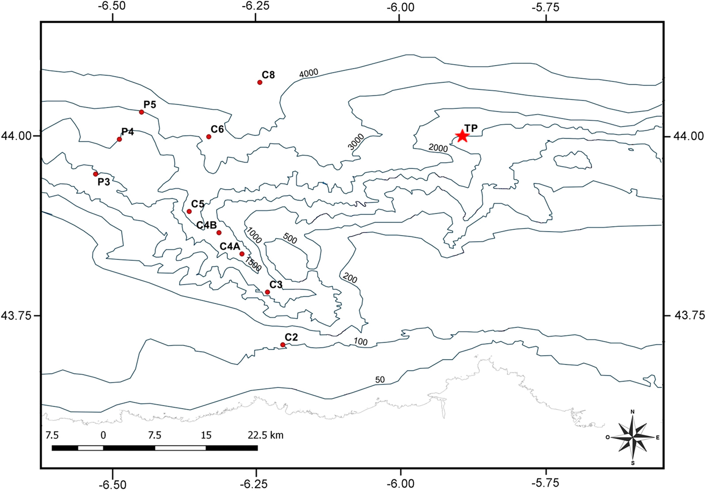
Holotype: MNCN 29.04/148, BIOCANT station: TP (44.03°N 5.87°W) 1500 m, over corals, 11 March 2012.
Paratypes: eight specimens (MNCN 29.04/149 to MNCN 29.04/156), same data as the holotype.
Non-type material: 11 specimens (MNCN 29.04/157), same data as the holotype.
TYPE LOCALITY
North-western Atlantic, Bay of Biscay, central Cantabrian continental slope in the boundaries of La Gaviera and El Corbiro Canyons (Avilés Canyon System), 44.03°N 5.87°W, 1500 m (Figure 1).
Map with the sampling stations of BIOCANT cruise. The station with presence of Psolus rufus sp. nov. is marked with a star.

COMPARATIVE MATERIAL
Psolus squamatus: 2 specimens (MNCN/29.04/32 and MNCN/29.04/37), Sweden, Atlantic Ocean.
DIAGNOSIS
Ovoid and red coloured body, dorsal area covered by large multi-layered imbricating plates; 10 triangular plates of similar size surrounding mouth, 10 arborescent tentacles; no dorsal papillae; tube feet in two rows in the ventrolateral radii but one single row in the medial third of the body; ossicles big dorsal plates and small plates, smooth, irregular and perforated in the sole.
DESCRIPTION
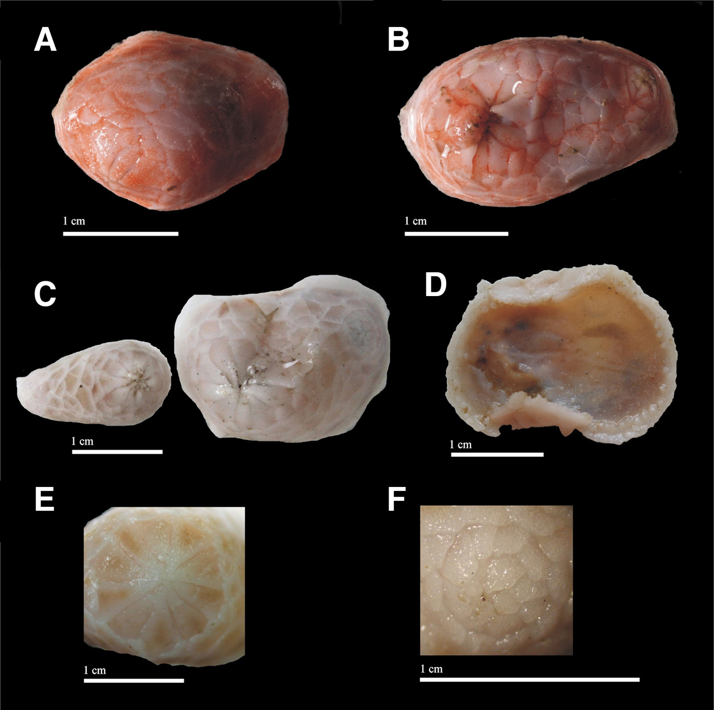
Preserved specimens 1–3 cm in length. Ovoid body shape, dorsally convex. Mouth surrounded by 10 distinct oral valves of equal size; anal opening covered by a variable number of small, irregular, non-differentiated anal valves arranged in several layers (Figure 2E, F). Ten tentacles, arborescent and reddish. Dorsal area covered by imbricated large thick scales covered with tiny tubercles in their surface (Figure 2C). Marginal dorsolateral scales smaller than dorsal scales. Ventral side sole-like, lacking large scales, thin and semitransparent, internal organs partly visible (Figure 2D). Sole midventrally bare, tube feet small, restricted to the sole, gathered in two outer zig-zag series, becoming a single row in the medial third of the body.
Psolus rufus sp. nov. (A, B), dorsal view (live specimens); (C), dorsal view (fixed specimens); (D), ventral view (fixed specimens); (E), detail of the oral valves; (F), detail of the anus. Scale bars 1 cm.

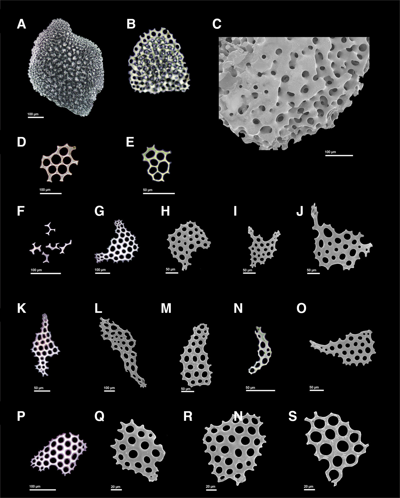
Dorsal ossicles large, multi-layered scales up to 6.5 mm long (Figure 3A–C), covering completely the dorsal side of the animal; dorsolateral scales up to 3 mm long. Sole ossicles single-layered, flat, thin, smooth perforated plates up to 140 µm long, with a variable number of perforations (Figure 3D, E), and circular perforated plates in the tube feet. Ossicles scarce in the sole. Tentacle with single-layered, perforated plates and rods up to 200 µm long, sometimes slightly curved, sometimes flat, variably thick or thin, with a variable number of holes (Figure 3O–S). Other tentacle ossicles half-moon-shaped ossicles with two arm-like prolongations (sometimes incomplete and in progress) up to 250 µm long (Figure 3G–J), and perforated, flat smooth rods up to 750 µm long (Figure 3K–N).
Psolus rufus sp. nov. (A, B), Dorsal multi-layered scales; (C), detail of the layers of the scale; (D, E), ventral perforated plates; (F), small ventral rods; (G–J), half-moon-shaped ossicles with arm-like prolongations from the tentacles; (K–S), perforated plates from the tentacles. Scale bars (A, C, D, F, G, L, P) 100 µm. Scale bars (E, H–K, M–O) 50 µm. Scale bars (Q–S) 20 µm.

ETYMOLOGY
The specific name ‘rufus’ comes from the Latin and means ‘red’. It refers to the colour of live specimens, which are brightly red (Figure 2A, B).
REMARKS
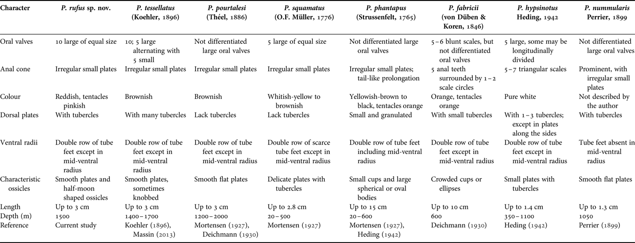
Psolus rufus sp. nov. is similar to P. tessellatus (Table 1); however, both species can be easily distinguished by the size and arrangement of the oral valves: P. rufus sp. nov. has 10 oral valves of the same size while P. tessellatus features 5 large alternated with 5 small oral valves. The possession of 10 equal-sized oral valves is also the easier diagnostic feature to differentiate P. rufus sp. nov. from the other species of Psolus occurring in the Atlantic Ocean (Table 1).
Differences among Psolus species reported for the North-eastern Atlantic Ocean.

DISTRIBUTION AND ECOLOGY
This species is only known for the central Cantabrian Sea, specifically in the continental slope in the boundaries of La Gaviera and El Corbiro Canyons (secondary canyons of the Avilés Canyon System). Psolus rufus sp. nov. was only collected in a single station (44.03°N 5.87°W) at 1500 m over coral outcrops. That area has plenty of reef-forming dark corals (Leiopathes Haime, Reference Haime1849 and Antipathes Pallas, Reference Pallas1766), white corals (Lophelia pertusa (Linnaeus, Reference Linnaeus1758) and Madrepora oculata (Linnaeus, Reference Linnaeus1758)), the sea fan Callogorgia verticillata (Pallas, Reference Pallas1766) and lithistid sponges (IEO, 2014). All these species constitute a habitat which is protected under the European Union's Habitats Directive.
GENETIC RESULTS
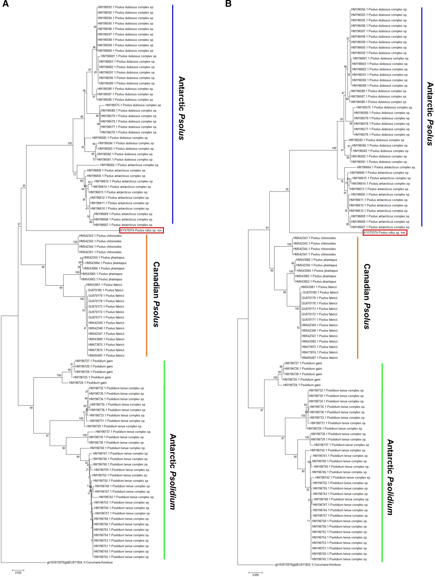
A 574-bp COI fragment was successfully obtained from Psolus rufus sp. nov. The obtained sequence of P. rufus sp. nov. was deposited in GenBank under accession number KY579374. Phylogenetic relationships of the available sequences (102) from different species within the family Psolidae (Supplementary Table S1) based on Neighbour–joining and Maximum likelihood estimations of COI sequence data show two main groups of species corresponding to the genera Psolus and Psolidium (Figure 4A, B). Within the clade of Psolus, there are three clearly separated groups: the Antarctic species complexes (P. dubiosus complex and P. antarcticus complex, with 100% bootstrap value), the Canadian species (Psolus chitonoides Clark, Reference Clark1901 from the North-east Pacific Ocean, P. phantapus and P. fabricii from the North-west Atlantic Ocean, with 99% bootstrap value), and then the novel Psolus rufus sp. nov., which is located near the clade of the Antarctic Psolus species complexes but with a low per cent bootstrap value in both different grouping methods (Figure 4A, B). This suggests P. rufus sp. nov. holds its own identity and could be an Evolutionary Significant Unit (ESU) belonging to a poor genetically studied Psolus species complex inhabiting the east Atlantic.
Phylogenetic analysis containing the COI sequences of Psolidae species. The analysis involved 103 nucleotide sequences. The percentage of replicate trees in which the associated taxa clustered together in the bootstrap test (1000 replicates) are shown next to the branches (per cent bootstrap values). There were a total of 332 positions in the final dataset. (A) Neighbour–joining method based on the Tamura–Nei model. (B) Maximum likelihood method based on the Tamura–Nei model.

DISCUSSION
Psolus rufus sp. nov. is a good representative of the genus Psolus, being consistent with the diagnosis given by O'Loughlin & Whitfield (Reference O'Loughlin and Whitfield2010): the body is dorsally and laterally enclosed by large imbricating scales while the sole is thin and lacks large scales; tube feet are restricted to the sole and the tentacles, and it possesses 10 dendritic tentacles.
Seven species of the genus Psolus have been reported in the North-east Atlantic Ocean to date: P. fabricii, P. hypsinotus, P. pourtalesi, P. phantapus, P. squamatus (reported in northern areas like Iceland, Greenland, Norway, Denmark, UK or Sweden), P. tessellatus (found in the Bay of Biscay) and P. nummularis (in Morocco and the Canary Islands) (Koehler, Reference Koehler1896; Mortensen, Reference Mortensen1927; Deichmann, Reference Deichmann1930; Hansson, Reference Hansson1999). Only P. phantapus and P. tessellatus have been reported from the Bay of Biscay (Koehler, Reference Koehler1896; Louzao et al., Reference Louzao, Anadón, Arrontes, Álvarez-Claudio, Fuente, Ocharan, Anadón and Acuña2010; Massin, Reference Massin2013).
The species of Psolus attach with their ventral sole – acting as a sucker – to hard substrates, nooks and quite inaccessible places (Mortensen, Reference Mortensen1927), making them harder to collect. This and the low sampling intensity at depths of 1000–1700 m (Massin, Reference Massin2013) may explain their scarceness in the Bay of Biscay.
Psolus rufus sp. nov. resembles P. tessellatus, but deeper examination revealed strong differences between these two species (Table 1). According to Koehler (Reference Koehler1896), the number, size and arrangement of the oral valves is a determining character to distinguish between P. tessellatus and other Psolus spp. Psolus rufus sp. nov. differs from P. tessellatus in the size and the arrangement of oral valves, which are 10 of equal size in P. rufus sp. nov. while P. tessellatus has five large valves alternated with five small ones (Koehler, Reference Koehler1896; Massin, Reference Massin2013). Other remarkable differences are the shape of the ossicles (P. rufus sp. nov. has smooth plates and half-moon shaped ossicles from the sole, while P. tessellatus only presents smooth plates, sometimes knobbed, from the sole) and the colour of the body of freshly collected specimens (reddish in P. rufus sp. nov. and brownish in P. tessellatus). Psolus rufus sp. nov. is similar to P. fabricii in the colour of fresh specimens (reddish and orange, respectively), but they differ in most of the other features, like the oral valves, anal cone or sole ossicles (Table 1). The presence of the half-moon shaped ossicles, together with 10 oral valves of equal size and reddish colour, seem to be distinct features of P. rufus sp. nov. to distinguish it from the other species from the Atlantic Ocean.
The molecular study of the COI gene supports the morphological results, grouping P. rufus sp. nov. with other members of the genus. However, the new species is genetically distinct from species of the Antarctic and Canadian groups for which sequences are available. The absence of COI sequences for the East Atlantic Psolus species generated some gaps in the genetic analysis and when drawing conclusions. The genus Psolus seems to be monophyletic according to the available genetic data. However, the bootstrap values for supporting the taxa – using two different grouping methods – were low (Figure 4A, B). The evolutionary history of the genus seems to be complex, pointing to different speciation processes in different geographic locations, as other authors have pointed out before (O'Loughlin et al., Reference O'Loughlin, Paulay, Davey and Michonneau2011). In our case, it is clear that further research and analyses including more than one gene and also several species of Psolus are needed for obtaining a more accurate phylogenetic location of the new species within the genus.
KEY TO THE SPECIES OF PSOLUS FROM NORTH-EAST ATLANTIC OCEAN:
1. Presence of differentiated oral valves2
— Absence of differentiated oral valves5
2. With 10 oral valves3
— With 5 oral valves4
3. All oral valves of equal sizeP. rufus sp. nov.
— 5 large oral valves alternating with 5 small onesP. tessellatus
4. With irregular small plates in anal cone; no tubercles in dorsal scalesP. squamatus
— 5–7 triangular plates in anal cone; 1–3 tubercles in dorsal scalesP. hypsinotus
5. Without tubercles on dorsal scalesP. pourtalesi
— With tubercles on dorsal scales6
6. With tube feet in mid-ventral radius; tail-like anal prolongationP. phantapus
— Without tube feet in mid-ventral radius; not tail-like anal prolongation7
7. Ossicles crowded cups or ellipsesP. fabricii
— Ossicles smooth flat platesP. nummularis
SUPPLEMENTARY MATERIAL
The supplementary material for this article can be found at https://doi.org/10.1017/S0025315417001138
ACKNOWLEDGEMENTS
We thank F.J. Sánchez Almazán and F. Yagüe (MNCN of Madrid) for the generous loan of the comparative material. This is a contribution from the Marine Observatory of Asturias (OMA).
FINANCIAL SUPPORT
This work was supported by the project DOSMARES (reference: CTM2010-21810-C03-02) and RES-14-CI-058, University of Oviedo.