Introduction
Prevalence of dementia is expected to rise tremendously in an aging population over the coming decades (Alzheimer's Association, Reference Association2015). Since the interventions and treatments for dementia are unfavorable when the disease progresses to a later stage, it is crucial to identify the specific marker to differentiate healthy aging from the conditions associated with high risks of progressive neurodegeneration. Apolipoprotein E (APOE) ε 4 allele, one of the well-replicated genetic risk factor for sporadic AD (Corder, et al., Reference Cendes, Andermann, Frcp, Gloor and Evans1993; Saunders et al., Reference Saunders1993; Liu et al., Reference Liu, Kanekiyo, Xu and Bu2013), has been reported to participate the formulation of β-amyloid (Aβ) (Mahley et al., Reference Mahley, Weisgraber and Huang2006). Given the evidence of positron emission tomography (PET) studies, the Aβ deposition in specific brain regions is a pathological hallmark of Alzheimer's disease (AD) (Buckner et al., Reference Buckner2005; Bero et al., Reference Bero2011).
Notably, the organizations under these specific anatomical regions are referring to a crucial modularity, that is the default mode network (DMN) (Fox et al., Reference Fox2005; Sperling et al., Reference Sperling2009). DMN refers to a set of brain regions that demonstrates reduced activity during attention-demanding tasks and increased activity during resting state (Buckner and Vincent, Reference Buckner and Vincent2007). Beyond the network level, the DMN subregions present two classes of features: (1) the first class is that the default network regions, coordinating as a system, engage in various cognitive processes (Beckmann et al., Reference Beckmann, DeLuca, Devlin and Smith2005; Smith et al., Reference Smith2009) and interact with other brain network (i.e. salience network) (Uddin et al., Reference Uddin2015); (2) the second class is that each DMN subregions plays critical roles in specific cognitive functions. For instance, dorsolateral prefrontal cortex (DLPFC) involved in decision making (Szczepanski and Knight, Reference Szczepanski and Knight2014), hippocampus formation (HF) involved in memory (Wikenheiser and Redish, Reference Wikenheiser and Redish2012), CG involved in cognitive control and attention (Leech and Sharp, Reference Leech and Sharp2014). This “two-level” feature of DMN raises the question of whether APOE ε 4 allele affects the cortical morphometry of default network regions generally or regionally? A number of VBM studies has shown that hippocampus (Lemaître et al., Reference Lemaître2005), medial temporal cortex (Honea et al., Reference Honea, Vidoni, Harsha and Burns2009; Taylor et al., Reference Taylor2014), and frontotemperal region (Wishart et al., Reference Wishart2006) are vulnerable to APOE ε 4 – related pathology. Further, enhanced investigation of cortical morphology has benefited from the advances of SBM analysis. Similar to VBM findings, cortical reduction has been found in entorhinal cortex (Shaw et al., Reference Shaw2007), anterior cingulate, and lateral temporal cortices (Alexander et al., Reference Alexander2012) in young APOE ε 4 carriers (i.e. age range 25–45). However, some studies focusing on the middle-aged APOE ε 4 carriers (i.e. age range 44–48) have reported no APOE ε 4 effects on cortical thickness of DMN subregions (Bunce et al., Reference Bunce2012), reduced cortical thickness in frontal regions (Fennema-Notestine et al., Reference Fennema-Notestine2011), or even a thicker cortical thickness in temporal regions (Espeseth et al., Reference Espeseth2008). It seems that the interactive dynamics between APOE ε 4 and brain morphometry present an age-specific property. Indeed, some studies have reported that APOE ε 4 has an impact on accelerating the cortical reduction in healthy aging elderly (Chen et al., Reference Chen2007; Espeseth et al., Reference Espeseth2008; Shi et al., Reference Shi2014).
Considering the inevitable brain changes in aging (Walhove et al., Reference Walhovd2011), it is not easy to answer whether APOE has a specific effect on DMN subregions in elderly carriers. Besides of age, the poor-matched baseline in terms of gender and education could strongly influence the brain morphometry and lead to the mixed results as well. Hence, the present study has two aims: (1) To assess the global volumetric difference between APOE ε 4 carriers and matched HC. (2) To quantitatively evaluate and compare the GMV and GMT of DMN subregions. To achieve the objectives, VBM would be used to detect the global brain volume, and SBM would be conducted to evaluate the regional GMV and GMT. Based on previous findings, we would hypothesize that APOE ε 4 carriers would present cortical reduction of DMN subregions. Moreover, we also expected that the defaults network regions with morphometric changes in APOE ε 4 carriers would be associated with clinical features.
Materials and methods
Participants
Thirty-eight community-dwelling Chinese older adults aged from 65–80 years were recruited from another cohort study that aims to establish a detailed characterization of cognitive and healthy profiles of Chinese older adults and to determine the diagnostic markers for preclinical dementia (Lam et al., Reference Lam2015). Potential participants 65 and older were firstly scheduled for a 1.5-hour in-person interview and APOE genotyping at the research center. Further, the eligible participants were invited to participate the MRI scanning. During the on-site interview, the participants received a structured questionnaire of medical history and a neuropsychological battery. Ethics approval was obtained from the Joint Chinese University of Hong Kong-New territories East Cluster Clinical Research Ethics Committee. Written informed consent from all participants was obtained before the cognitive assessments and APOE genotyping.
Apolipoprotein E genotyping
All participants provided a sample of DNA for APOE genotyping. Genomic DNA was extracted from peripheral blood samples using DNA extraction kit according to the instruction (Qiagen, US). The presence of APOE ε 2, APOE ε 3, and APOE ε 4 alleles were analyzed and performed using PCR-RFLP, which was well validated and described in our previous work (Ma et al., Reference Ma, Tang, Lam and Chiu2005).
Cognitive assessments and clinical evaluation
A standardized neuropsychological battery was used to evaluate the global cognition and major domains of cognition (Lu et al., Reference Lu, Fung, Chan and Lam2015). The battery was administered by trained research assistants and the scores were calculated by two independent raters. Cantonese Mini-Mental State Examination (CMMSE) and Alzheimer's Disease Assessment Scale (ADAS) (Chu et al., Reference Chu2000) were used as a global measure of cognitive function. Delayed recall of words: the number of recalled words was used as the performance of episodic memory. Chinese verbal fluency test (CVFT): the average number of the unique words within a semantic category in 1 minute was used as a measure of executive function. Trail making tests (TMT): the participants had to connect either numbers (A), or numbers and letters in an alternate fashion (B) as fast as possible; the time of completing in seconds was used as a measure of attention (TMT-A) and executive function (TMT-B). Digit span tests: the participants had to repeat the numbers they heard, either forward or backward; the number of correct repeated numbers was used as a measure of attention (DSF) and a measure of working memory (DSB) (Lam et al., Reference Lemaître2005). All the measures were conducted with Chinese instructions.
Magnetic resonance imaging acquisition
3D high-resolution MR images were acquired in Prince of Wales Hospital (Hong Kong) with a 3.0 Tesla Philips scanner (Philips Medical System, Best, the Netherlands). The scanning time for each participant was 8.5 minute. T1-weighted MRI data set was configured with the following acquisition parameters: axial acquisition with a 256 × 256 × 192 matrix, thickness = 1 mm, no gap, field of view (FOV) = 230 mm, TR = 2070 ms, TE = 3.93 ms, flip angle = 15o. The sequence yielded high quality isotropic images with the voxel size of 1 mm × 1 mm × 1 mm.
Voxel-based morphometry (VBM) analysis
Quality check of T1 images, pre-processing, and optimal VBM analysis were performed by VBM8 toolbox (http://dbm.neuro.uni-jena.de/vbm/) running on MATLAB (MATLAB R2012b, The Math Works Inc. Natick, MA) with default parameters (Good et al., Reference Good2001). The processing included the following steps: (1) All anatomical images were reoriented by anterior and posterior (AC-PC) line manually; (2) Images were bias-corrected, tissue classified, and registered using linear (12-parameter affine) and non-linear transformations (warping), within a unified model (Ashburner and Friston, Reference Ashburner and Friston2005); (3) The segmentation procedure from the VBM8 toolbox produced gray matter (GM), white matter (WM) and registered rigid-body aligned tissue segments for each image in the Montreal Neurological Institute (MNI) space; (4) Modulation of different tissue segments by nonlinear normalization parameters to account for the differences of individual brain size globally; (5) The normalized and modulated GMV were smoothed with a Gaussian kernel of 8 mm full width at half maximum (FWHM).
Surface-based morphometry (SBM) analysis
Cortical morphometry, including GMV and GMT, were calculated by BrainSuite 14.0 (http://brainsuite.org/). BrainSuite is an automatic cortical surface identification integrated package with the updated version of Brain Surface Extraction (BSE) and friendly interface (Shattuck and Leahy, Reference Shattuck and Leahy2002; Pantazis et al., Reference Pantazis2010), which are widely used in dementia and aging studies (Frisoni et al., Reference Frisoni, Prestia, Rasser, Bonetti and Thompson2009; Apostolova et al., Reference Apostolova2010; Hwang et al., Reference Hwang2013).
To calculate regional GMV and GMT individually, we followed the standard pipeline and default parameters (Shattuck et al., Reference Shattuck2008). The SBM analysis was obtained by submitting each participant's MRI to a series of steps, including: (1) motion correction; (2) intensity normalization; (3) removal of non-brain voxels; (4) segmentation of GM, WM, and cerebrospinal fluid (CSF); (5) tessellation of the GM/WM boundary, and automated topology correction. At each step, the results were visually inspected and manual interventions were performed when required to correct topological defects. Cortical image processing was conducted by the investigators blinded to subjects’ demographics, APOE ε 4 statuses, and cognitive performance.
Statistical analysis
First, group differences of demographics, cognitive performance, voxel-wise GM differences were tested either with χ2 for categorical variables or with independent two samples t-test for continuous variables. Second, regional GMV and GMT were calculated individually. We followed the Cendes methods (Cendes et al., Reference Cendes, Andermann, Frcp, Gloor and Evans1993) to adjust the heterogeneity of head size and conducted a correction of individual variance of total intracranial volume (TIV) through the following formula: Corrected SV = (MBV × SV)/IBV. MBV is the mean brain volume in the group (which is a constant), SV is the regional volume of GM, and IBV is the individual brain volume. Third, the group-wise comparisons of GMV and GMT were conducted using the code embedded in MATLAB (http://neuroimage.usc.edu/neuro/Resources/BST_SVReg_Utilities). Multiple comparison correction was performed by the above code using false discovery rate (FDR) estimation (Reiter et al., Reference Reiter2012). Person's correlation was used to detect the relationship between age, cognitive functions, and regional morphometry of DMN subregions. Bonferroni correction was used to reduce chances of obtaining false-positive results of correlation analysis (Jiang et al., Reference Jiang, He, Zang and Weng2004; Watanabe et al., Reference Watanabe2013). Given the six pairs of ROI, the statistical significant level would be 0.05/12 = 0.0042. The χ2 test, two sample t-test, and Person's correlation were performed by IBM SPSS Statistics (Version 20).
Results
Demographics and Cognitive performance
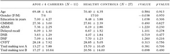
As shown in Table 1, the demographics in term of age, gender, years of education were similar between APOE ε 4 carriers and healthy group. The global cognition and cognitive functions cross the main domains between two groups were also comparable. While, there was a trend that APOE ε 4 carriers presented worse cognitive performance than HC.
Demographics and cognitive performance of APOE ε 4 carriers and healthy controls

Note: Data are raw scores and presented as mean ± SD. Abbreviations: CMMSE = Cantonese Mini-Mental State Examination; ADAS = Alzheimer's Disease Assessment Scale; DSB = Digit span backward; DSF = Digit span forward; CVFT = Chinese verbal fluency test.
VBM results
With a threshold of uncorrected p < 0.001, there was no voxel left. Although the APOE ε 4 carriers demonstrated less GMV (APOE ε 4 carriers: 544.99 ± 38.06; HC: 557.90 ± 65.08, t = 0.613, p = 0.543) and WMV (APOE ε 4 carriers: 445.21 ± 52.27; HC: 459.50 ± 51.66, t = 0.771, p = 0.446), no significant difference was found between two groups.
SBM results
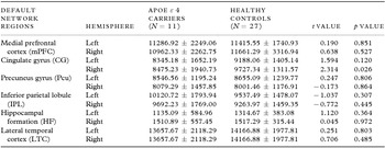
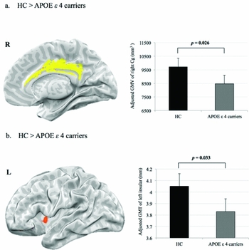
Based on the well-established neural underpinnings of DMN (Fox et al., Reference Fox2005; Buckner et al., Reference Buckner, Andrews-Hanna and Schacter2008), six pairs (left/right) of brain regions are selected as regions of interest (ROIs), including medial prefrontal gyrus (mPFC), CG, precuneus gyrus (Pcu), inferior parietal lobe (IPL), hippocampal formation (HF), and lateral temporal cortex (LTC). As Table 2 shown, APOE ε 4 carriers demonstrates a global cortical atrophy, but significant decreased GMV was found in right CG (Figure 1a) (APOE ε 4 carriers: 8475.23 ± 1940.73 mm3, HC: 9727.34 ± 1311.57 mm3, t = 2.314, p = 0.026, 95% CI [154.65, 2349.57], and reduced GMT was found in left insular (Figure 1b) (APOE ε 4 carriers: 3.83 ± 0.37 mm, HC: 4.05 ± 0.25 mm, t = 2.197, p = 0.033, 95% CI [0.007, 0.427]).
Comparisons of GMV of default network regions in APOE carriers and healthy controls

Note: Data are raw scores and presented as mean ± SD.
Inflated surface maps of the morphometric difference between APOE ε 4 and healthy groups. (a) depicts the medial view of right cingulate gyrus (Cg) and (b) depicts the lateral view of left insular.

Associations between age, cognitive functions, and DMN subregions
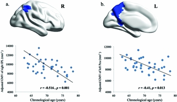
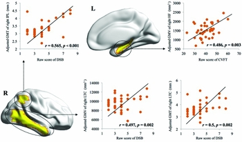
Using gender, education and TIV as covariates, significant negative correlations (Figure 2) were found between age and subregions of DMN, including GMV of right IPL (2a. r = −0.516, p = 0.001) and left Pcu (2b. r = −0.41, p = 0.013). The results suggest that the cortical atrophy of DMN subregions is coupled with advancing age. With age, gender, and education as covariates, extensive associations were found between scores of cognitive functions and cortical morphometry of DMN subregions (Figure 3). Short-term memory measured by DSB was positively correlated with right LTC (GMV: r = 0.497, p = 0.002; GMT: r = 0.5, p = 0.002) and right IPL (GMT: r = 0.565, p < 0.001). Executive function measure by CVFT was positively associated with right hippocampus (GMV: r = 0.486, p = 0.003).
Scatterplot of the correlation between age and cortical morphometry.

Scatterplot of the correlation between scores of cognitive functions and cortical morphometry.

Discussion
The current study was the first attempt to combine both VBM and SBM analysis to identify the morphometric changes of default network regions in Chinese senior adults with genetic risk for AD. Generally, the present study highlights the reduced cortical volume and thickness of DMN subregions in cognitively normal senior APOE ε 4 carriers.
Does APOE ε 4 have a preferential influence on DMN in old age?
It is a challenge to postulate the characteristic changes of brain morphometry in APOE ε 4 carriers with normal cognition. With regard to the accelerating effect of APOE ε 4, cortical changes were more pronounced in senior APOE ε 4 carriers than the younger ones (Papenberg, Lindenberger, and Bäckman, Reference Papenberg, Lindenberger and Bäckman2015). Evidence from functional MRI (fMRI) studies also confirmed the stronger APOE ε 4 effects on multiple DMN subregions in senior adults (Filippini et al., Reference Filippini2011). It seems that APOE ε 4 has the differential effects on brain in young and old adults.
Even though it is very difficult to identify the pure APOE ε 4 effects, the well-matched baseline is necessary in the comparison and explanation of morphometric properties accounting for APOE ε 4. Thus, the question we addressed as whether APOE ε 4 has a differential effect on cortical properties of DMN subregions maybe revised as does APOE ε 4 have a preferential effect on DMN subregions in elderly carriers. In the continuum from cognitively normal APOE ε 4 carriers to preclinical stage of AD, DMN is vulnerable to be affected by healthy and pathological aging. Aligned with previous studies (Shiino et al., Reference Shiino2006; Filippini et al., Reference Filippini2009; Alexander et al., Reference Alexander2012), it is not surprising that the prominent age-related atrophy was found in DMN subregions (Figure 2). In the comparison between APOE ε 4 carriers and HC, the significant morphometric difference was detected by SBM instead of VBM analysis. With particular interest, pronounced decreased GMV of right CG and reduced GMT of left insular have been found in APOE ε 4 carriers. The results indicate at least two things: (1) Morphometric property in terms of volumetric and cortical thickness may change in a nonlinear fashion in late adulthood (Westlye et al., Reference Westlye, Grydeland, Walhovd and Fjell2011). (2) APOE ε 4 may influence the cortical morphometric specifically rather than globally in the non-symptom stage.
APOE ε 4-related effects on cortical morphometry
Indeed, the mechanism that contributes to the regional changes within default mode regions in APOE ε 4 carriers has not been fully understood. The possible explanations could be postulated from two perspectives. Firstly, from the perspective of pathological vulnerability, the APOE ε 4 related brain regions with Aβ deposition are coordinated with the pathological process proposed by Braak's (Braak and Braak, Reference Braak and Braak1991; De Leon et al., Reference De Leon2001). Two hallmarks of AD-related pathology, neurofibrillary tangles (NFTs) and Aβ, are largely distributed in anterior insular context (Bonthius et al., Reference Bonthius, Solodkin and Van Hoesen2005) and CG (Sperling et al., Reference Sperling2009) and at the early stage of AD. Besides the volumetric changes, reduced cortical thickness (Gutiérrez-Galve et al., Reference Gutiérrez-Galve2009) and WM lesions (Zhuang et al., Reference Zhuang2012) of CG have also been found in APOE 4 ε carriers. Furthermore, evidence from PET and magnetic resonance spectroscopy (MRS) studies also confirmed the decreased metabolism of CG (Reiman et al., Reference Reiman2012; Gomar et al., Reference Gomar2014) and insular (Yao et al., Reference Yao2015) in APOE 4 ε carriers, even several years before the onset of cognitive impairment. Second, from the perspective of functional anatomy, the multifaceted roles of CG and insular have been reported to engage across cognitive, affective, and behavioural contexts. CG, as a member of cortical midline structures (CMS), acts as a “hub” connecting multiple brain regions (Buckner and Krienen, Reference Buckner and Krienen2013). Insular is one of the most frequently identified regions with degenerative change in AD. However, the heterogeneity within insular makes it very difficult to discuss the results across studies. Recent studies employing various methods appear to converge on the functional anatomy of insula into at least three distinct subregions (Chang et al., Reference Chang, Yarkoni, Khaw and Sanfey2013). One of them is the dorsoanterior region of insular, associating with higher cognitive processing and coordinating the interplay between DMN and salience network (Uddin et al., Reference Uddin2015), which is compatible with the region presented reduced GMT in our results (Figure 1b).
As to the brain-behavior correlations, although the cortical morphometry were positively correlated with cognitive functions (Figure 3), we have not detected the relationship between CG, insular, and cognitive functions. The potential explanation might be due to the comparable scores of cognitive and clinical evaluation between APOE 4 ε and HC. Besides, the non-linear fashion of morphometric properties in late adulthood may affect the correlation results as well (Westlye et al., Reference Westlye, Grydeland, Walhovd and Fjell2011). In summary, our results highlight the difference between different cortical measures and suggest that the cortical reduction of CG and insular maybe a potential neuroimaging marker for APOE 4 ε senior carriers.
Limitations and future works
The findings of this study should be circumspectly interpreted with its limitations. First, the cortical properties of cingulate cortex subregions have not been launched due to the brain template embedded in BrainSuite. Second, cross-sectional design with a small sample size may limit the generalizability of the findings. Third, some cerebrovascular risk factors, such as blood pressure and cholesterol level, are not included in this study, which may affect the cortical morphometry as well. Fourth, only APOE genotype was tested as the genetic risk factor. Fifth, all participants were from Chinese community, which may not give an accurate reflection of the features of senior adults with various cultural backgrounds.
Given the lateralized feature of APOE ε 4-related change, it would be intriguing to investigate the potential APOE-related effects on cortical lateralization in two completely independent cohorts with healthy and pathological aging. Besides, it is crucial to validate our findings in a longitudinal cohort. Finally, the APOE ε 4-related regions in our finding could be used as the seeds to test the functional connectivity within DMN in the future studies by using resting state fMRI.
Conflict of interest
None.
Description of authors’ roles
Hanna Lu, Sandra S.M. Chan, and Linda C.W. Lam conceived and designed this study. Hanna Lu performed the VBM and SBM analysis. Suk Ling Ma conducted the Apolipoprotein E genotyping of this study. Regarding to this paper, Hanna Lu drafted, Suk Ling Ma, Sandra S.M. Chan, and Linda C.W. Lam discussed and agreed the final version.
Acknowledgments
This research was supported by the Direct Grant from the Chinese University of Hong Kong. The authors would like to thank all the participants and their relatives that kindly agreed to participate in the study.