INTRODUCTION
Solar ultraviolet radiation (UVR) (wavelengths; UVB: 280–320, UVA: 320–400 nm) has always been a strong selective force in aquatic communities (Williamson et al. Reference Williamson, Neale, Grad, De Lange and Hargreaves2001; Sommaruga, Reference Sommaruga, Helbling and Zagarese2003; Hansson and Hylander, Reference Hansson and Hylander2009). As an integral part of a complex array of fluctuating environmental factors in these ecosystems, UVR varies considerably in time (e.g. seasonal) and space (e.g. latitude, water depth; Tedetti and Sempere, Reference Tedetti and Sempere2006). Although life on earth has evolved in the presence of UVR, the recent and on-going stratospheric depletion in ozone has altered the selective pressure that UVR, especially UVB, may exert (Lesser and Barry, Reference Lesser and Barry2003 and references therein). Moreover, on-going and predicted climate changes are expected to further exacerbate exposure of organisms to UVB in aquatic ecosystems (Haeder et al. Reference Haeder, Helbling, Williamson and Worrest2011).
UVR, especially UVB, is predominantly known for its potentially deleterious effects, damaging biological macromolecules and cellular structures including enzymes, membranes, DNA and RNA (e.g. Haeder et al. Reference Haeder, Kumar, Smith and Worrest1998; Vincent and Neale, Reference Vincent, Neale, de Mora, Demers and Vernet2000; Day and Neale, Reference Day and Neale2002; Dahms and Lee, Reference Dahms and Lee2010) (see also Paul and Gwynn-Jones, Reference Paul and Gwynn-Jones2003). Ecologically relevant effects at the whole-organism level include decreases in fecundity, growth, development, mobility and survival rate, all of which can translate into changes in species composition at the community and ecosystem level (e.g. Bothwell et al. Reference Bothwell, Sherbot and Pollock1994; Haeder et al. Reference Haeder, Kumar, Smith and Worrest1998; Bancroft et al. Reference Bancroft, Baker and Blaustein2007; Hansson and Hylander, Reference Hansson and Hylander2009). As a consequence, organisms have evolved a range of behavioural (e.g. migration), physiological (e.g. accumulation of UV-absorbing compounds) and molecular mechanisms (e.g. DNA damage repair), to avoid, minimize or repair damage induced by exposure to UVR (Roy, Reference Roy, de Mora, Demers and Vernet2000; Sinha and Haeder, Reference Sinha and Haeder2002; Hansson and Hylander, Reference Hansson and Hylander2009; Dahms and Lee, Reference Dahms and Lee2010 and references therein).
Biological and ecological effects of UVR have been well studied in aquatic systems (e.g. Haeder et al. Reference Haeder, Kumar, Smith and Worrest1998; de Mora et al. Reference de Mora, Demers and Vernet2000; Helbling and Zagarese, Reference Helbling and Zagarese2003; Bancroft et al. Reference Bancroft, Baker and Blaustein2007; Hansson and Hylander, Reference Hansson and Hylander2009). Organisms vary considerably in their susceptibility to UVR (e.g. Bancroft et al. Reference Bancroft, Baker and Blaustein2007). Hence, it is very likely that at least some of their interactions with other organisms would be influenced by UVR (see examples in Paul and Gwynn-Jones, Reference Paul and Gwynn-Jones2003; Sommaruga, Reference Sommaruga, Helbling and Zagarese2003). However, studies on the effects of UVR on aquatic organisms have largely ignored species interactions, particularly those between parasites and their hosts. Despite the fact that parasitism is known to be of high ecological relevance (e.g. Hudson et al. Reference Hudson, Dobson and Newborn1998; Lafferty et al. Reference Lafferty, Dobson and Kuris2006; Mouritsen and Poulin, Reference Mouritsen and Poulin2010), knowledge on the effects of UVR on parasites and diseases in aquatic systems has remained limited (see Sommaruga, Reference Sommaruga, Helbling and Zagarese2003).
The sensitivity of a range of parasites from aquatic environments to UVR, however, has been acknowledged (see Marcogliese, Reference Marcogliese2001 and references therein). UVR is expected to be particularly detrimental in clear, shallow waters and for parasites with small, delicate free-living stages that are extremely susceptible to environmental conditions (MacKenzie et al. Reference MacKenzie, Williams, Williams, McVicar and Siddall1995). Sublethal and lethal effects of UVR on transmission stages of important human parasites (e.g. Schistosoma spp., Cryptosporidium parvum) have been investigated using artificial lights (Standen and Fuller, Reference Standen and Fuller1959; Ghandour and Webbe, Reference Ghandour and Webbe1975; Prah and James, Reference Prah and James1977; Ariyo and Oyerinde, Reference Ariyo and Oyerinde1990; Connelly et al. Reference Connelly, Wolyniak, Williamson and Jellison2007; Ruelas et al. Reference Ruelas, Karentz and Sullivan2006, Reference Ruelas, Karentz and Sullivan2007, Reference Ruelas, Karentz and Sullivan2009). Results from these studies consistently confirm strong negative effects of UVR on survival as well as infectivity of parasites.
However, most of these studies generally do not consider ecologically relevant interactions between a parasite and its host(s). Using a more inclusive approach, Ruelas et al. (Reference Ruelas, Karentz and Sullivan2006, Reference Ruelas, Karentz and Sullivan2007, Reference Ruelas, Karentz and Sullivan2009) investigated the effects of UVR on the transmission of Schistosoma mansoni as well as the parasite's aquatic snail host (Biomphalaria glabrata). Their results indicated that not only the parasite, but also juvenile and infected snails were vulnerable to UVR exposure, but that both snail and parasite were also able to repair UV-induced DNA damage. This clearly highlights the importance of direct and indirect effects of UVR on the interaction between parasites and hosts, suggesting that the overall effect of UVR on a particular system may be highly complex and thus difficult to predict.
UVR effects may be particularly relevant in intertidal ecosystems, where conditions for organisms at low tide are almost equivalent to atmospheric conditions (Kramer, Reference Kramer, Beukema, Wolff and Brouns1990; Karentz, Reference Karentz, McClintock and Baker2001), and even more so for those in the Southern hemisphere where incident UVR levels are greatest (Seckmeyer and McKenzie, Reference Seckmeyer and McKenzie1992; McKenzie et al. Reference McKenzie, Conner and Bodeker1999). In intertidal ecosystems, trematode parasites are ubiquitous and important ecological components (e.g. Lauckner, Reference Lauckner1984; Sousa, Reference Sousa1991; Mouritsen and Poulin, Reference Mouritsen and Poulin2002, Reference Mouritsen and Poulin2010). In the present study, the transmission process of the intertidal trematode Maritrema novaezealandensis was examined. Like most trematodes, M. novaezealandensis has a complex life cycle involving several members of an intertidal community (Martorelli et al. Reference Martorelli, Fredensborg, Mouritsen and Poulin2004). The species in this life cycle are all specific to New Zealand and hence are exposed to the particularly high UVB levels occurring in this region compared to Northern hemisphere regions of the same latitude (Seckmeyer and McKenzie, Reference Seckmeyer and McKenzie1992; McKenzie et al. Reference McKenzie, Conner and Bodeker1999), and to the transient high levels of UVB associated with high latitude ozone depletion in the Southern hemisphere (McKenzie et al. Reference McKenzie, Bjorn, Bais and Ilyasd2003, Reference McKenzie, Aucamp, Bais, Bjorn and Ilyas2007).
The different stages of the parasite throughout a trematode's life cycle experience vastly different conditions in terms of the solar radiation regimes to which they are exposed. Maritrema novaezealandensis, for example, lives as an adult worm in the intestine of mudflat affiliated birds (environment with no direct solar irradiation). Eggs pass out with the bird's feces and persist in the environment until ingested by a first intermediate snail host (Zeacumantus subcarinatus) foraging on mudflats. Within a snail's opaque shell, a miracidium hatches from an egg and develops into a sporocyst, probably shielded from solar irradiance. Within the sporocysts, asexual reproduction occurs and large numbers of larval transmission stages (cercariae) are produced, which emerge from an infected snail under optimal conditions in order to infect a second intermediate crustacean host. Cercariae are small (approx. 170–200 μm including tail, Martorelli et al. Reference Martorelli, Fredensborg, Mouritsen and Poulin2004; Koehler and Poulin, Reference Koehler and Poulin2010), short-lived (<24 h), non-feeding transmission stages and in the case of M. novaezealandensis, they are also translucent. These cercariae are directly exposed to and influenced by environmental conditions. After infecting a second intermediate crustacean host, such as the amphipod Paracalliope novizealandiae used in this study, the parasite experiences different levels of exposure to solar radiation depending on the opaqueness or transparency of the carapace of its crustacean host. Within a crustacean, the parasite develops into an encysted stage (metacercaria). The life cycle is completed when a crustacean harbouring a mature metacercaria is ingested by a definitive bird host and successfully establishes in its intestine.
In this study, we investigated the effects of ecologically relevant levels of UVR on the transmission process of M. novaezealandensis from its first intermediate snail host (Z. subcarinatus) to its second intermediate amphipod host (P. novizealandiae). In laboratory and outdoor experiments, the effects of UVR on (1) the emergence of cercariae from snail hosts, (2) the survival of cercariae, (3) the infectivity of cercariae, (4) the susceptibility of the amphipod host to infection, and (5) the survival of infected and uninfected amphipods were assessed (for a conceptual figure summarizing these steps see Studer et al. Reference Studer, Thieltges and Poulin2010). Our aim was to identify steps in this transmission process that are particularly sensitive to UVR, and to evaluate overall net effects of this environmental factor on the transmission of M. novaezealandensis from its first to its second intermediate host.
MATERIALS AND METHODS
Parasite and host material
First intermediate snail hosts (Z. subcarinatus) were collected from a high prevalence site (Lower Portobello Bay, Otago Harbour) and screened for infections with M. novaezealandensis (see Studer et al. Reference Studer, Thieltges and Poulin2010 for details). For all experimental infections, uninfected second intermediate amphipod hosts (P. novizealandiae) were collected from Hooper's Inlet (Otago Peninsula) several days prior to an experiment in order to allow for acclimatization to laboratory conditions. At Hooper's Inlet, first intermediate snail hosts are absent and therefore, amphipods are not infected naturally (neither by M. novaezealandensis nor by any other trematode metacercariae) (Fredensborg et al. Reference Fredensborg, Mouritsen and Poulin2004; Bryan-Walker et al. Reference Bryan-Walker, Leung and Poulin2007). After their use in experiments (see below), amphipods were stored in small plastic containers containing 300 ml of aerated seawater and a strip of sea lettuce (Ulva sp.) until measured (size classes: 2·5, 3·0, 3·5, 4·0, 4·5±0·25 mm), sexed and dissected under a dissecting microscope to assess the number of parasites present (infection status and intensity). All experimental infections as well as the cercarial survival experiment were conducted using 96-well plates (wells 7×10 mm; total volume 320 μl). For all experimental infections, a cercarial mixture from 40 infected snails was obtained (see details in Studer et al. Reference Studer, Thieltges and Poulin2010).
Experimental design and laboratory set-up
All experiments except the amphipod survival experiment (see details below) were conducted as 3×2 designs including 3 UV treatments (i.e. exposure to photosynthetically active radiation (PAR) only (no UV; 400–700 nm), PAR+UVA (UVA; 320–700 nm) or PAR+UVA+UVB (UVA+B; 280–700 nm) and 2 exposure durations (i.e. doses). UV treatments were achieved using Plexiglas filters transmitting PAR (81%), only minimal UVA (5·2%) and no UVB (0·0%) (no UV treatment); PAR (77·9%), UVA (46·5%), and only minimal UVB (0·1%) (UVA treatment); PAR (84·5%), UVA (84·6%) and UVB (80·6%) (UVA+B treatment) (see Lister et al. Reference Lister, Lamare and Burritt2010 for transmission profiles of filters).
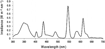
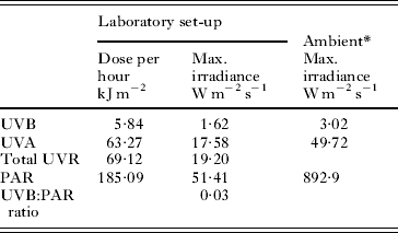
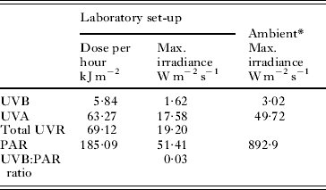
The laboratory set-up included 1 UVB (Atlas 07-29006), 2 UVA (Dr. Kern® excellent RA 80W), and 3 full spectrum light tubes (Phillips Alto TLD 58W/840), which were suspended 45 cm above the bench top and which were wrapped in cellulose acetate to absorb any UVC radiation being emitted. The spectral output of the lamps was measured with a LiCor Li-1800UW spectroradiometer (Table 1, Fig. 1). Data given in Table 1 do not include reductions by filters and only provide lamp output irradiances from 300–700 nm. Compared to ambient local levels of solar irradiance during midday peaks in summer, maximum irradiances from our laboratory set-up were relatively low for UVB, UVA and PAR (Table 1). All doses administered in each experiment can be calculated based on the dose per hour given in Table 1. All low dose exposures (shorter exposures) were administered during the second half of the high dose treatments (longer exposure) and all laboratory experiments were conducted at 20°C. Statistical analyses are described in each of the following subsections. We checked for differences between replicates and transformed data where necessary to ensure the assumptions of the statistical tests were met.
Spectral output (300–700 nm) of all light tubes combined in the laboratory set-up (W m−2 nm−1) as measured by the LiCor Li-1800UW spectroradiometer.

Irradiances in the laboratory set-up: dose per hour (kJ m−2) and maximum irradiance (W m−2 s−1) for UVB, UVA and PAR
Output of Maritrema novaezealandensis cercariae from first intermediate snail hosts
Output of cercariae of M. novaezealandensis from infected Z. subcarinatus snails was assessed as a short-term response by counting the number of cercariae that emerged from snail hosts during exposure to the different UV treatments at 20°C. There were 2 trials each with 16 replicate snails per treatment. For the exposures, snails were individually placed in 1·5 ml Eppendorf tubes filled with 1 ml of filtered seawater. Tubes were left open, covered with the respective filters and then exposed for 3 or 6 h. After incubation and removal of the snails, tubes were centrifuged (5 min, 20 817 g) and 900 μl of the seawater exchanged with 70% ethanol to preserve the samples. The supernatant was checked for the presence of cercariae before disposal. Samples were counted under a dissecting microscope. A General Linear Model (GLM) was used to test for the effects of the trial, UV treatment, trial×UV treatment and dose on the number of emerged cercariae (log transformed).
Survival of cercariae
For this experiment, the survival and activity of cercariae was monitored when exposed to the different UV treatments and doses. At the start of the experiment, 40 μl from a cercarial mixture (average age of cercariae 30 min) were added to 24 replicate wells on two 96-well plates per treatment corresponding to an addition of approximately 45 cercariae per well. Cercariae were then exposed for either 2 or 4 h to one of the UV treatments. After exposure, well plates were placed under visible light (Phillips 40 W) for further monitoring. Survival and activity was categorized visually as fully active, sluggishly motile or immotile/dead. Cercariae were checked at an average age of 3, 5, 7, 9 and 12 h. A repeated measures ANOVA was used to test for the effect of the UV treatment and dose on the proportion of fully active cercariae (arcsine-square root transformed) at an average age of 3, 5, 7 and 9 h. Due to violation of the assumption of sphericity, multivariate results for within-subject effects are reported. Additionally, LD50 values were calculated to assess the time in each treatment for 50% of cercariae to lose their full functional activity. This was done by fitting logistic functions to the proportion of fully active cercariae data of each treatment and dose and then calculating the inflection point.
Infectivity of cercariae
Infectivity, i.e. the proportion of cercariae that successfully infects second intermediate amphipod hosts (P. novizealandiae), was assessed after exposure of cercariae to the UV treatments and doses. For this, 48 uninfected amphipods (⩾2·25 mm body size) per treatment were put individually into wells on two 96-well plates containing approximately 75 μl of filtered seawater. A cercarial mixture was obtained which was divided into wells of 12-well plates (4 ml per well; 2 wells per treatment) and then exposed for either 30 min or 1 h to respective UV treatments. Based on aliquot counts prior to exposure, 50 μl of the irradiated cercarial mixtures were added to each amphipod, which corresponded to an addition of approximately 18 cercariae per amphipod. Amphipods and irradiated cercariae were then incubated for 2 h at 25°C under constant illumination with visible light (Philips 40 W). After incubation, amphipods were transferred into plastic containers filled with seawater and dissected under a dissecting microscope 7 days after the experiment. A GLM was used to test for the effect of UV treatment, dose, UV×dose, as well as sex and size of amphipods on the proportion of parasites successfully infecting amphipods (arcsine-square root transformed).
Susceptibility of amphipods to infections
Susceptibility of amphipod hosts was studied by exposing amphipods to the UV treatments before adding non-irradiated cercariae, and then comparing the infection success of the cercariae in these hosts. This experiment consisted of 2 separate trials. In the first trial, amphipods were exposed for 30 and 60 min. In the second trial, amphipods were exposed for 3 and 6 h. In each trial, 48 amphipods per treatment were transferred individually into wells of two 96-well plates per treatment (in approximately 75 μl natural seawater) prior to exposure. A cercarial mixture was prepared which was then added to individual amphipods at the end of the exposure period (first trial: 60 μl of the cercarial mixture corresponding to approximately 13 cercariae per amphipod; second trial: 30 μl corresponding to approximately 20 cercariae per amphipod). Well plates containing irradiated amphipods and cercariae were then incubated for 2 h at 25°C under visible light. After incubation, amphipods were transferred into plastic containers and dissected 7 days later. The two trials were analysed separately using GLM's assessing the effect of the UV treatment, dose, UV×dose, as well as size and sex of amphipods on the proportion of cercariae (arcsine-square root transformed) successfully infecting the amphipods. In the second trial, this was followed by a Tukey's post-hoc comparison between the UV treatments.
Survival of infected and uninfected amphipods
This experiment was conducted under ambient conditions and assessed the effect of UVR on the survival of uninfected and infected amphipods using the same filters as specified above (treatments: no UV, UVA, UVA+B). The experiment was conducted over a 24 d period at the Portobello Marine Laboratory, Otago Harbour, New Zealand in austral summer. UVA and UVB levels during the experiment were recorded at the laboratory's weather station (UVA SKU 420 and UVB SKU 430 sensors, Skye Instruments Ltd). Mean total daily doses during this period were 1938·67±131·63 kJ m−2 of UVA and 113·98±8·12 kJ m−2 of UVB, with maximum irradiances of 44·1 (UVA) and 2·83 W m−2 s−1 (UVB). For the experimental infections, 48 uninfected amphipods per replicate container (3 containers per treatment) were individually put into wells of 96-well plates. A cercarial mixture was obtained, of which 50 μl were added to half of the amphipods (corresponding to an addition of approximately 28 cercariae per amphipod), whereas the same volume of filtered seawater was added to controls. Amphipods were then incubated for 2 h at 25°C under constant illumination with visible light. Infected and uninfected amphipods were left in separate plastic containers (1 l; aerated seawater) overnight to allow the parasites to complete infection. Infected and uninfected amphipods (24 each per container, sexes matched) were then distributed into white opaque rectangular plastic containers (1 l) provided with a small strip of sea lettuce (Ulva sp.) and covered with the respective filter. Containers were placed outdoors and received filtered flow-through seawater. Survival of amphipods was subsequently monitored and container positions rotated twice a day. Dead amphipods recovered were measured, sexed and dissected under a dissecting microscope to assess the number of parasites present in each amphipod. After 24 days, all remaining amphipods were dissected. The risk of dying during the experimental period was analysed with a Cox proportional hazard regression model with UV treatment, sex, size and infection status of amphipods as predictor variables.
Net effect on transmission
In order to validate the conclusion that UVR, by affecting both the survival of cercariae as well as the susceptibility of amphipods, may overall have a neutral net effect (i.e. the decreased survival of cercariae being compensated by an increased susceptibility of amphipods; see Results and Discussion sections), an additional outdoor experiment was conducted assessing the success of cercariae at infecting amphipods under natural light conditions. For this, cercariae (approximately 25 per amphipod; pooled from 40 infected snails) and amphipods (64 per treatment) were exposed together in wells of 2 replicate 96-well plates to no UV, only UVA or UVA+B for 2 h on a clear summer day using the same filters as described above (total UVB dose 1·607 Jcm−2). Amphipods were dissected the following day and the number of parasites infecting each amphipod was assessed. A GLM was used to assess the effect of UV treatment, size and sex of amphipods on the proportion of parasites successfully infecting the amphipods (arcsine-square root transformed).
RESULTS
Cercarial output from snails
There was substantial variability of cercarial output among individual snails (range 0–2738 cercariae per snail per incubation), with a mean output (± standard error as in all following results) of 152±31 cercariae per snail (at 20°C). Thus, no clear pattern was found for the effect of the UV treatment or dose on the number of cercariae emerging from infected snails (log number of cercariae; GLM, UV treatment: F2,185=1·44, P=0·239; dose: F1,185=1·14, P=0·287). While there was also no significant difference between trials (F1,185=3·29, P=0·072), there was a significant interaction between trial and UV treatment (F2,185=3·45, P=0·034), indicating an inconsistency of the UVR effects between trials. The sizes of snails used did not differ between the trials or UV treatments (ANOVA, trials: F1,191=1·16, P=0·284; UV treatment: F2,191=1·18, P=0·309).
Cercarial survival
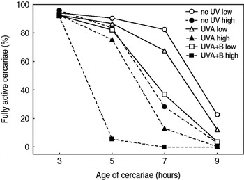
UVR negatively influenced the survival of cercariae in a dose-dependent manner. Mortality was highest for cercariae exposed to a high dose of both, UVA+B (all dead at an average age of 7 h), and lowest for the cercariae exposed to a low dose of no UV (23% still fully active at an average age of 9 h (Fig. 2)). Survival after administration of high doses was reduced in all cases when compared to the low dose (Fig. 2), even for exposure to visible light only. This effect was exacerbated when cercariae were exposed to UVR with survival under UVA+B < UVA. The repeated measures ANOVA carried out on the proportions of fully active cercariae (arcsine-square root transformed) indicated a significant effect of all predictor variables and their interactions (between subjects; UV treatment: F2,138=301·76; dose: F1,138=774·62; UV×dose: F2,138=28·82; within subjects; time: F3,414=3566·28; time×UV: F6,414=93·68; time×dose: F3,414=270·32; time×UV×dose: F6,414=92·77; for all P<0·001). LD50 values calculated from these data revealed the following times (i.e. ages) at which 50% of cercariae lost their full functional activity: 8·1 h (low no UV dose), 6·3 h (high no UV dose), 7·6 h (low UVA), 5·7 h (high UVA), 6·5 h (low UVA+B) and 3·9 h (high UVA+B).
Survival (i.e. full activity as a percentage) of Maritrema novaezealandensis cercariae at an average age of 3, 5, 7 and 9 h post-emergence from infected snails, when exposed to either a low or a high dose of no UV, UVA or UVA+B radiation.

Infectivity
While UV treatment and dose did not have a significant effect on the proportion of parasites successfully infecting the amphipods (arcsine-square root transformed proportion of successful parasites, GLM; UV treatment: F2,253=2·65, P=0·073; dose: F1,253=0·13, P=0·725), their interaction did (F2,253=8·39, P< 0·001). Results indicated that for cercariae exposed to no UV or only UVA, the proportions of successful parasites were higher after a high dose than a low dose (no UV: 0·11±0·02 (low) to 0·13±0·02 (high); UVA: 0·08±0·02 (low) to 0·13±0·02 (high), whereas the proportions for cercariae exposed to UVA+B decreased (from 0·11±0·01 (low) to 0·06±0·01 (high)). While the size of the amphipods did not have a significant effect, sex did, with males being more infected than females (F1,253=4·11, P=0·044). In total, 264 amphipods were dissected of which 74% were infected. The mean number of parasites per infected amphipod was 1·6±0·2, indicating only low levels of successful transmission overall.
Susceptibility of amphipods to infections
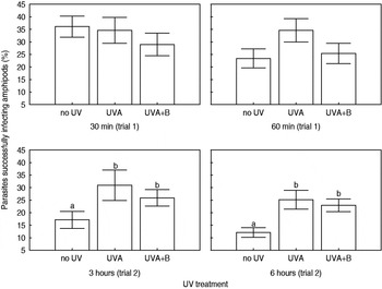
UVR influenced the susceptibility of amphipods in terms of the proportion of cercariae successfully infecting the amphipods, but only after several hours of exposure to UVR (Fig. 3). The pattern observed suggests that, after reaching a certain threshold of exposure to UVR, susceptibility of amphipods increases. After only relatively short exposures (30 and 60 min, trial 1), UV treatment or dose did not have a significant effect on the susceptibility of amphipods (Fig. 3). In this trial, 94% of a total of 155 amphipods dissected were infected (average number of parasites per amphipod: 4·0±0·2). After 3 and 6 h (trial 2), there was a significant effect of the UV treatment, but not of any other factor tested (arcsine-square root transformed proportion of successful parasites, GLM; UV treatment: F2,165=6·84, P=0·001, dose: F1,165=1·01, P=0·316; UV×dose: F2,165=0·20, P=0·822; sex: F1,165=0·17, P=0·683, size: F4,165=0·42, P=0·794). The Tukey's post-hoc test revealed a significant difference between the no UV treatment and the UVA and UVA+B treatments (P< 0·05), but not between the UVA and UVA+B treatments (Fig. 3). In total, 176 amphipods were dissected in this second trial of which 86·5% (no UV, n=52), 96·3% (UVA, n=54), 91·4% (UVA+B, n=70) were infected. The average number of parasites per amphipods was 3·0±0·4 (no UV), 5·5±0·7 (UVA) and 4·9±0·4 (UVA+B). Due to differences in the number of cercariae originally added to amphipods in the two trials, the resulting proportions are not directly comparable between trials.
Susceptibility of Paracalliope novizealandiae amphipods to infection with Maritrema novaezealandensis, measured as the proportion of parasites successfully infecting the amphipods after a low or a high dose exposure of amphipods to no UV, UVA or UVA+B radiation (mean±s.e.). Exposure times in trial 1 were 30 and 60 min. In trial 2, amphipods were exposed for 3 and 6 h. Trials are not directly comparable due to differences in numbers of cercariae originally added. The letters denote significant differences between treatments according to Tukey's post-hoc test (significance level P<0·05).

Amphipod survival
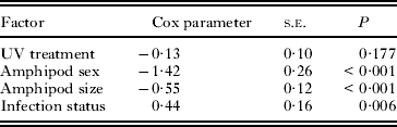
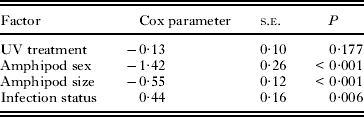
There was no significant difference in the risk of dying for amphipods exposed to different UV treatments during the 24-day outdoor experiment (Table 2; model χ 2=38·58, d.f.=4, P<0·001). In contrast, sex, size and infection status had significant effects with infected amphipods, male and larger amphipods having a higher risk of dying than uninfected ones, females or smaller size classes. There was no significant correlation between the number of days alive and the number of parasites present in infected amphipods (Spearman's P=−0·21, P=0·063). In total, 293 amphipods were dissected (139 females, 150 males, 4 unknown) of which 43% were infected (no UV: 38% (n=116), UVA: 48% (n=83), UVA+B: 44% (n=88) with a mean infection intensity of 5·1±0·4 parasites per infected amphipod.
Results of the Cox proportional hazard model assessing the effects of the UV treatment (no UV, UVA, UVA+B), sex, size and infection status of Paracalliope novizealandiae amphipods on the risk of dying during the outdoor experiment
Net effect on transmission
There was no significant difference in the proportion of parasites successfully infecting amphipod hosts for cercariae and amphipods simultaneously exposed to ambient conditions under the different UV filters. Moreover, neither size nor sex of amphipods had a significant effect (arcsine-square root transformed proportion of successful parasites, GLM; UV treatment: F2,177=1·30, P=0·276, size: F3,177=0·44, P=0·722, sex: F1,177=1·77, P=0·185). Proportions of successful parasites were 0·20±0·02 (no UV), 0·20±0·01 (UVA) and 0·24±0·02 (UVA+B). In total, 185 amphipods were dissected (n=63, 59 and 63 for the no UV, UVA and UVA+B treatments, respectively), which harboured on average 5·1±0·38 (no UV), 5·1±0·35 (UVA) and 6·0±0·43 (UVA+B) parasites.
DISCUSSION
The transmission of parasites in situ is taking place under exposure to prevailing environmental conditions, including incident solar irradiance. These conditions can influence the parasite, but also the host, and may thus affect their interaction. In the present study, 2 steps of the transmission process investigated were affected by UVR: the survival of the cercarial transmission stages of M. novaezealandensis and the susceptibility of amphipod hosts to infection. This indicates that UVR should be considered an important ecological modulator of parasite transmission in natural systems.
Effects on the parasite
There was a strong negative dose-dependent effect of exposure to UVR on the survival of cercariae. This result is supported by an additional experiment run under ambient conditions on a warm sunny day which confirmed the marked decline in survival and activity when comparing cercariae exposed to no UV, only UVA or UVA+B: after 2 h of exposure in 96-well plates, 97% (no UV), 84% (only UVA) and 14% (UVA+B) of cercariae were fully active, whereas after 3 h of exposure, only 61% (no UV), 4% (only UVA) and 0% (UVA+B) were still fully active. In the laboratory experiment, even exposure to a high dose of no UV (i.e. PAR only) reduced the survival of cercariae when compared to a low dose. This suggests that once cercariae emerge into the environment, an event which does not itself seem to be conclusively influenced by UVR (see Results section), one of the factors determining the life span of these cercariae will be the amount of solar radiation and particularly of UVR that prevails at that time. The high vulnerability and the reduced survival of M. novaezealandensis cercariae are consistent with other studies reporting an increased mortality of parasitic transmission stages after exposure to UVR (e.g. Prah and James, Reference Prah and James1977; Ariyo and Oyerinde, Reference Ariyo and Oyerinde1990), and with the general notion of large, negative effects of UVR on the survival of a range of organisms (e.g. Bancroft et al. Reference Bancroft, Baker and Blaustein2007).
In contrast to the clear effect on cercarial survival, results regarding the output and infectivity of cercariae were less straightforward. The effect of UVR on cercarial output from snails may only become apparent after longer exposures, potentially in the order of weeks or months, and we can only conclude here that short-term exposure of the snail host had no clear immediate effect. Regarding infectivity, we expected to find a reduced capacity of cercariae to complete penetration of amphipod hosts after exposure to UVR, similar to what has been described for miracidia (parasite transmission stage hatching from eggs) of Schistosoma sp. These were shown to have an impaired host penetration capability due to the loss of activity after exposure to UVR, whereas the developmental potential of those that successfully infected a host was normal (Prah and James, Reference Prah and James1977). Our findings indicated that exposure to a high dose of UVA+B results in lower proportions of cercariae capable of infecting amphipods when compared to cercariae exposed to high doses of no UV or UVA only. The significant interaction was based on the fact that proportions of successful parasites were higher for no UV and UVA-exposed cercariae and lower for UVA+B-exposed cercariae compared to the low dose treatments. This may indicate a stimulating effect of PAR and UVA on cercarial infectivity promoting their successful transmission to the next host when exposed to ambient conditions, whereas a high dose of UVA+B has detrimental effects on the cercariae. However, we have been unable to confirm the responses observed in the present experiment in any subsequent trial conducted (A. Studer, unpublished data) and thus no consistent effect of UVR has been found on the subtle functional aspect of infectivity of M. novaezealandensis. It is possible that UVR may not harm cercariae in their functionality until a certain threshold is reached, after which not just infectivity, but survival as such is impaired. However, different approaches may be needed to reach more conclusive results on these aspects of the transmission process.
Effects on the host
The susceptibility of P. novizealandiae amphipods to infection was increased after a prolonged exposure to UVR compared to prolonged exposure to no UV. A range of mechanisms could account for this observation. For example, exposure to UVR may exert immediate behavioural or physiological stress reactions that allow for easier location of the host and subsequent penetration by the cercariae. Alternatively, UVB has been shown to cause epidermal tissue damage in fish (e.g. McArdle and Bullock, Reference McArdle and Bullock1987; Ewing et al. Reference Ewing, Blazer, Fabacher, Little and Kocan1999; McFadzen et al. Reference McFadzen, Baynes, Hallam, Beesley and Lowe2000; Sommaruga, Reference Sommaruga, Helbling and Zagarese2003 and references therein). This may increase a host's susceptibility to bacterial or parasitic infections which may cause further tissue damage, increasingly weakening the functionality of the epidermal tissue as a physical barrier (Kramer, Reference Kramer, Beukema, Wolff and Brouns1990 and references therein). Similar effects may facilitate penetration of cercariae in organisms such as crustaceans. Alternatively, UVR is known to be immunosuppressive in some organisms (e.g. Patz et al. Reference Patz, Epstein, Burke and Balbus1996; Salo et al. Reference Salo, Aaltonen, Markkula and Jokinen1998); thus stress reactions in crustacean hosts due to the exposure to UVR may suppress immune responses and thus limit the ability of a host to deal with parasites after their successful penetration.
Various species of zooplankton and benthic invertebrates are known to be sensitive to UVR (e.g. Bothwell et al. Reference Bothwell, Sherbot and Pollock1994; Leech and Williamson, Reference Leech and Williamson2000). UVR has also been shown to affect the survival, fecundity and sex ratio in some intertidal copepods, but these effects were highly species-specific (Chalker-Scott, Reference Chalker-Scott1995). Amphipods are translucent to UVR and therefore intertidal species are thought to be particularly prone to stress due to the fluctuating exposure levels (Obermueller et al. Reference Obermueller, Karsten and Abele2005). Nonetheless, no effect of UVR was found on the survival of P. novizealandiae amphipods in our study. Infected amphipods had, however, an increased risk of dying during this experiment. Organisms have evolved strategies to cope with UVR and the occurrence of these mechanisms often depends on the radiation levels to which organisms are normally exposed to (e.g. Siebeck et al. Reference Siebeck, Vail, Williamson, Vetter, Hessen, Zagarese, Little, Balseiro, Modenutti, Seva and Shumate1994; Gleason and Wellington, Reference Gleason and Wellington1995; Helbling et al. Reference Helbling, Zaratti, Sala, Palenque, Menchi and Villafane2002b). Paracalliope novizealandiae lives in high UVR environments, i.e. shallow intertidal soft-sediment habitats and tidal pools during low tide, and thus should be adapted to high levels of UVR. Several protective mechanisms may contribute to this. Vegetation present in this habitat allows for behavioural adaptations (i.e. seek protective shading). Amphipods may be capable of photo-repairing damage therefore mitigating the effect of UVR on their survival (e.g. Obermueller et al. Reference Obermueller, Karsten and Abele2005). Also, these amphipods contain photo-protective compounds such as mycosporine-like amino acids (A. Studer and V. Cubillos, unpublished data) and it is likely that other protective compounds (e.g. carotenoids) or repair mechanisms are present, similar to what has been described for other marine, herbivore amphipod species (e.g. Helbling et al. Reference Helbling, Menchi and Villafane2002a; Obermueller et al. Reference Obermueller, Karsten and Abele2005).
Net effects
The net effect of UVR on the transmission process of a parasite from one host to the next depends on the differential response and sensitivity of each species and step involved. Based on our results, we suggest that the negative effect of UVR on the survival of M. novaezealandensis cercariae is the most pronounced response, but that this negative effect on cercarial survival might be compensated, at least to some degree, by the increased susceptibility of amphipod hosts to infection. While this research was not designed to quantify total net effects, it is possible that no clear impact of UVR on the transmission process would be expected, a conclusion which was supported by the additional outdoor experiment conducted to assess the net effect of UVR on the transmission process (see Results section).
UVR does not act in isolation under natural conditions (see e.g. Przeslawski et al. Reference Przeslawski, Davis and Benkendorff2005). Our study is therefore limited by the fact that interactive effects with other environmental factors, especially temperature, were not incorporated and that many experiments were only conducted under laboratory conditions. For example, during optimal conditions for the transmission of M. novaezealandensis in the field, i.e. during low tide on warm sunny days when water in the shallow tide pools warms up, temperature and UVR may synergistically affect the organisms and their interaction differently than what has been described here. In particular, cercarial survival is likely to be strongly negatively affected by synergistic effects of UVR and temperature; however, increased infectivity at an optimum temperature level (Studer et al. Reference Studer, Thieltges and Poulin2010) may, on the other hand, counterbalance this reduced survival of cercariae. Such interactions, as well as many other aspects of the ecological role of UVR for parasitism and disease transmission in marine ecosystems (as well as other ecosystems), remain to be elucidated. Appropriate experiments should be conducted in laboratory settings closely matching relevant ambient field conditions, and preferably, whenever feasible, should be repeated under natural conditions.
In summary, we found that UVR negatively influenced both the parasite and its amphipod host. The survival of cercariae was reduced and the susceptibility of amphipod hosts to infection increased. UVR should therefore be considered an important ecological component in the transmission process of intertidal and possibly other parasites. Although it remains unclear how these effects may manifest in nature, the overall net effect of UVR on the host-parasite system studied here may be considered neutral, with the negative effect on cercarial survival being compensated by the increased susceptibility of amphipods.
ACKNOWLEDGEMENTS
We are grateful to an anonymous reviewer for constructive comments that have considerably improved the manuscript. We would also like to thank the staff at the Portobello Marine Laboratory for their technical and logistical support for this study. We also like to thank David J. Burritt for advice and helpful discussions. This research was funded by the University of Otago, New Zealand, and the Department of Zoology for postgraduate research by A.S.A.S. also acknowledges the support from a Postgraduate Publishing Bursary for the preparation of this manuscript.