Aortic arch anomalies constitute 15–20% of CHDsReference Dubey, Gupta and Kothari 1 ; they are secondary to developmental errors of the branchial arches, including the persistence of normally involuting structures. In the absence of a vascular ring, patients are typically asymptomatic. A right aortic arch with complete isolation of the left innominate artery is rare and sometimes associated with genetic conditions, including additional intracardiac defects.Reference Dubey, Gupta and Kothari 1 This is the first report of right aortic arch with complete isolation of the left innominate artery associated with global cerebral white matter atrophy.
Case
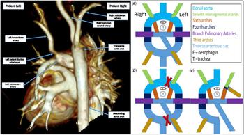
Our patient was born at term gestation via caesarean section to a 28-year-old nulliparous mother; the pregnancy was complicated by type 2 diabetes requiring metformin and insulin therapy. A prenatal ultrasound showed right lateral cerebral ventriculomegaly but was otherwise normal. The delivery and immediate postnatal course were unremarkable. Given the history of maternal diabetes, a transthoracic echocardiogram was obtained after birth, which demonstrated a right aortic arch with isolated origin of the left innominate artery from the left pulmonary artery via a small left-sided ductus arteriosus. There was bidirectional flow in the left ductus arteriosus (Fig 1, second panel).
The first panel shows the patient’s initial chest CTA showing a right aortic arch with isolated left innominate from the aortic arch. The left innominate is connected to the pulmonary artery by a left patent ductus arteriosus. The second panel shows ( a ) the initial Edward’s hypothetical double aortic with ( b ) regression of the left truncus arteriosus sac or ventral aortic root proximal to the left common carotid artery and the dorsal aorta distal to the left subclavian artery leading to ( c ) a right aortic arch with isolated left innominate. CTA=CT angiography.

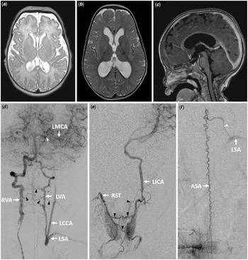
CT angiography of the chest, head, and neck confirmed this diagnosis. The base of the left innominate artery was approximately 7 mm away from the transverse aortic arch (Fig 1), a distance that increased to 23 mm at 14 months of age. There was adequate perfusion of the patient’s left arm, with blood pressure measurements consistently 20–30 mmHg lower than in the right arm. Given a concern for cerebral perfusion, a brain MRI was performed at 7 days of life, showing mild dilation of the lateral ventricles and thinning of the corpus callosum, consistent with mild global volume loss without evidence of stroke (Fig 2a). Although surgical intervention was considered in this patient, it was determined at that time that the distance between the isolated innominate artery and the aortic arch complicated reimplantation with potential need for a conduit and future upsizing with somatic growth over time. Close outpatient follow-up with neurodevelopmental surveillance was arranged.
( a ) Axial T2-weighted magnetic resonance image obtained at 7 days of life reveals mild dilation of the supratentorial ventricular system and diffusely thin corpus callosum. ( b ) Axial T2-weighted and ( c ) sagittal T1-weighted images obtained at 13 months of age show progression of the diffuse white matter volume loss and commensurate ventricular dilatation with diffuse thinning of the corpus callosum. ( d ) digital subtraction angiography (DSA), right vertebral artery injection, with collateral flow to the left vertebral, subclavian, and carotid arteries, also involving the cervical segment of the anterior spinal artery (cervical anterior spinal artery steal). Note the opacification of the left middle cerebral artery via the left posterior communicating artery. ( e ) DSA, selective injection of the right superior thyroid artery, collateral flow to the left carotid artery via the left superior thyroid artery (thyroid steal). ( f ) DSA, right T8 injection, documenting a prominent anterior spinal artery supplying the left subclavian artery via an enlarged left C7 radiculomedullary artery. ASA=anterior spinal artery; DSA=digital subtraction angiography; LCCA=left common carotid artery; LICA=left internal carotid artery; LMCA=left middle cerebral artery; LSA=left subclavian artery; LVA=left vertebral artery; RST=right superior thyroid artery; RVA=right vertebral artery.

The patient met appropriate developmental milestones with no focal neurologic deficits. Interestingly, the arm circumference was approximately 1 cm greater on the right, with a persistent 20–30 mmHg difference in systolic blood pressure measurements. As part of her neurologic surveillance, a contrast-enhanced head ultrasound was performed at 4 months of age, showing homogenous and symmetric perfusion. At 13 months of age, her parents reported intermittent dragging of her left foot, though her neurologic examination was normal at that time. Brain MRI at that time demonstrated global cerebral white matter atrophy, thinning of the corpus callosum, and commensurate ex-vacuo ventricular dilation, a progression from the initial scan (Fig 2b and c). MR perfusion studies showed a mildly prolonged time to peak in the left middle cerebral artery territory but symmetric cerebral blood flow, volume, and mean transit time. Cerebral angiography showed multiple collateral channels supplying the left innominate artery, with evidence of a subclavian steal phenomenon (i.e., retrograde flow from the left vertebral artery opacifying both the left subclavian and internal carotid arteries) (Fig 2d). Spinal angiography was also performed due to concerns for transient gait deficit and abnormal spinal vessels on non-invasive studies. It revealed a thyroid steal phenomenon with collateral flow to the left internal carotid artery via the left superior thyroid artery (Fig 2e). It also showed a dilated thoracolumbar anterior spinal artery, best opacified from a right T8 injection and supplying the left subclavian artery via a prominent left C7 radiculomedullary artery, consistent with an angiographic spinal steal phenomenon (Fig 2f). Genetic and metabolic testing investigating alternative causes of cerebral white matter atrophy were negative. Of note, plasma lactate level drawn at 15 months of age was elevated at 3.4, consistent with hypoperfusion.
Discussion
A right aortic arch with isolated left innominate artery is a rare aortic arch anomaly. In 1948, Edwards proposed a model of aortic arch development that included a hypothetical double aortic arch system, with corresponding third, fourth, and sixth embryonic arches and bilateral ductus arteriosi.Reference Gil-Jaurena, Ferreiros, Zabala and Cuenca 2 , Reference Türkvatan, Büyükbayraktar, Öl̈сer and Cumhur 3 Based on this model, the resorption and/or persistence of portions of the arches would lead to either a normal aortic arch or an aortic arch anomaly. An isolated left innominate artery is theoretically formed by the regression of the ventral aortic root proximal to the left common carotid artery and the dorsal aortic root distal to the left subclavian arteryReference Gil-Jaurena, Ferreiros, Zabala and Cuenca 2 , Reference Türkvatan, Büyükbayraktar, Öl̈сer and Cumhur 3 (Fig 1, panel 2).
With no direct connection of the isolated innominate artery to the aortic arch, blood flow to the left innominate artery is supplied retrogradely from the circle of Willis and from cervical collateral pathways. It is known from cases of aortic coarctation repaired by a left subclavian flap technique that, despite the sacrifice of the left subclavian artery, the left upper extremities of these patients remain adequately perfused secondary to collateral flow via the left vertebral artery, and the thyrocervical and costocervical trunks.Reference Diemont, Chemla and Pierre 4 However, cases of left upper extremity claudication during left arm exercise have been reported, typically after the first decade of life, not requiring intervention.Reference Diemont, Chemla and Pierre 4 Despite the subtle difference between our patient’s left and right arm circumferences, there was no difference in strength, tone, or reflexes.
Global cerebral white matter atrophy documented by MRI is concerning for a cerebral steal phenomenon. Serial brain MRIs showed no progression of atrophy between 13 and 16 months of age, but long-term effects, notably regarding neurodevelopmental and cognitive outcomes, are unknown. Spinal angiography documented an anterior spinal artery steal phenomenon, which has been characterised by bilateral spastic paraparesis and dissociated sensory deficits but preserved proprioception in adults.Reference Mohassel, Wesselingh, Katz, McArthur and Gailloud 5 Surgery was discussed but thought to be challenging due to the 23 mm distance between the left innominate artery and the aortic arch. It is uncertain that surgical intervention would prevent further cerebral white matter loss and alter the natural history of this rare disease. After extensive discussions with the patient’s family and several multidisciplinary conferences involving providers from cardiology, neurology, cardiothoracic surgery, and interventional neuroradiology, it was decided to proceed with conservative management including close neurodevelopmental surveillance.
Conclusion
A right aortic arch with an isolated left innominate artery is a very rare aortic arch anomaly, with an unclear neurodevelopmental prognosis. While the presence of global cerebral white matter atrophy raises the concern for future neurodevelopmental deficits, surgical options remain challenging with unclear long-term benefits.
Author ORCIDs
Edem Binka 0000-0002-9940-2506
Acknowledgments
None.
Financial Support
E.B. was supported by the T32 HL125239 from NIH/NHLBI while conducting this study.
Conflicts of Interest
None.