Introduction
Endolymphatic hydrops is a typical marker of Ménière's disease. This has been demonstrated in autopsy specimens,Reference Fraysse, Alonso and House1–Reference Morita, Kariya, Farajzadeh Deroee, Cureoglu, Nomiya and Nomiya4 and in vivo using 3 T magnetic resonance imaging (MRI) following intratympanic or intravenous gadolinium administration.Reference Nakashima, Naganawa, Sugiura, Teranishi, Sone and Hayashi5, Reference Nakashima, Naganawa, Teranishi, Tagaya, Nakata and Sone6 In particular, MRI is a reliable method for advancing our knowledge of the clinical manifestation of Ménière's disease related to endolymphatic hydrops.Reference Fiorino, Pizzini, Beltramello and Barbieri7, Reference Fiorino, Pizzini, Beltramello and Barbieri8 Imaging of endolymphatic hydrops relies on the enlargement of the space occupied by non-enhancing endolymph-filled membranous structures, at the expense of visualisation of enhancing perilymphatic compartments.Reference Nakashima, Naganawa, Sugiura, Teranishi, Sone and Hayashi5, Reference Nakashima, Naganawa, Teranishi, Tagaya, Nakata and Sone6 The fact that increased severity of perilymphatic enhancement defects of cochlear and vestibular structures is associated with increased duration of Ménière's disease indicates that endolymphatic hydrops is a progressive degenerative phenomenon.Reference Fiorino, Pizzini, Beltramello and Barbieri7 Endolymphatic hydrops progression, as a function of Ménière's disease duration, does not preclude the possibility of observing spontaneous or drug-induced fluctuations of the hydrops. Anecdotal cases of altered endolymphatic hydrops severity on MRI during remission of the disease,Reference Sone, Naganawa, Teranishi, Nakata, Katayama and Nakashima9 or following medical treatment with osmotic diuretics,Reference Miyagawa, Fukuoka, Tsukada, Oguchi, Takumi and Sugiura10 have been reported.
Endolymphatic hydrops depletion tests have been clinically used to improve the diagnosis of Ménière's disease. Typically, acute glycerol administration is used; it is believed that this acts on the cochlear hydrops, as it improves auditory thresholds and reduces summating potentials on electrocochleography.Reference Kimura, Aso and Watanabe11 However, the frusemide test, associated with caloric stimulation and vestibular-evoked myogenic potentials, has been used to reveal vestibular hydrops.Reference Futaki, Kitahara and Morimoto12–Reference Seo, Node, Yukimasa and Sakagami15
The present paper investigates the acute modifications induced by the intravenous administration of frusemide on endolymphatic hydrops, as revealed by three-dimensional fluid-attenuated inversion recovery (3D-FLAIR) sequence imaging conducted in a 3 T MRI unit, following intratympanic gadolinium administration. The study aimed to assess the efficacy of frusemide in reducing endolymphatic hydrops, and to contribute to our understanding of the role of endolymphatic hydrops in Ménière's disease.
Materials and methods
Twelve patients (5 males and 7 females, aged 19–74 years) with Ménière's disease (diagnosed according to the Committee on Hearing and Equilibrium guidelines16) were examined with 3D-FLAIR MRI using intratympanic gadolinium as a contrast medium. The inclusion criteria were: unilateral disease, recurrent vertigo attacks, the absence of disorders associated with secondary endolymphatic hydrops, and no history of middle-ear or neurological disorders.
Disease duration ranged from 0.5 to 8 years, with a frequency of 0.5 to 6 vertigo spells per month, as calculated in the last 6 months. The pure tone average at 500 to 3000 Hz ranged from 20 to 60 dB HL in the affected ear. The contralateral ear showed a pure tone average equal to or better than 15 dB HL.
Most patients had been treated with different medical therapy regimens, mainly based on a low-salt diet and diuretics; these were discontinued at least one month prior to the MRI examination. However, symptomatic drugs, such as benzodiazepine and anti-emetics, were occasionally administered after an acute vertigo spell during this period.
Intratympanic gadolinium solution was administered to the affected ear of all patients 24 hours before the MRI investigation (as per Nakashima et al.Reference Nakashima, Naganawa, Sugiura, Teranishi, Sone and Hayashi5). The hospital medical management team was informed regarding the ‘off-label’ intratympanic use of gadolinium, and all patients gave written informed consent. The method of intratympanic gadolinium administration has been reported in previous articles.Reference Fiorino, Pizzini, Beltramello and Barbieri8, Reference Fiorino, Pizzini, Beltramello, Mattellini and Barbieri17 Briefly, 0.6 ml of 1:7 gadobutrol (1 mmol/ml) was injected through the tympanic membrane, using a 22-gauge spinal needle. The patient was kept with their head rotated 45 degrees contralaterally for 30 minutes after the injection.
The MRI was performed with a 3 T unit (Allegra; Siemens, Erlangen, Germany), using a four-channel, receive-only, phased-array coil. The 3D-FLAIR sequences were acquired using the generalised auto-calibrating, partially parallel acquisition imaging technique, with an acceleration factor of 4. The sequence parameters were as follows: voxel size = 0.4 × 0.4 × 2 mm, signal-to-noise ratio = 1, scan time = 20 minutes, 12 slices, repetition time = 9000 ms, echo time = 128 ms, inversion time = 2500 ms, flip angle = 180 degrees, slice thickness = 2 mm, echo train length = 23, field of view = 16 cm and matrix size = 384 × 384.
Frusemide 20 mg was given intravenously immediately after the completion of MRI. The MRI investigation was then repeated after 1 hour, using the same parameters and sequence as for the first investigation.
The MRI scans were evaluated by a neuroradiologist with long-standing experience (FBP), who was blinded to patient identity and clinical data. Each portion of the labyrinth (i.e. the basal, middle and apical turns of the cochlea; the vestibule; and the superior, posterior and lateral semicircular canals) was evaluated separately and judged to have either normal, reduced or absent enhancement. Enhancement was considered absent when the examined structure was not visible. When the endolymphatic content of the considered structure was clearly visible, the ratio of its area to the total area was calculated, and a value exceeding one-third was attributed to the endolymphatic hydrops and classified as reduced enhancement.Reference Fiorino, Pizzini, Beltramello, Mattellini and Barbieri17 If there was no visual evidence of separation between the endolymphatic and the perilymphatic compartments, the contrast-to-noise ratio of the considered structure was calculated, according to methods reported in a previous paper.Reference Fiorino, Pizzini, Beltramello, Mattellini and Barbieri17 The signals of the different portions of the inner ear were considered reduced when inferior to the tolerance limit of 54.1 contrast-to-noise ratio.Reference Fiorino, Pizzini, Beltramello, Mattellini and Barbieri17
Results
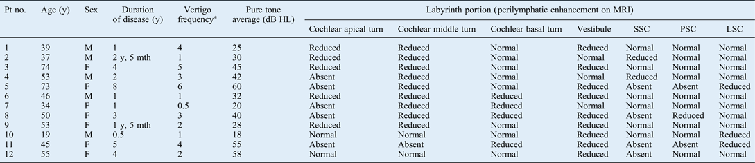
All Ménière's disease patients showed impaired perilymphatic enhancement of variable degrees in the affected inner ear, with absent or reduced signals at the level of vestibular and/or cochlear sites (Table I). The number of involved sites for each single ear spanned from 2 to 7. None of the patients showed MRI changes 1 hour after intravenously administered frusemide.
Ménière's disease patients' characteristics and 3D-flair mri findings
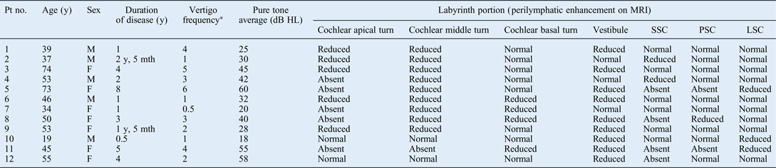
No modification of perilymphatic enhancement could be observed 1 hour after the intravenous administration of frusemide. *Number of spells per month. 3D-FLAIR = three-dimensional fluid-attenuated inversion recovery; MRI = magnetic resonance imaging; Pt no. = patient number; y = years; SSC = superior semicircular canal; PSC = posterior semicircular canal; LSC = lateral semicircular canal; M = male; mth = months; F = female
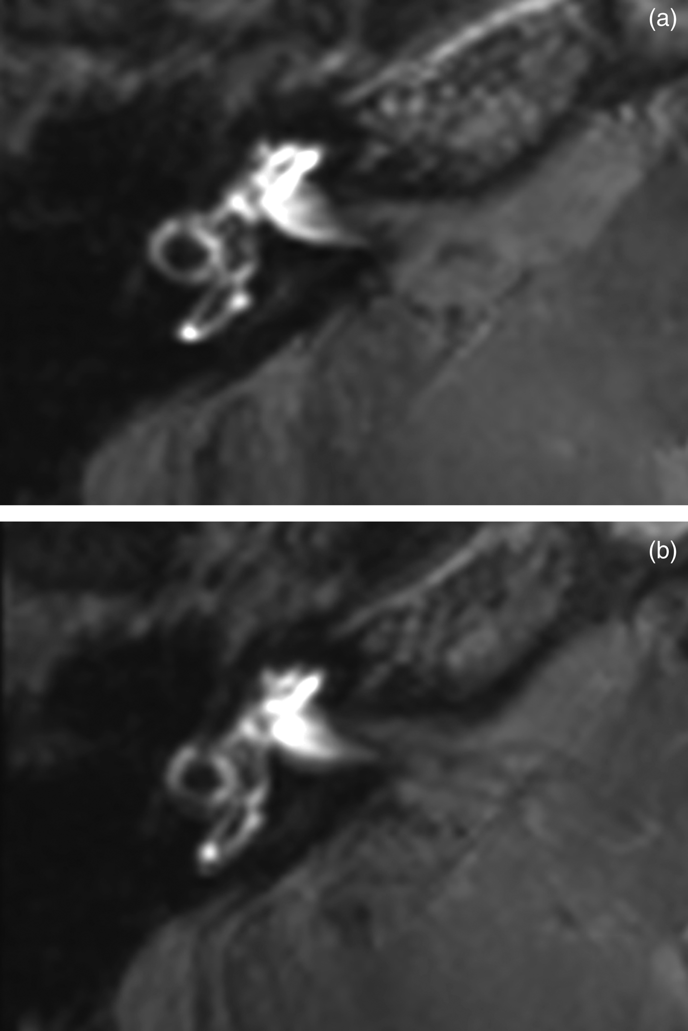
Figure 1 shows the MRI findings observed 24 hours after intratympanic gadolinium administration in a patient with right-sided Ménière's disease (patient 12). Enhancement of the cochlea, and lateral and posterior semicircular canals was normal. Perilymphatic enhancement was absent in the superior semicircular canal and markedly reduced in the vestibule, suggesting the presence of endolymphatic hydrops (Figure 1a). The imaging picture 1 hour after frusemide administration was identical to that observed prior to frusemide administration (Figure 1b).
Three-dimensional fluid-attenuated inversion recovery magnetic resonance imaging scans taken 24 hours after intratympanic gadolinium injection in a 55-year-old female with right-sided Ménière's disease (patient 12). A comprehensive axial view was obtained by processing the images passing through the inner ear by maximum intensity projection. (a) Basal images obtained before frusemide administration. Perilymphatic enhancement was absent in the superior semicircular canal and markedly reduced in the vestibule. In particular, endolymphatic compartments occupied 62 per cent of the vestibule. (b) Images obtained 1 hour after intravenously administered frusemide. No differences could be observed between the two (pre- and post-frusemide) sequences.

Discussion
Endolymphatic hydrops is consistently linked to Ménière's disease, although it is not completely clear whether the former is a cause or a mere epiphenomenon of the latter.Reference Foster and Breeze18, Reference Merchant, Adams and Nadol19 Originally, the so-called ‘central dogma’ implied a close correspondence between endolymphatic hydrops and symptoms and signs of Ménière's disease, with endolymphatic hydrops believed to cause clinical manifestations such as episodic vertigo and hearing loss. However, temporal bone studies have evidenced that endolymphatic hydrops may occur in the absence of typical Ménière's disease symptoms.Reference Foster and Breeze18, Reference Alpay and Linthicum20 In addition, experimental investigations have shown that the dysregulation of inner-ear fluids caused by cytochemical and ultrastructural disruption of the fibrocytes of the spiral ligament and hair cells precedes endolymphatic hydrops, which may therefore be just a consequence of the alterations leading to Ménière's disease.Reference Ichimiya, Adams and Kimura21, Reference Nadol, Adams and Kim22 This has prompted some authors to consider endolymphatic hydrops an epiphenomenon or marker of Ménière's disease.Reference Merchant, Adams and Nadol19 Recently, however, a comprehensive review of articles containing descriptions of temporal bones from patients with endolymphatic hydrops and/or a history of Ménière's disease has revalued the hypothesis that endolymphatic hydrops is a causative factor, mainly based on the consistent presence of endolymphatic hydrops in virtually all temporal bones of Ménière's disease patients.Reference Foster and Breeze18
The frequent use of treatments aimed at relieving hydrops in Ménière's disease patients supports the hypothesis that endolymphatic hydrops causes clinical manifestations of Ménière's disease. For example, osmotic and loop diuretics, and carbonic anhydrase inhibitors, are currently utilised to reduce acute manifestations of Ménière's disease and/or prevent vertigo attacks, as they are believed to decrease the volume of the endolymphatic space.Reference Martin Gonzàlez, Gonzalez, Trinidad, Ibáñez, Pinilla and Martínez Ruiz-Coello23 However, definite evidence of the efficacy of these therapeutic solutions is lacking.Reference Burgess and Kundu24 Based on the supposed depletive effect on endolymphatic hydrops, diuretics administration is also used as a diagnostic tool to confirm the presence of endolymphatic hydrops, by assessing: improvements in hearing threshold, caloric excitability and vestibular-evoked myogenic potentials, and reductions in the summating potential on electrocochleography. Frusemide or glycerol tests are often carried out for this purpose.Reference Futaki, Kitahara and Morimoto12–Reference Seo, Node, Yukimasa and Sakagami15
On the basis of these assumptions, an imaging technique, such as 3D-FLAIR MRI performed after intratympanic gadolinium administration, should be able to visualise the depletive effect of diuretics on endolymphatic hydrops, in terms of improved perilymphatic enhancement. In particular, frusemide should lead to reduced enhancement defects in the vestibule and semicircular canals, based on its presumed prevalent activity on these portions of the inner ear.Reference Kimura, Aso and Watanabe11–Reference Seo, Node, Yukimasa and Sakagami15 Frusemide is a sulfamoyl anthranilic acid with a potent and rapid diuretic effect. It depresses the reabsorption of sodium, water and chloride in both the proximal and distal tubules, including the ascending limb of the loop of Henle.Reference Stason, Cannon, Heinemann and Laragh25 Frusemide also influences extracellular fluids, decreasing blood pressure, and cerebrospinal fluid and ocular pressure. In accordance with these properties, frusemide may influence endolymphatic hydrops as a consequence of systemic diuresis. An alternative hypothesis is the reduced production of endolymph based on the effect of loop diuretics on the stria vascularis. This structure undergoes significant but reversible changes following the administration of loop diuretics, such as shrinkage of the marginal cells and swelling of intermediate cells and intrastrial space.Reference Pike and Bosher26, Reference Santi and Lakhani27
In the present investigation, 12 patients with unilateral, active Ménière's disease were examined with 3D-FLAIR MRI performed after intratympanic gadolinium injection. Contrary to expectations, no subject showed improvement in endolymphatic hydrops 1 hour after the intravenous administration of frusemide. This is quite surprising as the frusemide test is reported to have a sensitivity of approximately 50 per cent in Ménière's disease, as detected by improvements in vestibular-evoked myogenic potential amplitudeReference Seo, Node, Yukimasa and Sakagami15 or caloric reflectivity.Reference Ito, Watanabe, Shojaku, Kobayashi, Aso and Mizukoshi13, Reference Tsunoda, Fukaya and Komatsuzaki14 The positive rate depends on Ménière's disease activity being higher for ears with active vestibular disease than for those with inactive disease.Reference Seo, Node, Yukimasa and Sakagami15 The lack of significant modification of endolymphatic hydrops after frusemide administration may be attributable to inefficacy of the drug in reducing hydrops, or to limited sensitivity of the MRI technique. Further developments of imaging may shed light on this issue in the near future. The present results are, however, in agreement with previous imaging investigations which showed that endolymphatic hydrops is a progressive degenerative phenomenon,Reference Fiorino, Pizzini, Beltramello and Barbieri7 and is scarcely correlated with acute manifestations of Ménière's diseaseReference Fiorino, Pizzini, Beltramello and Barbieri8 and the effect of treatment.Reference Fiorino, Pizzini, Barbieri and Beltramello28, Reference Gurkov, Flatz, Keeser, Strupp, Ertl-Wagner and Krause29 For example, there is no evidence of a reduction of endolymphatic hydrops following the administration of intratympanic gentamicin or oral betahistine.Reference Fiorino, Pizzini, Barbieri and Beltramello28, Reference Gurkov, Flatz, Keeser, Strupp, Ertl-Wagner and Krause29
In accordance with histopathological investigations that demonstrated a relationship between Ménière's disease duration and increased volume of the cochlear duct and saccule,Reference Fraysse, Alonso and House1, Reference Okuno and Sando3, Reference Morita, Kariya, Farajzadeh Deroee, Cureoglu, Nomiya and Nomiya4 imaging studies have shown that endolymphatic hydrops severity increases as a function of the time elapsed since symptom onset.Reference Fiorino, Pizzini, Beltramello and Barbieri7, Reference Fiorino, Pizzini, Beltramello and Barbieri8 The enhancement defect of the vestibule on MRI is precocious and becomes severe at five years, whereas progression of endolymphatic hydrops in the other inner-ear portions slows with time. For example, progression of endolymphatic hydrops from the apex to the base of the cochleaReference Fiorino, Pizzini, Beltramello and Barbieri7 is in line with the well-known phenomenon of low-tone hearing loss in the initial stages of Ménière's disease, followed by pantonal involvement in more advanced stages of the disease. In vivo imaging techniques have also shown a significant correlation between endolymphatic hydrops and other otoneurological parameters that are expressions of inner-ear degenerative phenomena, such as abnormal vestibular-evoked myogenic potentials, and disease duration and stage.Reference Fiorino, Pizzini, Beltramello and Barbieri8, Reference Gurkov, Flatz, Louza, Strupp, Ertl-Wagner and Krause30 Similarly, temporal bone studies have shown that a permanent threshold shift in the ears of patients with late-stage endolymphatic hydrops is related to the degeneration of sensory structures in the cochlea.Reference Vasama and Linthicum31 However, no correlation was found between endolymphatic hydrops severity, as revealed on MRI, and some parameters linked to acute manifestations, such as the frequency of vertigo attacks, the time interval since the last attack and the functional level scale.Reference Fiorino, Pizzini, Beltramello and Barbieri8
These outcomes suggest that the symptomatological complex characterising Ménière's disease attacks cannot be related to acute variations of endolymphatic hydrops, and that this pathological aspect of Ménière's disease is not subject to undergo significant spontaneous or drug-induced rapid modifications. In a recent study, both cochlear and vestibular endolymphatic hydrops became negative or reduced in four of seven patients who underwent endolymphatic sac surgery with sac incision, gelatine film insertion and steroid application.Reference Uno, Imai, Watanabe, Tanaka, Kitahara and Horii32 Interestingly, the number of vertigo spells was reduced in all 7 patients at 6–12 months after surgery, indicating that acute symptomatology is not fully dependent on endolymphatic hydrops.
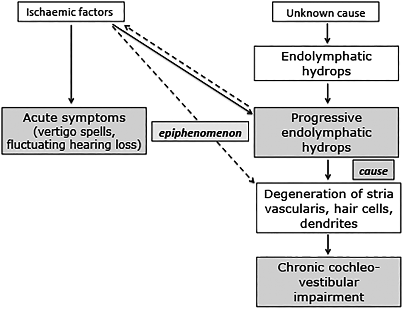
On the basis of these observations, we present a first hypothesis to answer the old question of whether endolymphatic hydrops is a cause or an epiphenomenon of Ménière's disease (Figure 2). The consistent presence of endolymphatic hydrops in Ménière's disease strongly indicates a fundamental role of this pathological marker in the manifestations of the disease. Hydrops develops from a process that is still elusive, despite numerous hypotheses indicating increased production or decreased absorption of endolymph. Endolymphatic hydrops may remain mild and asymptomatic for an indefinite period of time, being necessary but not sufficient to cause Ménière's disease.Reference Foster and Breeze18 It is possible that endolymphatic hydrops acquires a progressive course with the contribution of additional factors, such as ischaemic injury.Reference Foster and Breeze33 This may be compatible with the imaging observation of limited hydropic enlargement in the initial forms of Ménière's disease.Reference Fiorino, Pizzini, Beltramello and Barbieri7 The progression of endolymphatic hydrops with time, and the correlation of its extent and severity with some degenerative signs, as evidenced on otoneurological investigation,Reference Fiorino, Pizzini, Beltramello and Barbieri7, Reference Fiorino, Pizzini, Beltramello and Barbieri8 makes it conceivable that endolymphatic hydrops is a cause of the degenerative counterpart of Ménière's disease. A direct mechanic effect, impairing microcirculation, with degeneration of the stria vascularis, hair cells and dendrites, may be hypothesised.Reference Foster and Breeze33 Direct injury of the inner-ear structures by the same microcirculation defect may cause endolymphatic hydrops evolution and impaired autoregulation of blood flow to the inner ear provoked by the endolymphatic hydrops.Reference Foster and Breeze33–Reference Nakashima36
Schematic representation of the possible role of endolymphatic hydrops in the development of symptoms and signs of Ménière's disease. See main text for full description.
Ischaemic events contributing to endolymphatic hydrops and its pathological consequences may also be independent factors that trigger acute manifestations of Ménière's disease, such as fluctuating hearing loss and vertigo spells. The vascular theory of Ménière's disease attacks has recently been revalued by Foster and Breeze, who hypothesised that Ménière's disease spells occur when perfusion pressure has been lowered to just above the ischaemic threshold in an ear with pre-existing endolymphatic hydrops.Reference Foster and Breeze33 From this point of view, the endolymphatic hydrops should be considered as an epiphenomenon of Ménière's disease, as the same pathological processes result in both Ménière's disease symptoms and endolymphatic hydrops evolution independently, and the progression of endolymphatic hydrops does not significantly influence acute manifestations of the disease.Reference Fiorino, Pizzini, Beltramello and Barbieri7, Reference Fiorino, Pizzini, Beltramello and Barbieri8
• Endolymphatic hydrops is a typical marker of Ménière's disease
• This has been demonstrated in autopsy specimens, and in vivo using magnetic resonance imaging (MRI) after intratympanic or intravenous gadolinium injection
• Depletion tests using glycerol or frusemide are currently employed to reveal hydrops
• The present study did not show any short-term endolymphatic hydrops modification, as revealed by MRI, after intravenously administered frusemide
• The reported effects of frusemide on Ménière's disease symptoms and signs probably cannot be attributed to a reduction of endolymphatic hydrops
Conclusion
The present investigation did not show any modification of endolymphatic hydrops 1 hour after the intravenous administration of frusemide. The reported effects of this drug on the symptoms and signs of Ménière's disease probably cannot be attributed to a reduction of endolymphatic hydrops. It must be stressed, however, that a systematic review failed to demonstrate an effect of diuretics on vertigo, hearing loss, tinnitus or aural fullness in clearly defined Ménière's disease.Reference Stason, Cannon, Heinemann and Laragh25 In addition, Pirodda et al. have recently outlined the possibility of adverse effects of diuretics on inner-ear function associated with an abrupt lowering of blood pressure, with exaggerated vasomotor response inducing local ischaemia.Reference Pirodda, Ferri, Raimondi and Borghi37 Therefore, diuretics in Ménière's disease, which are today used on a purely empirical basis, must be reconsidered.