Introduction
Childhood stress and trauma significantly influence the risk of adult psychopathology. Early life stress (ELS) exposure lowers the threshold for depressive reactions to stressors later in life (Harkness et al. Reference Harkness, Bruce and Lumley2006), while the intensity of ELS predicts symptom severity of mood episodes (Martins et al. Reference Martins, Von Werne Baes, Tofoli and Juruena2014). ELS exposure can have long-lasting effects on hypothalamic–pituitary–adrenal (HPA) axis regulation (McEwen, Reference McEwen2003), which may in part explain the relationship between ELSs and depression. It is currently unclear whether depression is associated with exposure to specific stressors or whether any childhood trauma could increase vulnerability to depression. Although stressors in childhood or adolescence contribute to a wide range of adulthood psychopathology, some studies associate major depressive disorder (MDD) with exposure to certain types of ELSs including sexual abuse (Kaplow & Widom, Reference Kaplow and Widom2007), emotional abuse (Martins et al. Reference Martins, Von Werne Baes, Tofoli and Juruena2014) and family conflict (Kessler & Magee, Reference Kessler and Magee1994). In contrast, others concluded there is insufficient evidence associating specific childhood adversities with specific psychiatric disorders (Gershon et al. Reference Gershon, Sudheimer, Tirouvanziam, Williams and O'Hara2013). Such potential ELS–disorder specificity is probably related to more than just the occurrence of the stressor and may be related to the intensity, duration, developmental stage of the victim and physiological stress response at the time of the stressor. Presumably only stressors resulting in significant or sustained stress responses characterized by HPA axis or other immune system activity would influence vulnerability to psychiatric illnesses. Such stressors would also be expected to be associated with cognitive performance and magnetic resonance imaging (MRI) differences.
Poorer cognitive performance is increasingly recognized as an important aspect of MDD, characterized by poor performance on measures of executive function, processing speed and episodic memory (Snyder, Reference Snyder2013). ELS exposure is associated with poorer adult cognitive function in memory domains and executive function in populations with and without psychopathology (Navalta et al. Reference Navalta, Polcari, Webster, Boghossian and Teicher2006). However, not all studies associate ELS exposure with poorer cognitive performance and much of this work focuses on post-traumatic stress disorder. It is thus unclear whether ELS exposure influences cognitive performance in MDD.
More consistently, even in individuals without psychiatric disorders, neuroimaging studies associate ELS exposure with volumetric and functional alterations in brain regions including the anterior cingulate cortex (ACC) (Cohen et al. Reference Cohen, Grieve, Hoth, Paul, Sweet, Tate, Gunstad, Stroud, McCaffery, Hitsman, Niaura, Clark, McFarlane, Bryant, Gordon and Williams2006; Udo et al. Reference Udo, Anja, Victoria, Peter, Thomas, Dominik, Katharina, Christa, Patricia, Jochen, Christian, Christian, Carsten, Volker, Walter, Thomas and Harald2012), medial prefrontal cortex (van Harmelen et al. Reference van Harmelen, van Tol, van der Wee, Veltman, Aleman, Spinhoven, van Buchem, Zitman, Penninx and Elzinga2010), caudate (Cohen et al. Reference Cohen, Grieve, Hoth, Paul, Sweet, Tate, Gunstad, Stroud, McCaffery, Hitsman, Niaura, Clark, McFarlane, Bryant, Gordon and Williams2006) and insula (Baker et al. Reference Baker, Williams, Korgaonkar, Cohen, Heaps and Paul2013). In contrast, depressed patients exposed to ELSs exhibit smaller volumes of the orbitofrontal cortex (OFC) and prefrontal cortex (Frodl et al. Reference Frodl, Reinhold, Koutsouleris, Reiser and Meisenzahl2010; Udo et al. Reference Udo, Anja, Victoria, Peter, Thomas, Dominik, Katharina, Christa, Patricia, Jochen, Christian, Christian, Carsten, Volker, Walter, Thomas and Harald2012) and the hippocampus (Cohen et al. Reference Cohen, Grieve, Hoth, Paul, Sweet, Tate, Gunstad, Stroud, McCaffery, Hitsman, Niaura, Clark, McFarlane, Bryant, Gordon and Williams2006; Udo et al. Reference Udo, Anja, Victoria, Peter, Thomas, Dominik, Katharina, Christa, Patricia, Jochen, Christian, Christian, Carsten, Volker, Walter, Thomas and Harald2012). Jointly, these findings suggest that ELS effects on brain structure may differ between healthy and depressed populations. Although it is challenging to disentangle the effects of ELS v. the effects of depression itself, such population-specific findings may provide clues related to depression vulnerability or resilience.
We hypothesized that specific ELSs are associated with a diagnosis of MDD in adulthood. We further hypothesized that those ELSs associated with MDD would also be associated with poorer performance on cognitive tests and structural alterations in brain regions involved in mood regulation. As those ELSs by definition would increase the risk of MDD, we also tested for statistical interactions between ELS and MDD diagnosis to determine whether the effect of ELS exposure on cognition and brain structure differed between depressed and non-depressed groups.
Method
Subjects
Subjects were between 20 and 50 years of age and enrolled at Duke University (n = 112) and Vanderbilt University (n = 17) between April 2008 and December 2013. Depressed subjects had a Diagnostic and Statistical Manual of Mental Disorders, 4th edition (DSM-IV) diagnosis of recurrent MDD, as assessed by the Mini-International Neuropsychiatric Interview (M.I.N.I., version 5.0) (Sheehan et al. Reference Sheehan, Lecrubier, Sheehan, Amorim, Janavs, Weiller, Hergueta, Baker and Dunbar1998) and interview with a psychiatrist. Additional inclusion criteria included onset of first depressive episode before age 35 years and a Montgomery–Åsberg Depression Rating Scale (MADRS) (Montgomery & Åsberg, Reference Montgomery and sberg1979) score of 15 or greater. Inclusion criteria specified no psychotropic medication use in the last month; most subjects reported no use for at least 3 months or longer. Eligible control subjects had neither a history of psychiatric disorders nor a history of psychotropic medication use. Although not an entry criterion, medical co-morbidity was quantified using the Cumulative Illness Rating Scale (CIRS) (Miller et al. Reference Miller, Paradis, Houck, Mazumdar, Stack, Rifai, Mulsant and Reynolds1992).
Exclusion criteria included other lifetime DSM-IV Axis I disorders including substance abuse or dependence, although co-morbid anxiety symptoms occurring in context of depressive episodes were allowable. Subjects were excluded for Axis II disorders assessed by the Structured Clinical Interview for DSM-IV Axis II Personality Disorders (SCID-II) (First et al. Reference First, Gibbon, Spitzer, Williams and Benjamin1997). Additional exclusion criteria included: history of psychosis, acute suicidality, use of illicit substances in the last month, electroconvulsive therapy in the last 6 months, a family history of bipolar disorder, any unstable medical condition, any history of neurological illness or head injury, or MRI contraindications.
Both the Duke University and the Vanderbilt University Institutional Review Boards approved this study. All subjects provided written informed consent.
Assessment of ELS and estimation of total time depressed
Exposure to childhood stressors was assessed using the self-report Early Life Stress Questionnaire (ELSQ). The ELSQ was developed from the Child Abuse and Trauma Scale (Sanders & Becker-Lausen, Reference Sanders and Becker-Lausen1995), which has strong internal consistency, validity, and test–retest reliability. The ELSQ consists of 19 traumatic items answered yes or no occurring during childhood and adolescent ages up to 17 years. We modified the ELSQ to ask about the ages during the time exposed for each stressor, allowing us to estimate the duration of exposure in years.
Following procedures similar to past reports (Sheline et al. Reference Sheline, Sanghavi, Mintun and Gado1999), duration of lifetime depression was assessed through detailed clinical interview and acquisition of medical records. We used a life-charting approach to anchor each episode, applying diagnostic criteria to each episode.
Neuropsychological testing
A trained psychometric technician supervised by a licensed neuropsychologist administered neuropsychological testing. Similar to our approach in geriatric depression (Sheline et al. Reference Sheline, Pieper, Barch, Welsh-Bohmer, McKinstry, MacFall, D'Angelo, Garcia, Gersing, Wilkins, Taylor, Steffens, Krishnan and Doraiswamy2010), we created rationally constructed composite domain variables from a broad test battery. To combine tasks, we created Z-scores for each measure based on the performance of all subjects, then averaged the Z-scores for all tests within each domain. Internal consistency for each domain was assessed using Cronbach's coefficient α (CoA). This resulted in four composite neuropsychological measures: (a) episodic memory (logical memory 1 and 2; Benton Visual Retention Test, number correct; Rey's Verbal Learning Test, total I–V and total VII; CoA = 0.87); (b) executive function (Controlled Oral Word Association Test, total score; Trail Making B time, reverse scored time to completion; verbal fluency, total phonological and semantic; Stroop color–word interference condition, number completed; CoA = 0.75); (c) processing speed (symbol–digit modality, number completed; Trail Making A, reverse scored time to completion; Stroop color naming condition, number completed; CoA = 0.70); and (d) working memory (digit span forward, number of trials correctly completed; digit span backward, number of trials correctly completed; CoA = 0.75).
MRI acquisition
Due to differences in MRI manufacturers, only MRI data acquired at Duke University were included in analyses. Cranial MRI was performed using the eight-channel parallel imaging head coil on a whole-body MRI system (Trio, Siemens Medical Systems, USA). Parallel imaging was employed with an acceleration factor of 2. Duplicate T1-weighted image sets were acquired during the scan session using a sagittal MPRAGE sequence with repetition time/echo time = 2300/3.46 ms, a 240 Hz/pixel bandwidth, a 256 × 256 matrix, a 240 mm diameter field of view, 160 slices with a 1.2 mm slice thickness, yielding an image with voxel sizes of 0.9 × 0.9 × 1.2 mm. In eight cases, subjects did not complete MRI or scan quality was not suitable for image processing.
Structural MRI analyses
Volumetric MRI analyses
Regional volumes and cortical thickness were calculated using FreeSurfer (version 5.1) software. The FreeSurfer methods used to derive cortical and subcortical brain volumes have been previously described (Dale et al. Reference Dale, Fischl and Sereno1999; Fischl et al. Reference Fischl, Salat, Busa, Albert, Dieterich, Haselgrove, van der Kouwe, Killiany, Kennedy, Klaveness, Montillo, Makris, Rosen and Dale2002, Reference Fischl, Salat, van der Kouwe, Makris, Segonne, Quinn and Dale2004a , Reference Fischl, van der Kouwe, Destrieux, Halgren, Segonne, Salat, Busa, Seidman, Goldstein, Kennedy, Caviness, Makris, Rosen and Dale b ). Cortical parcellation used an anatomical mask derived from the Desikan–Killiany Atlas (Desikan et al. Reference Desikan, Ségonne, Fischl, Quinn, Dickerson, Blacker, Buckner, Dale, Maguire, Hyman, Albert and Killiany2006); in each hemisphere, this method identified 33 cortical and seven subcortical gray matter regions (nucleus accumbens, amygdala, caudate, hippocampus, pallidum, putamen and thalamus). Intracranial volume was assessed using the method implemented in FreeSurfer. We visually inspected the data by overlaying the surfaces and subcortical segmentations over the T1 data. Individual slices in each orientation were assessed for errors. No manual corrections were needed.
Thickness analyses
This was an exploratory approach to test for differences not captured using our atlas-based comparisons. We tested for differences in cortical thickness using FreeSurfer's QDEC module. We used a general linear model (GLM) to test for differences in cortical thickness between groups exposed or not exposed to ELSs, including age as a nuisance variable. In this method, cortical thickness is computed as the shortest distance between any point on the pial surface and the gray/white boundary and vice versa; these two values are averaged (Fischl & Dale, Reference Fischl and Dale2000). Maps were smoothed using the standard Gaussian kernel of 10 mm. We used a GLM to test for differences in cortical thickness between diagnostic groups, including age as a nuisance variable. Correction for multiple comparisons was carried out using the Monte Carlo simulation method using an initial cluster threshold set at p < 0.01. Data were tested against an empirical null distribution of maximum cluster size by running 10 000 synthesized Gaussian noise simulations, producing clusters fully corrected for multiple comparisons. Right and left hemispheres were tested separately.
Statistical analyses
All analyses were conducted using SAS 9.4 (USA). We tested for univariate differences between diagnostic groups in demographic and clinical variables using χ2 tests for categorical variables and two-tailed t tests for continuous variables. Initial tests for differences in the report of ELSs between depressed and non-depressed cohorts were conducted using χ2 tests, or Fisher's exact test when cell sizes were low.
ELSs that differed between groups in univariate tests were incorporated into GLMs predicting diagnosis (MDD or non-depressed) while controlling for age, sex, education and medical morbidity (CIRS score). Retaining these demographic variables, we conducted backward regression to develop a parsimonious model, removing each ELS item based on its statistical significance level of p = 0.05. The remaining ELS items that subsequently predicted a MDD diagnosis were termed ‘predictive ELSs’.
We next planned a hypothesis-driven approach to reduce the number of comparisons. For neuropsychological analyses, we consolidated individual test results into domain scores as described above. For MRI analyses, we selected a priori regions associated with ELS in existent literature: the ACC (Cohen et al. Reference Cohen, Grieve, Hoth, Paul, Sweet, Tate, Gunstad, Stroud, McCaffery, Hitsman, Niaura, Clark, McFarlane, Bryant, Gordon and Williams2006; Udo et al. Reference Udo, Anja, Victoria, Peter, Thomas, Dominik, Katharina, Christa, Patricia, Jochen, Christian, Christian, Carsten, Volker, Walter, Thomas and Harald2012), OFC (Frodl et al. Reference Frodl, Reinhold, Koutsouleris, Reiser and Meisenzahl2010; Udo et al. Reference Udo, Anja, Victoria, Peter, Thomas, Dominik, Katharina, Christa, Patricia, Jochen, Christian, Christian, Carsten, Volker, Walter, Thomas and Harald2012), amygdala (Tottenham et al. Reference Tottenham, Hare, Quinn, McCarry, Nurse, Gilhooly, Millner, Galvan, Davidson, Eigsti, Thomas, Freed, Booma, Gunnar, Altemus, Aronson and Casey2010), hippocampus (Sheline et al. Reference Sheline, Wang, Gado, Csernansky and Vannier1996; Cole et al. Reference Cole, Costafreda, McGuffin and Fu2011; Teicher et al. Reference Teicher, Anderson and Polcari2012) and caudate (Cohen et al. Reference Cohen, Grieve, Hoth, Paul, Sweet, Tate, Gunstad, Stroud, McCaffery, Hitsman, Niaura, Clark, McFarlane, Bryant, Gordon and Williams2006; Udo et al. Reference Udo, Anja, Victoria, Peter, Thomas, Dominik, Katharina, Christa, Patricia, Jochen, Christian, Christian, Carsten, Volker, Walter, Thomas and Harald2012). For exploratory analyses of ELS effects on other regions identified by FreeSurfer, we controlled for multiple comparisons using false discovery rate (FDR), implemented within SAS.
We first examined the effect of the total ELS exposure (defined as total ELSQ score) on cognitive domains and brain volumes. For models examining cognitive domains, we controlled for diagnosis, age, sex and education. For models examining MRI variables, we controlled for diagnosis, age, sex and intracranial volume. A similar approach was used for examining the effects of predictive ELSs on cognition and brain structure, with participants dichotomized as exposed or not exposed.
To determine whether ELS effects differed between diagnostic groups, we added a term coding for an interaction between ELS and diagnosis. This was done for analyses of both total ELSQ score and predictive ELS exposure. If the interaction did not reach statistical significance, this suggested that there was no significant difference in the relationship between ELSs, cognition and brain morphology between diagnostic groups.
Finally, for predictive ELSs, we conducted exploratory analyses using models similar to those described above, but examining the effect of duration of stressor exposure. For these analyses, individuals who denied each stressor were scored as a duration of zero. Due to the high number of people reporting the absence of trauma, we utilized a log transformation of the years of reported duration plus 1.
Results
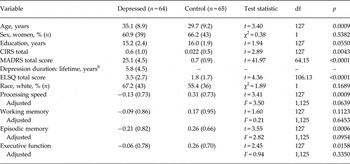
We examined 129 subjects: 64 with MDD and 65 non-depressed controls. The diagnostic groups differed in age and medical morbidity (Table 1), with the depressed group being older, having more medical illnesses, and higher total ELSQ score (range: depressed 0–11, non-depressed 0–8). In univariate analyses, depressed groups exhibited poorer performance in episodic memory, executive function and processing speed. However, these were no longer statistically significant after controlling for age and education level (Table 1).
Demographics and cognitive function a

Data are given as mean (standard deviation) unless otherwise indicated.
df, Degrees of freedom; CIRS, Cumulative Illness Rating Scale; MADRS, Montgomery–Åsberg Depression Rating Scale; ELSQ, Early Life Stress Questionnaire.
a Comparison of demographic variables utilized pooled, two-tailed t tests for continuous measures with equal variances and Satterthwaite's t tests for unequal variances. Comparison of categorical variables utilized χ2 tests. Cognitive measures were Z-transformed and presented both as unadjusted (pooled t tests) and adjusted for age and education level (general linear models with F values).
b Note n = 52 for depression duration as not all participants could provide data for past episodes.
ELSs: predicting MDD diagnosis
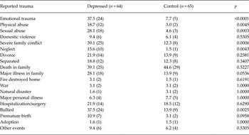
Depressed patients reported significantly higher rates of six ELSs (Table 2). While controlling for covariates, we incorporated those ELSs into a model predicting MDD diagnosis. After backwards regression, in the final parsimonious model three ELS variables significantly and independently predicted a diagnosis of MDD: emotional trauma (F 1,121 = 6.79, p = 0.0103), sexual abuse (F 1,121 = 6.00, p = 0.0157) and severe family conflict (F 1,121 = 7.85, p = 0.0059).
Reported ELS exposure between depressed and non-depressed participants a

Data are given as percentage (number) of subjects exposed to each trauma type.
ELS, Early life stress; df, degrees of freedom.
a Due to small cell sizes, ELSs were compared using Fisher's exact test except χ2 tests were used for death in family (χ2 = 0.41, 1 df) and hospitalization/surgery (χ2 = 0.23, 1 df).
We next dichotomized the sample based on whether they reported one or more of those three ELSs: emotional abuse, sexual abuse and severe family conflict (‘predictive’ ELSs). Of the study population, 40% (approximately 75% of the depressed sample and 33% of the non-depressed sample) reported one or more of these predictive ELSs (χ2 = 24.34, 1 degree of freedom, p < 0.0001). Women were more highly represented in the group reporting predictive ELSs (78.4%, compared with 53.9% of those denying predictive ELSs; χ2 = 8.05, 1 degree of freedom, p = 0.0046). These groups did not otherwise significantly differ on age, education, medical morbidity or duration of depression (online Supplementary Table S1).
Effect of ELS on cognition
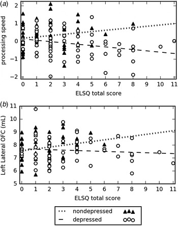
We found no significant main effect of total ELSQ score on any Z-transformed cognitive domain. After controlling for covariates of age, sex and education, we observed an interaction between total ELSQ score and diagnosis on processing speed (F 1,122 = 5.28, p = 0.0232) and a trend for an effect on working memory (F 1,122 = 3.64, p = 0.0588), but not episodic memory or executive function. On analysing the interaction, depressed patients exhibited worsening processing speed with increasing ELSQ score (Fig. 1a ), while non-depressed subjects exhibited faster processing speed performance with increased ELSQ score.
Relationship of Early Life Stress Questionnaire (ELSQ) total score with processing speed and lateral orbitofrontal cortex (OFC) volume. The figures show how exposure to increasing numbers of early life stressors (ELSs; as total ELSQ score) have different effects on processing speed performance and lateral OFC volume between individuals with and without depression. (a) While an increased number of ELSs resulted in progressively poorer performance in the Z-transformed process speed domain in depressed patients, a greater number of ELSs is associated with better process speed performance in non-depressed controls. (b) With increasing ELS exposure, non-depressed subjects showed relative increases in OFC volume. Conversely, depressed subjects exhibited a slight decline in OFC volume with increasing ELS exposure.

In contrast, exposure to predictive ELSs was associated with progressively poorer performance on working memory (F 1,123 = 5.08, p = 0.0260) and processing speed (F 1,123 = 7.74, p = 0.0062), but not executive function or episodic memory. To better elucidate these relationships, we found that increasing predictive ELS exposure was associated with worsening performance on mean Z-transformed cognitive domain scores of working memory (0 predictive ELS = 0.24, s.d. = 0.93; 1 ELS = −0.13, s.d. = 0.96; 2 ELS = −0.28, s.d. = 0.64; 3 ELS = −0.70, s.d. = 0.50) and processing speed mean (0 predictive ELS = 0.24, s.d. = 0.68; 1 ELS = 0.09, s.d. = 0.77; 2 ELS = −0.26, s.d. = 0.83; 3 ELS = −0.70, s.d. = 0.68). Importantly, we found no statistically significant interactions between diagnosis and predictive ELS exposure, suggesting that predictive ELSs affect cognition comparably regardless of depression.
Finally, in exploratory analyses, we examined the relationship between domain scores and log-transformed duration of predictive ELS exposure. Poorer processing speed performance was associated with longer childhood exposure to emotional (F 1,121 = 5.74, p = 0.0183) and sexual abuse (F 1,121 = 4.98, p = 0.0277) in both cohorts. We did not find statistically significant associations between predictive ELS duration and other cognitive domains.
Effect of ELS on brain structure
Due to differences in MRI manufacturers, MRI analyses included only data gathered at Duke University, resulting in a sample of 51 depressed and 53 non-depressed individuals. After controlling for age, sex and intracranial volume, we found no significant differences between diagnostic groups in a priori brain regions (online Supplementary Table S2). We also found no direct effects of total ELSQ score on a priori brain regions. However, we observed an interactive effect between total ELSQ score and diagnosis on the left lateral OFC (F 1,97 = 4.05, p = 0.0469). Greater numbers of ELSs are associated with increasing OFC volumes in non-depressed subjects, but minimal differences in depressed subjects (Fig. 1b ). In exploratory analyses, after controlling for multiple comparisons we did not observe significant direct or interactive effects of total ELSQ score on other brain regions.
Of the MRI sample, 40% (32 depressed and 10 non-depressed subjects) reported one or more predictive ELSs. In analyses of a priori regions, predictive ELSs were associated with smaller left lateral OFC (F 1,98 = 5.11, p = 0.0260) and smaller right caudate volumes (F 1,98 = 6.19, p = 0.0145). We found a significant interaction between diagnosis and predictive ELS exposure only for the hippocampus, with predictive ELSs being associated with smaller left hippocampus volume (F 1,98 = 4.98, p = 0.0280) and a trend for smaller right hippocampus volume (F 1,98 = 3.35, p = 0.0705), but only in the depressed group. After controlling for multiple comparisons there were no statistically significant findings in other brain regions.
In exploratory analyses, we examined the relationship between the volumes of the a priori regions and duration of exposure to the predictive ELSs. A statistically significant relationship was only observed between exposure to sexual abuse and caudate volume, wherein a greater log-transformed duration of exposure was associated with smaller caudate volumes (left: F 1,96 = 4.92, p = 0.0292; right: F 1,96 = 5.02, p = 0.0276).
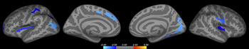
Finally, we tested for relationships between predictive ELS exposure and cortical thickness. After controlling for sex and diagnosis, predictive ELS exposure was associated with reduced cortical thickness in several regions, including the bilateral insula, frontal and parietal lobes (Fig. 2 and online Supplementary Table S3). We found no association between predictive ELS exposure and increased cortical thickness.
Cortical thickness differences related to predictive early life stress (ELS) exposure. Whole-brain vertex-wise display shows the direct effect of reported predictive ELSs (emotional abuse, sexual abuse or severe family strife) on cortical thickness. Analyses controlled for diagnosis (major depressive disorder or non-depressed) and sex. Lighter blue color reflects areas where ELS exposure is associated with thinner cortex. ELS exposure was not significantly associated with increased cortical thickness in any region.

Discussion
Similar to past literature (Wise et al. Reference Wise, Zierler, Krieger and Harlow2001), depressed individuals report more ELSs than non-depressed individuals. However, only emotional abuse, sexual abuse and severe family conflict significantly predicted adult MDD. Exposure to these childhood stressors is associated with poorer cognitive performance and alterations in brain morphology that did not differ between diagnostic groups. Additionally, we found that exposure to predictive ELSs was associated with smaller hippocampal volumes, but only in depressed individuals. In contrast, when defined broadly, overall ELS exposure has a different relationship with processing speed and OFC volumes between depressed and non-depressed adults.
Childhood trauma has prominent effects on adult mental health (Navalta et al. Reference Navalta, Polcari, Webster, Boghossian and Teicher2006). Past work shows altered HPA axis regulation and secondary regional brain structure changes in children exposed to emotional abuse, sexual abuse and aggressive families (McEwen, Reference McEwen2003). Emotional abuse is common in childhood and adversely affects self-esteem, interpersonal skills and personal autonomy and integrity (Vietze et al. Reference Vietze, Falsey, Sandler, O'Connor and Altemier1980). Childhood sexual abuse has a worldwide prevalence of 20% (Jakubczyk et al. Reference Jakubczyk, Klimkiewicz, Krasowska, Kopera, Slawinska-Ceran, Brower and Wojnar2014) and is associated with multiple psychiatric disorders including MDD, addictions and increased suicide risk (Jakubczyk et al. Reference Jakubczyk, Klimkiewicz, Krasowska, Kopera, Slawinska-Ceran, Brower and Wojnar2014). Significant family strife also interferes with normal development and creates a vulnerability to maladjustment and internalizing personal problems (Luebbe & Bell, Reference Luebbe and Bell2014). Admittedly, while these specific stresses predicted adulthood MDD, others propose that any significant childhood stress may increase the risk of depression (Brown & Harris, Reference Brown and Harris1978). Such effects may depend on stressor severity and chronicity, age of exposure and positive support (Brown & Harris, Reference Brown and Harris1978).
Effects of ELS on cognition
ELS exposure also affected cognitive performance. When defined broadly using the total ELSQ score, ELS exhibited different effects between depressed and non-depressed subjects on processing speed (Fig. 1a ). We propose that these group differences reflect different long-term adaptations to stress that are related to either vulnerability to or resilience to developing later depression. In contrast, the effect of predictive ELS exposure on processing speed and working memory was independent of diagnosis. Similarly, duration of emotional and sexual abuse exposure was associated with slower processing speed.
Our results did not support prior literature associating poorer performance on executive function measure or episodic memory with ELSs (Polak et al. Reference Polak, Witteveen, Reitsma and Olff2012; Brewin, Reference Brewin2014). The difference between our results and past work associating ELS with episodic and semantic memory impairment (Parks & Balon, Reference Parks and Balon1995) could be related to heterogeneity in samples or measures used to examine episodic memory. While prior studies used the word-cueing technique and the Logical Memory test (Parks & Balon, Reference Parks and Balon1995), we additionally used the Benton Visual Retention and Rey's Verbal Learning Tests.
When considering the relationship between ELS and processing speed, we may be observing an inverted U-shaped curve. In this model, stress exposure broadly defined is associated with improved processing speed, but exposure to more severe (and potentially more chronic) ELSs results in impaired performance and vulnerability to depression. This is concordant with studies in older adults associating childhood trauma with better processing speed (Feeney et al. Reference Feeney, Kamiya, Robertson and Kenny2013). In our non-depressed population, it is possible that less severe stresses result in improved processing speed and contribute to a resiliency mechanism (Wu et al. Reference Wu, Feder, Cohen, Kim, Calderon, Charney and Mathe2013). However, subjects predisposed to depression may have pre-existing circuit dysfunction where neurobiological changes related to even milder stresses may result in poorer cognitive performance.
A similar model may apply to working memory, although we observed a relationship only with the more severe predictive stressors that did not differ between diagnostic groups. Past work supports negative effects of childhood stressors on working memory (Navalta et al. Reference Navalta, Polcari, Webster, Boghossian and Teicher2006). This may be related to altered function of stress-sensitive systems, as working memory deteriorates with increased allostatic stress load (Evans & Schamberg, Reference Evans and Schamberg2009).
Effects of ELS on brain structure
ELS exposure was also associated with altered volumes of several regions involved in emotional regulation (Udo et al. Reference Udo, Anja, Victoria, Peter, Thomas, Dominik, Katharina, Christa, Patricia, Jochen, Christian, Christian, Carsten, Volker, Walter, Thomas and Harald2012). In parallel with our observations on processing speed, we observed diagnostic group differences on the relationship between total ELSQ score and lateral OFC volume (Fig. 1b ). A similar inverted U-shaped model may also apply to this relationship. This theory is concordant with a primate study examining early-life maternal separation (Parker et al. Reference Parker, Buckmaster, Justus, Schatzberg and Lyons2005). This study associated separation with increased adult OFC volume, a finding thought to be related to stress resiliency by learning extinction of fear through top-down regulation (Lyons et al. Reference Lyons, Parker, Katz and Schatzberg2009). Although the underlying mechanism is unclear, the localization of our OFC finding to the left hemisphere is supported by prior studies suggesting higher left hemisphere sensitivity to emotional neglect during brain development (Frodl et al. Reference Frodl, Reinhold, Koutsouleris, Reiser and Meisenzahl2010).
Exposure to more severe predictive ELSs was associated with smaller OFC and caudate volumes in both cohorts. This is concordant with past work showing that physically and emotionally abused children exhibit smaller OFC volumes (De Brito et al. Reference De Brito, Viding, Sebastian, Kelly, Mechelli, Maris and McCrory2013) while domestic violence and sexual abuse are associated with smaller caudate volumes (Cohen et al. Reference Cohen, Grieve, Hoth, Paul, Sweet, Tate, Gunstad, Stroud, McCaffery, Hitsman, Niaura, Clark, McFarlane, Bryant, Gordon and Williams2006). Our results from cortical thickness analyses are in line with a study associating decreased insula thickness with ELS exposure (Baker et al. Reference Baker, Williams, Korgaonkar, Cohen, Heaps and Paul2013). As the insula plays a role in salience network regulation, reported abnormalities may explain deterioration in working memory and processing speed (Krishnadas et al. Reference Krishnadas, Palaniyappan, Lang, McLean and Cavanagh2014).
Smaller hippocampal volumes are reported in MDD (MacQueen & Frodl, Reference MacQueen and Frodl2011). We found that predictive ELSs were associated with hippocampal volume, but only in depressed individuals. Animal models demonstrate that controlled maternal separation results in decreased hippocampal volumes; however, those volumes may normalize in adulthood (Herpfer et al. Reference Herpfer, Hezel, Reichardt, Clark, Geiger, Gross, Heyer, Neagu, Bhatia, Atas, Fiebich, Bischofberger, Haas, Lieb and Normann2012). Extending that finding to our data, we may be observing a vulnerability mechanism wherein subjects who do not experience recovery of hippocampal neurogenesis are at increased risk of adult depression. This theory is concordant with past work demonstrating that depression itself contributes to hippocampal volume reduction (Sheline et al. Reference Sheline, Wang, Gado, Csernansky and Vannier1996) while smaller hippocampal volumes are also a risk factor for depression (Cole et al. Reference Cole, Costafreda, McGuffin and Fu2011). Moreover, our finding is consistent with past observations that the left hippocampus is more sensitive to stressful events than the right hippocampus (Teicher et al. Reference Teicher, Anderson and Polcari2012). Although the underlying cause for this difference is unclear, it is hypothesized that it is related to cortisol's effect on N-methyl-d-aspartate (NMDA) receptor function and hemispheric differences in NMDA subunit distributions (Teicher et al. Reference Teicher, Anderson and Polcari2012).
Limitations and conclusions
Despite the strength of a large, well-characterized sample, the study also has weaknesses. These include using a retrospective, self-report scale for ELS, which does not measure severity, chronicity, or include stressful events with positive connotations. Self-report is subject to a memory bias, which may be significant in depressed individuals, although others report consistency between retrospective accounts and documented events (Martins et al. Reference Martins, Von Werne Baes, Tofoli and Juruena2014). Further, our study is cross-sectional so cannot address longitudinal developmental effects of ELS exposure on brain volume or cognition. It also does not inform us if the observed cognitive and volumetric differences observed in the MDD population persist with successful depression treatment.
There are additional limitations specifically related to the depressed group. The depressed group is older than the never-depressed group, which is important as increased age is associated with changes on MRI and neuropsychological testing. Although this concern is ameliorated by controlling for age in statistical analyses and the lack of an observed age difference between individuals who were and were not exposed to predictive ELSs, study findings do need replication in a truly age-matched sample. Finally, it can be challenging to disentangle the effects of ELS from the occurrence of depression. As we did not observe significant differences in cognitive or MRI measures between groups after controlling for demographic variables (Table 1, online Supplementary Table S2), the predictive ELSs do not appear to be serving as a surrogate marker for depression diagnosis. However, it is possible that predictive ELS exposure may influence the duration or recurrence of depressive episodes, or be related to early antidepressant treatment. The complexity of this relationship may require prospective longitudinal studies to disentangle these effects and identify potential benefits for early intervention.
It is important to note that analyses included numerous comparisons. This included analyses of four Z-transformed cognitive domains, seven a priori brain regions measured bilaterally, and numerous other regions examined in exploratory analyses. For analyses of exploratory brain regions, we controlled for multiple comparisons using the FDR method. However, as examination of cognitive domains and our a priori regions were based on specific hypotheses and past literature, we did not control for multiple comparisons in those analyses. Despite this hypothesis-driven approach, this does increase the risk for false-positive findings. Adjusting those analyses for multiple comparisons would have limited cognitive findings to processing speed with no a priori brain region surviving correction. Thus our findings should be viewed cautiously and need replication in context of the broader literature.
This study supports that not all childhood stressors increase risk of depression in adulthood. We found a complex relationship between ELSs, cognitive function and regional brain structures that in some cases differed between diagnostic groups. As we defined predictive ELSs based on their relationship with MDD, these findings require replication in independent populations. Future longitudinal human studies are required to incorporate physiological measures of stress reactivity, investigate what factors contribute to the cognitive deficits observed with exposure to ELS, and to clarify the association with volumetric brain changes. Such studies should also examine the effect of emotional processes on cognitive performance, and how ELS exposure may influence those relationships.
Supplementary material
The supplementary material for this article can be found at http://dx.doi.org/10.1017/S0033291716002403
Acknowledgements
This project was supported by the National Institute of Mental Health (NIMH) grants R01 MH077745 and K24 MH110598; and the National Center for Advancing Translational Sciences (NCATS) award UL1TR000445. The authors would like to acknowledge Dr Hakmook Kang for guidance on statistical analyses. This project was conducted using the resources of the Advanced Computing Center for Research and Education at Vanderbilt University, Nashville, TN.
Declaration of Interest
None.