Introduction
Commercial poultry production is associated with various stresses leading to decrease of productive and reproductive performance of growing chickens, parent birds as well as commercial layers. A growing body of evidence has indicated that most of stresses in poultry production at the cellular level are associated with oxidative stress due to excess of free radical production or inadequate antioxidant protection. In the first (Surai and Fisinin, Reference SURAI and FISININ2016a) and second (Surai and Fisinin, Reference SURAI and FISININ2016b) parts of this review series, the technological, environmental, nutritional and internal stresses in poultry production were characterised and the aim of the following review is to describe the vitagene concept with special emphasis on their protective actions in commercially-relevant stress conditions.
Vitagene concept development
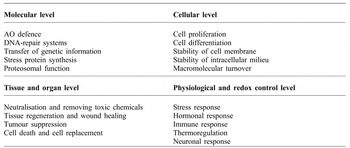
The term ‘vitagene’ was introduced in 1998 by Rattan who wrote ‘Our survival and the physical quality of life depends upon an efficient functioning of various maintenance and repair processes. This complex network of the so-called longevity assurance processes is composed of several genes, which may be called vitagenes’. The vitagene concept has been further developed in medical sciences (Calabrese et al., Reference CALABRESE, BOYD-KIMBALL, SCAPAGNINI and BUTTERFIELD2004; Reference CALABRESE, GUAGLIANO, SAPIENZA, PANEBIANCO, CALAFATO, PULEO, PENNISI, MANCUSO, BUTTERFIELD and STELLA2007; Reference CALABRESE, CORNELIUS, MANCUSO, BARONE, CALAFATO, BATES, RIZZARELLI and KOSTOVA2009a; Reference CALABRESE, SCAPAGNINI, DAVINELLI, KOVERECH, KOVERECH, DE PASQUALE, SALINARO, SCUTO, CALABRESE and GENAZZANI2014) and major pro-survival mechanisms controlled by the homodynamic vitagene network are shown in Table 1 . In accordance with Calabrese et al. (Reference CALABRESE, GUAGLIANO, SAPIENZA, PANEBIANCO, CALAFATO, PULEO, PENNISI, MANCUSO, BUTTERFIELD and STELLA2007; Reference CALABRESE, CORNELIUS, MANCUSO, BARONE, CALAFATO, BATES, RIZZARELLI and KOSTOVA2009a; Reference CALABRESE, SCAPAGNINI, DAVINELLI, KOVERECH, KOVERECH, DE PASQUALE, SALINARO, SCUTO, CALABRESE and GENAZZANI2014) the term vitagenes refers to a group of genes that are strictly involved in preserving cellular homeostasis during stress conditions and the vitagene family includes heat shock proteins (HSPs), including haem oxygenase-1 (HSP32, HO-1) and HSP70, the thioredoxin system and sirtuins. The list of potential candidates to the vitagene family was extended, and in particular SOD, a major inducible enzyme in the first level of antioxidant defence, has now been included (Surai, Reference SURAI2015a; Surai and Fisinin, Reference SURAI and FISININ2016c). The products of the genes actively operate in detecting and controlling diverse forms of stress and cell injuries. The co-operative mechanisms of the vitagene network have been reviewed in recently published papers (Calabrese et al., Reference CALABRESE, SCAPAGNINI, DAVINELLI, KOVERECH, KOVERECH, DE PASQUALE, SALINARO, SCUTO, CALABRESE and GENAZZANI2014; Trovato Salinaro et al., Reference TROVATO SALINARO, CORNELIUS, KOVERECH, KOVERECH, SCUTO, LODATO, FRONTE, MUCCILLI, REIBALDI, LONGO, UVA and CALABRESE2014) with the major conclusion indicating an essential regulatory role of the vitagene network in cell and whole organism adaptation to various stresses. Cellular stress responses are mediated via the regulation of pro-survival pathways and vitagene activation with the synthesis of a range of protective antioxidant molecules, which forms the central event in such an adaptation. The vitagene concept has helped in developing effective strategies to fight oxidative stress in various human diseases (Surai and Fisinin, Reference SURAI and FISININ2016c). The vitagene concept in relation to poultry production was further developed in previous publications (Fisinin and Surai, Reference FISININ and SURAI2011a; Reference FISININ and SURAI2011b; Surai and Fisinin, Reference SURAI and FISININ2012a; Reference SURAI and FISININ2012b; Surai, Reference SURAI2015a; Reference SURAI2015b; Reference SURAI2015c; Reference SURAI2015d). Therefore, possible protective roles of vitagene products in avian physiology and poultry production are considered in the following sections of this paper.
Major components of the vitagene network (adapted from Rattan, Reference RATTAN1998; Calabrese et al., Reference CALABRESE, GUAGLIANO, SAPIENZA, PANEBIANCO, CALAFATO, PULEO, PENNISI, MANCUSO, BUTTERFIELD and STELLA2007; Surai, Reference SURAI2015d).

Heat shock proteins
The protective roles of heat shock proteins (HSPs) in poultry production have been recently reviewed (Surai, Reference SURAI2015c) and can be summarised as being highly conserved families of proteins, first discovered in 1962 (Ritossa, Reference RITOSSA1962). It has since been realised that most HSPs have strong cytoprotective effects and are molecular chaperones for other cellular proteins. Indeed, the adaptation and survival of cells and organisms in various stress conditions require the ability to sense proteotoxic insults and to coordinate protective cellular stress response pathways and chaperone networks related to protein quality control (Calabrese et al., Reference CALABRESE, CORNELIUS, DINKOVA-KOSTOVA and CALABRESE2009b). In fact, in the case of oxidative stress, HSP network participates in detecting intracellular changes, protecting against protein misfolding and preventing activation of downstream events related to inflammation and apoptosis. Therefore, HSPs participate in the regulation of essential cell functions, such as protein translocation, refolding, assembly and the recognition, prevention of protein aggregation, renaturation of misfolded proteins and degradation of unstable proteins. In birds the HSP superfamily includes five broadly conserved families of proteins including HSP90, HSP70, HSP60, HSP40 and small HSP. Among them HSP70 and HSP32 (HO-1) are considered as vitagenes. Thus, HSPs play a significant role in cell/organism adaptation to various stresses being an integral part of the antioxidant protective network responsible for proteostasis maintenance (Surai, Reference SURAI2015c).
haem oxygenase (ho-1, hsp32)
HO-1, a 32-kDa protein, known as heat shock protein-32 (HSP32), is involved in the degradation of haem to generate carbon monoxide (CO), biliverdin and free iron. HO-1 is the stress-inducible isoform of the three HO isoforms described to date, serving as a critical protective mechanism in vertebrate systems responsible for adaptation to oxidative, inflammatory, and cytotoxic stress. In fact, HO-1 is expressed at a relatively low level in most tissues and it is induced in response to various oxidative stress-related insults including haem, ultraviolet light, heavy metals, cytokines, hydrogen peroxide, nitric oxide (NO) and glutathione depletion. The vital importance of HO-1 in stress adaptation has been confirmed in HO-1-deficient animal models showing atypical pro-inflammatory immune response with increased susceptibility to apoptosis (Surai, Reference SURAI2015c).
Data on HO-1 expression and its protective actions in poultry production are limited. In the early 1990s, HO-1 was purified from liver microsomes of chicks pretreated with cadmium chloride. It seems likely that HO-1 synthesis is under hormonal control, including glucagon (Bakken et al., Reference BAKKEN, THALER and SCHMID1972) and adrenocorticotropic hormone (Ryter et al., Reference RYTER, ALAM and CHOI2006) and, similar to mammals, birds HO-1 induction in stress conditions is mediated by various signalling pathways. Therefore, increased HO-1 expression in chicken embryos between internal (day 19) and external pipping (day 20) is an adaptive mechanism responsible for increased protection of tissues during this stressful period of the ontogenesis. In general, HO-1 is described in avian species, however, its response to different stresses in domesticated and wild birds are still not fully characterised (Surai, Reference SURAI2015c). Induction of HO-1 occurs as an adaptive and beneficial response and its upregulated activity plays an important role in animal/poultry adaptation to various commercially-relevant stresses.
hsp70
Among the HSPs, HSP70 is one of the most conserved and important protein families (Surai, Reference SURAI2015c). HSP70 refers to a family of 70 kDa chaperone proteins responsible for important house-keeping functions. These ATP-dependent chaperones represent central components of the cellular protein surveillance network and are involved in a large variety of protein-folding processes. Under various stress conditions additional synthesis of stress-inducible HSP70 enhances the ability of stressed cells to deal with increased concentrations of unfolded or denatured proteins. In 1986, Morimoto et al. (Reference MORIMOTO, HUNT, HUANG, BERG and BANERJI1986) determined organisation, nucleotide sequence, and transcription of the chicken HSP70 gene. They isolated a gene encoding a 70-kDa heat shock protein (HSP70) from a chicken genomic library and showed that the order and spacing of the sequences shared many features in common with the promoter for the human HSP70 gene. It was shown that the heat induced increase in HSP70 mRNA and protein in broiler chicken liver in vivo are time dependent, similar to that in mammals. The increased expression of HSP70 in broiler chicken embryos was affected not only by heat (40°C) but also by cold (32°C) stress, and is tissue- and age-dependent. Therefore, increased HSP70 expression is an adaptive mechanism to deal with oxidative stress-related changes in cell proteome.
HSP70 expression in post-natal chickens is tissue and allele-specific. HSP expression in the gut could be considered as an important mechanism of the antioxidant protection (Surai and Fisinin, Reference SURAI and FISININ2015). To summarise, increased HSP70 expression in chicken tissues is one of the most important protective responses to prevent or deal with, detrimental changes in protein structure and functions due to various stresses. However, there is a need for further research to understand molecular mechanisms of HSP70 regulation in avian species. Our analysis of recent data (Surai, Reference SURAI2015c) indicated that HSP70 and HO-1 expression effectively responds to various stresses including heat stress, heavy metal stress, Se deficiency, chicken transportation and increased stocking density. Furthermore, HSP expression can also be regulated by nutritional means, including vitamins E, C, D, carnitine, betaine and some phytochemicals including silymarin (Surai, Reference SURAI2015c; Reference SURAI2015d). Therefore these vitagenes (HO-1 and HSP70) are important elements responsible for adaptation to various stresses by maintaining cell proteostasis.
Thioredoxin system
The redox status of the cell is a major determinant of many different pathways including gene regulation (Reczek and Chandel, Reference RECZEK and CHANDEL2015). A thiol redox system consisting of the thioredoxin (thioredoxin/thioredoxin peroxidase (peroxiredoxins)/thioredoxin reductase) and glutathione systems (glutathione/glutathione reductase/glutaredoxin/glutathione peroxidase, Holmgren, Reference HOLMGREN1989) are believed to be major players in this regulation (Holmgren, Reference HOLMGREN2000; Gromer et al., Reference GROMER, URIG and BECKER2004). Together they supply electrons for deoxyribonucleotide formation, antioxidant defence, protein and DNA synthesis and repair and redox regulation of signal transduction, transcription, cell growth, differentiation and apoptosis (Mustacich and Powis, Reference MUSTACICH and POWIS2000). Indeed, the thioredoxin system not only plays a crucial role as thiol/disulphide redox controller, it is also essential for certain organisms as the only system ensuring redox homeostasis (Koháryová and Kollárová, Reference KOHÁRYOVÁ and KOLLÁROVÁ2015). Biological roles of the thioredoxin system are diverse and include (Gromer et al., Reference GROMER, URIG and BECKER2004), first of all, antioxidant defence by direct catalysis of several antioxidant reactions and by regeneration of other antioxidant enzymes such as peroxiredoxins or methionine sulphoxide reductase inactivated by oxidative stress; recycling dehydroascorbate to ascorbate and reduction of ubiquinone to ubiquinol. The thioredoxin system is involved in redox regulation of gene expression, various cellular functions, synthesis deoxyribonucleotides (DNA synthesis and repair), protein biosynthesis, hormone and cytokine action and apoptosis. (Lu and Holmgren, Reference LU and HOLMGREN2014). Some of the enzymatic reactions that appear to be modulated by thioredoxin similarly represent major control points for cellular metabolism.
thioredoxins
Thioredoxin (Trx), an approximately 12 kDa thiol/disulphide oxidoreductase, was first characterised in 1964 in E. coli and three years later it was described in rat hepatoma cells (Powis et al., Reference POWIS, MUSTACICH and COON2000). Thioredoxin, with a redox-active dithiol/disulphide, is an electron donor for essential enzymes including ribonucleotide reductase and a general protein disulphide reductase (Holmgren, Reference HOLMGREN2001). Trx represents an intracellular redox regulator that has been shown to be important for the regulation of redox-sensitive transcription factors and maintaining them in active form during oxidative stress. In fact, most, if not all, of the functions of Trx depend on the activity of TrxR. The cDNA sequence for chicken Trx predicts a protein of 105 amino acids with a molecular weight of 11,700 (Jones and Luk, Reference JONES and LUK1988). These authors showed that the sequence of the chicken Trx was very similar to the sequences of other thioredoxins. Comparison of the chicken Trx protein sequence with those from bacteria and plants indicates structural features that appear to be essential for activity. To investigate the biological significance of Trx2, chicken Trx2 cDNA was cloned and clones of the conditional Trx2-deficient cells were generated using chicken B-cell line, DT40. It was shown that chicken Trx2 is an essential gene and that Trx2-deficient cells undergo apoptosis upon repression of the Trx2 transgene, with an accumulation of intracellular ROS (Tanaka et al., Reference TANAKA, HOSOI, YAMAGUCHI-IWAI, NAKAMURA, MASUTANI, UEDA, NISHIYAMA, TAKEDA, WADA, SPYROU and YODOI2002; Wang et al., Reference WANG, MASUTANI, OKA, TANAKA, YAMAGUCHI-IWAI, NAKAMURA and YODOI2006). Increased Trx expression in chicken ovarian follicles was associated with high rates of egg production (Yang et al., Reference YANG, LIN, HUANG, LIOU, CHIEN, WU, HUANG, OU, CHEN, LEE, LIN, TANG, LEE, DING, CHENG and HUANG2008). Trx was found to be expressed in chicken jejunum (Xiao et al., Reference XIAO, POWER, MALLONEE, ROUTT, SPANGLER, PESCATORE, CANTOR, AO, PIERCE and DAWSON2012) and was shown to be important protein of the chicken seminal plasma (Marzoni et al., Reference MARZONI, CASTILLO, SAGONA, CITTI, ROCCHICCIOLI, ROMBOLI and FELICIOLI2013). Furthermore, chicken mitochondrial Trx2 possesses disulphide reductase activity in concentration-dependent manner showing protective effects on LPS-induced oxidative stress in chicken hepatocytes (Hu et al., Reference HU, YU, LI, LI, GUO and TANG2015).
peroxiredoxins
Peroxiredoxins (Prxs) are a family of highly conserved antioxidative proteins (non-seleno peroxidases), currently comprising six members in mammals and located in different parts of the cell. Prxs have a wide subcellular distribution and perform divergent biological functions (Poynton and Hampton, Reference POYNTON and HAMPTON2014). Its activity is based on a redox-active cysteine that is oxidised to a sulphenic acid by hydroperoxides including hydrogen peroxide, organic peroxides, peptide and protein hydroperoxides, and peroxynitrite. These cysteine-dependent peroxidases play major roles not only in peroxide detoxification, but also in regulating peroxide-mediated cell signalling. Prx importance is unarguable, as removal of highly expressed Prxs are associated with increased oxidative stress and reduced genome stability. Prx1 is the most ubiquitously expressed member of the peroxiredoxin family involved in antioxidant defence, cell differentiation and proliferation, immune responses, regulation of apoptosis and chaperone actions (Daly et al., Reference DALY, LEFEVRE, NICHOLAS, DEANE and WILLIAMSON2008). At least four different classes of Prx protein have been evolutionary conserved in chickens (Han et al., Reference HAN, SONG, SHIN, HAN, PARK, PARK, KIM, LILLEHOJ, LIM and KIM2005). Chicken Prx proteins demonstrate antioxidant activity similar to those of the mammalian enzymes and Prx expression in chickens are not tissue specific, indicating their essential role as a housekeeping gene in all tissues to protect against oxidative damage (Han et al., Reference HAN, SONG, SHIN, HAN, PARK, PARK, KIM, LILLEHOJ, LIM and KIM2005). Prx1 is expressed in chicken jejenum (Xiao et al., Reference XIAO, POWER, MALLONEE, ROUTT, SPANGLER, PESCATORE, CANTOR, AO, PIERCE and DAWSON2012), embryonic kidney (Cao et al., Reference CAO, HAN, SHAO, GENG, KONG and LIU2011) and macrophages (Lavric et al., Reference LAVRIC, MAUGHAN, BLISS, DOHMS, BENCINA, KEELER and NARAT2008), while Prx6 was shown to be expressed in chicken liver (Huang et al., Reference HUANG, RUAN, TANG, ZHANG, MA and ZOU2011) and gut (Lee et al., Reference LEE, LILLEHOJ, JANG, JEONG, KIM, XU, LEE, KIM, PARK, KIM and BRAVO2014).
thioredoxine reductase
Thioredoxin reductase (TrxR) was first characterised by Holmgren in 1977 from calf liver and thymus, and five years later was purified from rat liver cytosol by Luthman and Holmgren (Reference LUTHMAN and HOLMGREN1982). Fourteen years later it was shown that human TrxR is a selenoenzyme (Gladyshev et al., Reference GLADYSHEV, JEANG and STADTMAN1996). Therefore, TrxRs are involved in protein folding and critical protein-protein and protein-DNA interactions and mammalian TrxRs show increased activity with Se supplementation in the nutritional to supranutritional ranges (Surai, Reference SURAI2006). TrxR activity in cells is modulated by an intricate interplay, involving regulation by Se availability, posttranscriptional regulation and posttranslational inactivation by ROS. Both in vivo and in vitro studies demonstrated that Trx and TrxR have protective roles against cytotoxicity mediated by the generation of ROS (Calabrese et al., Reference CALABRESE, CORNELIUS, DINKOVA-KOSTOVA and CALABRESE2009b).
Smith et al. (Reference SMITH, MORRIS and LEVANDER2001) compared TrxR activity in mammals and chickens, finding chickens to have extremely low TrxR activities, probably reflecting low protein expression or being a result of differences between mammalian and chicken TrxR. Gowdy (Reference GOWDY2004) used Western blots and found TrxR protein expression at relatively low levels as well as differences in molecular weight of the chicken TrxR in comparison to the mammalian enzyme. Recently, TrxR activity has been detected in a range of tissues (liver, lung, heart, kidney, brain, breast muscle, bursa, thymus, spleen, RBC and plasma) in broiler chickens (Gowdy et al., Reference GOWDY, EDENS and MAHMOUD2015). Activity of chicken TrxR is selenium dependent. Subcellular distribution of TrxR activity was found in association with cytosolic, nuclear pellet and mitochondrial fractions. Compared with sodium selenite, organic Se-yeast or selenomethionine (SM) significantly increased the activity and Trx1 mRNA in the liver and kidney of broiler breeders and their offspring (Yuan et al., Reference YUAN, ZHAN and WANG2012). Selenium dietary supplementation (0.4 mg/kg diet) increased TrxR activity in duodenal mucosa, liver and in the kidney in chickens (Placha et al., Reference PLACHA, TAKACOVA, RYZNER, COBANOVA, LAUKOVA, STROMPFOVA, VENGLOVSKA and FAIX2014). Se deficiency was associated with a decreased expression of TrxR2 in chicken thyroids (Lin et al., Reference LIN, WANG, TAN, LIANG, YAO, ZHANG and XU2014). Similarly, Se deficiency in chickens was associated with a significant decrease in activity of TrxR1 (by 50%), TrxR2 (by 83%) and TrxR3 (by 36%) in the pancreas by the 55th day of the experiment (Zhao et al., Reference ZHAO, YAO, FAN, ZHANG and XU2014). Furthermore, TrxR activity decreased in chicken adipose tissues due to Se deficiency (Liang et al., Reference LIANG, LIN, WANG, YAO, ZHANG and XU2014). A low Se diet (0.028 mg.kg) or high Se diet (3 mg/kg) significantly reduced the TrxR activity in chicken kidney, with changes observed in mRNA levels. In particular, the low Se diet downregulated the mRNA expression of TrxR3 (Xu et al., Reference XU, ZHANG, CAO, ZHU, LI, SUN and LI2016). Therefore, Trxs, Prdxs and TrxRs are important vitagenes participating in cellular/organismal adaptation to stress and their upregulation is considered to be important in improving stress resistance in poultry.
Sirtuins
Sirtuins (SIRTs) are a highly conserved family of NAD+-dependent enzymes with deacetylases, deacylase, mono-ADP-ribosyltransferase and other activities, sharing a common 275-amino acid catalytic domain (Dali-Youcef et al., Reference DALI-YOUCEF, LAGOUGE, FROELICH, KOEHL, SCHOONJANS and AUWERX2007). The sirtuins are ubiquitously distributed from eubacteria to mammals. In particular, SIRTs have been linked to a number of cellular and metabolic processes playing a crucial role in cell plasticity mechanisms of adaptation to various stresses and have been found to be important for human health. In particular, SIRT1, SIRT2, SIRT3, and SIRT5 are shown to possess powerful protein deacetylase activity toward histones (Houtkooper et al., Reference HOUTKOOPER, PIRINEN and AUWERX2012). In fact, SIRTs regulate the biological functions of various molecules post-translationally by removing acetyl groups from protein substrates ranging from histones to transcription factors (Blander and Guarente, Reference BLANDER and GUARENTE2004). Activity of most SIRTs is controlled by post-translational modifications and by the availability of NAD. By affecting specific transcription factors SIRTs regulate redox balance in the cell and orchestrate cellular stress response by maintenance of genome integrity and protein stability (Radak et al., Reference RADAK, KOLTAI, TAYLOR, HIGUCHI, KUMAGAI, OHNO, GOTO and BOLDOGH2013) and playing a crucial role in a number of biological processes, including cell growth and differentiations, apoptosis, chromatin condensation, energy transduction and glucose homeostasis (Dali-Youcef et al., Reference DALI-YOUCEF, LAGOUGE, FROELICH, KOEHL, SCHOONJANS and AUWERX2007).
Sirtuins interact with and regulate a number of proteins, including different transcription factors, which are involved in numerous biological activities. Of all the sirtuins, SIRT1 has received the most attention. Recently the main twenty two cytosolic and nuclear targets of SIRT1 have been characterised (Nogueiras et al., Reference NOGUEIRAS, HABEGGER, CHAUDHARY, FINAN, BANKS, DIETRICH, HORVATH, SINCLAIR, PFLUGER and TSCHÖP2012), including histones, p53, DNA damage proteins, FOXO1, -3 and -4, HSF1, PPARγ, PPARα, UCP2, NF-κB and HIF1α. By targeting these proteins, SIRT1 regulates numerous important signalling pathways, including DNA repair and apoptosis, muscle and fat differentiation, neurogenesis, mitochondrial biogenesis, glucose and insulin homeostasis, hormone secretion, cell stress responses and circadian rhythms (Morris, Reference MORRIS2013; Hubbard and Sinclair, Reference HUBBARD and SINCLAIR2014). Acetyl-CoA synthase 2, glutamate dehydrogenase, p53, FOXO3a, succinate dehydrogenase, isocitrate dehydrogenase 2, Mn-SOD, proteins of the electron transport chain complex I are among important targets of SIRT3.
Sirtuins appear to be at the centre of various stress-related pathways within the complex signalling network that regulates stress response and restores homeostasis under stress conditions. SIRTs regulate gene silencing, DNA repair, rDNA recombination, mitochondrial function and metabolism, as well as programmed cell death (Pallàs et al., Reference PALLÀS, VERDAGUER, TAJES, GUTIERREZ-CUESTA and CAMINS2008). In this context, increasing SIRT expression has been found to protect cells against ROS-associated DNA damage, thereby reducing apoptotic death in vitro. Moreover, it has been demonstrated that over-expression of SIRTs by genetic or pharmacological means can rescue neurons affected by various diseases and elicit multiple metabolic benefits that protect human/animals from diet-induced disorders (Cencioni et al., Reference CENCIONI, SPALLOTTA, MAI, MARTELLI, FARSETTI, ZEIHER and GAETANO2015; Chen et al., Reference CHEN, ZANG, WANG, HUANG, HE, YAN, LIU and ZHENG2015). Similar effects were reported as a result of defects in the pathways controlled by SIRTs (Nogueiras et al., Reference NOGUEIRAS, HABEGGER, CHAUDHARY, FINAN, BANKS, DIETRICH, HORVATH, SINCLAIR, PFLUGER and TSCHÖP2012). Therefore, SIRTs have been proposed to contribute to increased stress resistance and increased antioxidant defence (Webster et al., Reference WEBSTER, LU, SACK and SCOTT2012; Bause and Haigis, Reference BAUSE and HAIGIS2013; Merksamer et al., Reference MERKSAMER, LIU, HE, HIRSCHEY, CHEN and VERDIN2013).
These findings clearly show the importance of SIRTs in the regulation of various physiological processes, being specific molecules in defence and stress adaptations. However, SIRTs role as epigenetic modulating agents await further investigation. SIRT expression remains relatively unexplored in birds, but accumulating evidence indicates that SIRTs are highly conserved among organisms (Hickey et al., Reference HICKEY, JÜLLIG, AITKEN, LOOMES, HAUBER and PHILLIPS2012). Indeed, SIRT1 activation in goose hepatocytes was shown to decrease fatty acids synthesis and cell proliferation, and increased the fatty acids oxidation, whereas SIRT1 inhibition had an opposite effect (Han et al., Reference HAN, WAN, MA, LIU, HE, WANG, PAN, LIU, LI, HE, XU, WEI and XU2014). In avian species, stress can activate SIRTs, e.g. in response to 48 h fasting there was an upregulation of SIRT1 in the chicken hypothalamus, liver, and muscle (Fang et al., Reference FANG, ZHU, CHEN, ZHANG, ZENG, DENG, PENG, YU, WANG, WANG, GAO, JIANG and SHU2014). Because of their roles in cellular stress responses, sirtuins (vitagene) would be expected to play important roles in adaptive responses of poultry to stress.
Superoxide dismutases (SODs)
Protective roles of the vitagene SOD in poultry production (Surai, Reference SURAI2015a) can be summarised from previous published work. SOD discovery by McCord and Fridovich (Reference MCCORD and FRIDOVICH1969) opened a new era in free radical research. At present, three distinct isoforms of SOD have been identified in mammals, and their genomic structure, cDNA, and proteins have been described. SOD1, or Cu, Zn-SOD, was the first enzyme of this family to be characterised, and is a copper and zinc-containing homodimer that is found almost exclusively in intracellular cytoplasma. It exists as a 32 kDa homodimer and is present in the cytoplasm and nucleus of every cell type examined. The second member of the family (SOD2) has manganese (Mn) as a cofactor and therefore called Mn-SOD. It is a 96 kDa homotetramer and located exclusively in the mitochondrial matrix, a prime site for superoxide radical production. Therefore the expression of Mn-SOD is considered to be essential for the survival of aerobic life and the development of cellular resistance to oxygen radical-mediated toxicity. Mn-SOD is an inducible enzyme and its activity is affected by cytokines and oxidative stress (Miao and St Clair, Reference MIAO and ST CLAIR2009). In 1982, a third SOD isozyme was discovered by Marklund et al. (Reference MARKLUND, HOLME and HELLNER1982) and termed extracellular superoxide dismutase (EC-SOD), due to its exclusive location. EC-SOD is a glycoprotein with a molecular weight of 135,000 kDa with high affinity for heparin. The expression pattern of EC-SOD is highly restricted to the specific cell types and tissues where its activity can exceed that of Cu, Zn-SOD or Mn-SOD. The fourth form of the enzyme is Fe-SOD which has been isolated from various bacteria but not found in animals (Surai, Reference SURAI2015a).
Analysis of the data related to SOD in poultry physiology and adaptation to stresses (Surai, Reference SURAI2015a) indicates that SOD as an important vitagene, is the driving force in cell/body adaptation to various stress conditions. Under stress conditions, additional synthesis of SOD is an adaptive mechanism to decrease ROS formation. However, if stress is too high, SOD activity is decreased and apoptosis is activated. There are tissue-specific differences in SOD expression which depends on the strength of such stress-factors including heat, exposure to heavy metals, mycotoxins and other toxicants. SOD is shown to provide an effective protection against lipid peroxidation in chicken embryonic tissues and in semen, as well as in heat and cold stress and other oxidative-stress related conditions in poultry production. There are complex interactions inside the antioxidant network of the cell/body to ensure an effective maintenance of homeostasis in stress conditions. Indeed, in many cases nutritional antioxidants (e.g. vitamin E, selenium and phytochemicals) in the feed can increase SOD expression. Nutritional means of additional SOD upregulation in stress conditions of poultry production and physiological and commercial consequences await investigation. In medical sciences, manipulation of SOD expression and usage of mimics are considered an important approach in disease prevention and treatment. Vitagene upregulation in stress conditions is emerging as an effective means for stress management.
Conclusions
From the data published to date, it is clear that the vitagene network plays a regulatory role in most important cellular processes in stress conditions. Indeed, HSPs, including HO-1 and HSP70, are responsible for protein homeostasis, while the thioredoxin system is the major player in maintaining redox status of the cell involved in protein and DNA synthesis and repair as well as in regulation of expression of many important genes. Furthermore, sirtuins regulate the biological functions of various molecules post-translationally by removing acetyl groups from protein substrates ranging from histones to transcription factors and orchestrate cellular stress response by maintenance of genome integrity and protein stability. Finally, SODs belong to the first level of antioxidant defence preventing lipid and protein oxidation at the very early stages. All the aforementioned vitagenes operate in concert building a reliable system of stress detection and response and are considered to be key elements in stress adaptation. Indeed, the vitagene concept was successfully transferred from medical sciences to poultry science (Fisinin and Surai, Reference FISININ and SURAI2011a; Reference FISININ and SURAI2011b; Surai and Fisinin, Reference SURAI and FISININ2012a; Reference SURAI and FISININ2012b; Surai, Reference SURAI2015a; Reference SURAI2015b; Reference SURAI2015c; Reference SURAI2015d; Surai and Fisinin, Reference SURAI and FISININ2015; Reference SURAI and FISININ2016c). However, further work is required to understand molecular mechanisms of the interactions of vitagenes with various signalling systems and transcription factors in the cell to build an adequate adaptive response to minimise detrimental consequences of commercially-relevant stresses in poultry production.