Introduction
Asbestos refers to a group of six fibrous silicate minerals, including amphibole-group minerals (anthophyllite, tremolite, actinolite, crocidolite, and amosite) and the serpentine-group mineral chrysotile (Gunter et al., Reference Gunter, Divi, Kulldorff, Vermeulen, Haverkos, Kuo and Sinha2007; Gunter, Reference Gunter2010; Strohmeier et al., Reference Strohmeier, Huntington, Bunker, Sanchez, Allison and Lee2010; Vignaroli et al., Reference Vignaroli, Ballirano, Belardi and Rossetti2014). Among these, amphibole asbestos types – especially crocidolite and tremolite – have been classified as Group I human carcinogens by major international health organizations (WHO, 1986; IARC, 1987; Kazan-Allen, Reference Kazan-Allen2005; IARC, 2012). In European regulations, respirable asbestos fibers are generally defined as particles which are longer than 5 μm, thinner than 3 μm, and with an aspect ratio (length divided by diameter) greater than 3:1 (Bloise et al., Reference Bloise, Fornero, Belluso, Barrese and Rinaudo2008). Numerous mineralogical, epidemiological, and environmental health studies have demonstrated that prolonged inhalation of asbestos fibers poses serious health risks (e.g. McDonald, Reference McDonald1985; Lippmann, Reference Lippmann1990; Honda et al., Reference Honda, Beall, Delzell, Oestenstad, Brill and Matthews2002; Niklinski et al., Reference Niklinski, Niklinska, Chyczewska, Laudanski, Naumnik, Chyczewski and Pluygers2004; Cattaneo et al., Reference Cattaneo, Somigliana, Gemmi, Bernabeo, Savoca, Cavallo and Bertazzi2012; Emmett, Reference Emmett2021). Exposure to asbestos has been associated widely with pleural thickening, asbestosis, mesothelioma, and lung cancer (Chew and Toyokuni, Reference Chew and Toyokuni2015; Smith, Reference Smith2015; Hiriart et al., Reference Hiriart, Deepe and Wessels2019; Toyokuni, Reference Toyokuni2019). These effects result from the transport of fibers either directly into the pulmonary airways or via the bloodstream to the alveoli (Böse-O’Reilly et al., Reference Böse-O’Reilly, Heudorf, Lob-Corzilius, Mühlendahl, Otto and Schmidt2007). Fibrous morphology is a key diagnostic feature of asbestos. Typically, asbestos fibers are long and thin, flexible, easily separable, and occur as parallel, bundle-like aggregates (Addison and McConnell, Reference Addison and McConnell2008; Ross et al., Reference Ross, Langer, Nord, Nolan, Lee, Van Orden and Addison2008). However, not all fibrous-appearing minerals are classified as asbestos (Strohmeier et al., Reference Strohmeier, Huntington, Bunker, Sanchez, Allison and Lee2010). All asbestiform amphibole and serpentine asbestos minerals are associated with an increased risk of lung cancer; nevertheless, amphibole asbestos minerals (e.g. crocidolite and amosite) are generally considered more hazardous than serpentine asbestos (chrysotile) (Bernstein et al., Reference Bernstein, Rogers and Smith2004; Pan et al., Reference Pan, Day, Wang, Beckett and Schenker2005; Blake et al., Reference Blake, Johnson and Harbison2009; Kakooei et al., Reference Kakooei, Yunesian, Marioryad and Azam2009; Bernstein et al., Reference Bernstein, Rogers, Sepulveda, Donaldson, Schuler, Gaering and Holm2010; Lahondère et al., Reference Lahondère, Cagnard, Wille and Duron2019). In contrast, antigorite and lizardite are non-asbestiform serpentine polymorphs and are not classified as asbestos.
Asbestos-related symptoms typically have a long latency period, ranging from 25 to 40 years for asbestosis and 15–30 years for lung cancer, depending on exposure levels (IARC, 2012). Nanoparticles with diameters of <100 nm are considered particularly toxic, capable of causing chronic bronchitis, asthma complications, respiratory infections, and even stroke (Samet, Reference Samet, Dominici, Curriero, Coursac and Zeger2000; IARC, 2012). During the extraction of ultramafic rocks such as talc, magnesite, and sometimes chromite, asbestos can be released as dust and mix with other minerals. Chronic inhalation of this dust can lead to severe health issues, including lung cancer and mesothelioma.
The health impact of asbestos is heavily influenced by fiber dimensions. Asbestosis is commonly associated with fibers which are >2 μm long and >0.15 μm in diameter; mesothelioma is associated with fibers which are >5 μm long and <0.1 μm in diameter; and lung cancer with fibers exceeding 10 μm in length (Lippmann, Reference Lippmann1990). For amphibole fibers, those >10 μm long pose the greatest risk for mesothelioma, followed by medium-length (6–10 μm) and short (<6 μm) fibers (Dodson et al., Reference Dodson, Atkinson and Levin2003).
In addition to asbestos minerals, certain fibrous zeolites – most notably erionite and fibrous ferrierite – have also been recognized as potential health hazards. Experimental studies have shown that these fibrous zeolites may occur as microcrystalline phases in minable rocks, display asbestiform morphologies, and release respirable-sized fibers into the air. Their high biodurability and the presence of surface-associated iron impurities may further enhance their pathogenic potential, making them comparable to fibrous erionite in terms of health risk (Gualtieri et al., Reference Gualtieri, Gandolfi, Passaglia, Pollastri, Mattioli, Giordani and Gualtieri2018). Distinguishing asbestos fibers from other hazardous fibrous minerals is therefore essential for accurate mineralogical identification and health-risk assessment.
In the study area, several villages are located directly on or adjacent to ultramafic rock units that are known to host asbestos-bearing lithologies. Previous studies have reported elevated incidences of asbestos-related lung diseases in parts of the region, including mesothelioma cases in nearby settlements (Canda, Reference Canda1989; Canda et al., Reference Canda, Bircan, Akın, Kabadayı and Kurt1988; Canda et al., Reference Canda, Giray, Topal and Görücü2001; Metintaş et al., Reference Metintaş, Batırel, Bayram, Yılmaz, Karadağ, Ak and Metintaş2017). Tremolite and chrysotile have been identified as minerals commonly associated with asbestos-related diseases. However, a definitive causal link between reported disease cases and specific asbestos exposure in the area remains unconfirmed.
Naturally occurring asbestos (NOA) refers to asbestos minerals occurring in their natural geological setting, rather than in manufactured or commercial products. Although NOA itself does not pose a health risk under undisturbed conditions, potential exposure may occur when asbestos-bearing rocks or soils are disturbed by natural processes or anthropogenic activities (Bloise et al., Reference Bloise, Ricchiuti, Lanzafame and Punturo2020). In the study area, agricultural and livestock practices such as soil tilling, grazing, and material collection may episodically disturb asbestos-bearing substrates, increasing the potential for airborne fiber release. Altered ophiolitic rocks containing asbestos have been used for decades in construction, plastering, and oven building within the study area. Despite these long-standing practices, the mineralogical characteristics and health relevance of the fibrous phases present have not been investigated systematically in recent decades. Given the limitations of X-ray diffraction in distinguishing serpentine polymorphs due to overlapping d-spacing values and low peak intensities, a multi-analytical approach is required. Accordingly, the objectives of this study are to: (1) identify asbestos minerals using complementary analytical techniques; (2) determine whether these minerals occur in an asbestiform habit; and (3) characterize asbestos-bearing rocks with respect to their potential environmental and health implications.
Geology
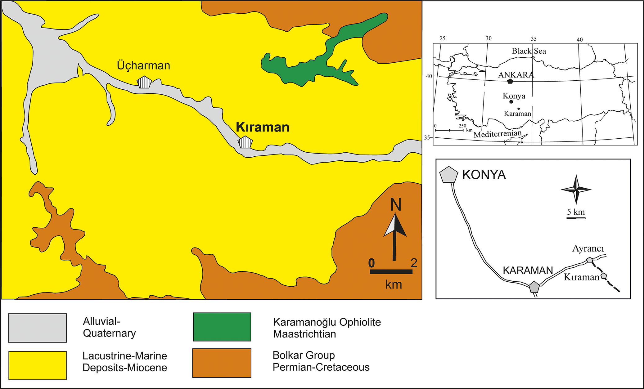
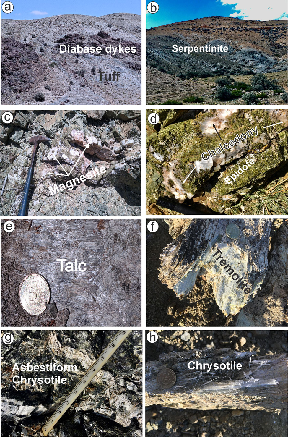
The study area, located ~20 km southwest of Karaman, comprises the Permian–Cretaceous Bolkar Group, consisting of quartzite, calc-schist, sericite-chlorite-micaschist, and marble units (Demirtaşlı et al., Reference Demirtaşlı1983; Pampal, Reference Pampal1987). These units are tectonically overlain by the Karamanoğlu ophiolitic mélange, commonly referred to as the Alihoca Ophiolite (Oktay, Reference Oktay1982) (Fig. 1). The Karaman ophiolitic mélange, emplaced during the Late Cretaceous, is composed of serpentinized ultrabasic and basic rocks (peridotite, harzburgite, diabase, and gabbro), radiolarite, chloritized tuff, and variously sized limestone blocks (Fig. 2a–c). Diabase is observed in different sizes and exhibits intense alteration, appearing in dark green, gray, or black colors, and displays a widespread fracture system. These fractures extend laterally for several meters and can be up to a few centimeters thick. Overlying the ophiolitic mélange with an unconformity is the Miocene Kıraman Formation, consisting of a basal conglomerate followed by claystone and marly limestone (Pampal and Meriç, Reference Pampal and Meriç1990). This unit is primarily composed of thinly bedded, pinkish-brown claystones. The Miocene-aged Karaisalı Formation, which consists locally of reefal limestones, was also defined by Pampal and Meriç (Reference Pampal and Meriç1990) as transitioning from the Kıraman Formation.
Location and geological map of the study area (simplified from Pampal and Meriç, Reference Pampal and Meriç1990).

Figure 1. Long description
The central panel is a color-coded geological map focused on Kiraman, with Üçharman to the northwest. Gray areas represent Alluvial-Quaternary deposits, yellow for Lacustrine-Marine Deposits-Miocene, green for Karamanoğlu Ophiolite-Maastrichtian, and brown for Bolkar Group-Permian-Cretaceous. Kiraman is labeled near the center, with a river running east-west. Üçharman is labeled to the northwest. A north arrow and scale bar (0 to 2 kilometers) are at the lower right of the main map. The upper right inset shows a simplified map of Turkey, with Konya and Karaman marked, and the Black Sea and Mediterranean labeled. The lower right inset is a local road map showing Konya, Karaman, Ayrancı, and Kiraman, with a 5 kilometer scale bar and north arrow. The legend at the bottom left explains the color codes for each geological unit.
Images of rocks and minerals formed as a result of alteration in ophiolitic rocks in the study area: (a,b) view of serpentinized-chloritized tuffs and diabase dykes in the ophiolitic mélange; (c) magnesite formation resulting from carbonation of ultrabasic rocks; (d) partially euhedral epidote and calcedony formed as a result of epidotization-carbonation-silicification; (e) pure, pale green-white talc; (f) fibrous, silky, pale green tremolite; (g,h) whitish-very light green, fibrous chrysotile developed in shear cracks of ultrabasic rocks.

Figure 2. Long description
The layout consists of eight labeled photos arranged in two rows of four. Panel a, top-left, shows a landscape with ‘Diabase dykes’ as dark bands cutting through lighter ‘Tuff’, both labels placed directly on the respective rock units. Panel b, top row second from left, displays a hillside labeled ‘Serpentinite’ across a greenish outcrop. Panel c, third from left, presents a close-up of ultrabasic rock with a hammer for scale and arrows pointing to white ‘Magnesite’ veins. Panel d, top-right, is a close view of a rock with arrows labeling ‘Chalcedony’ as white crystalline patches and ‘Epidote’ as greenish crystals. Panel e, bottom-left, shows a pale green-white fibrous mass labeled ‘Talc’ with a coin for scale. Panel f, second from left, features a fibrous, silky, pale green mineral labeled ‘Tremolite’ with a coin for scale. Panel g, third from left, displays a close-up of a rock with a ruler and the label ‘Asbestiform Chrysotile’ marking fibrous white veins. Panel h, bottom-right, shows a rock with a coin and the label ‘Chrysotile’ indicating white, fibrous mineralization in a shear crack. All text annotations are directly overlaid on the relevant features, and scale bars or coins are present in panels c, e, f, g, and h.
Advanced alteration (including serpentinization, talcification, and carbonation) is common in the ultrabasic rocks (Fig. 2a–h). Within gabbros, chalcedony bands and magnesite veins forming a network or filling fractures, along with serpentine minerals at lithological boundaries, have been identified. Magnesite veins typically range from 1 to 5 mm in thickness, occasionally reaching up to 10 cm in massive silica or chalcedony bands together with serpentinite (Fig. 2c). Serpentinization, resulting from extensive olivine alteration, is particularly widespread. Alteration in the ultrabasic rocks is generally concentrated along shear-fracture systems, with chrysotile, talc, and tremolite observed in veinlet-type structures (Fig. 2e–h). Serpentine and tremolite minerals are observed in light white-green needle-like, fibrous, and silky/glassy luster forms with platy textures. Asbestos formations are visible as shiny white fibers (Fig. 2g,h). In addition, alteration has produced characteristic mesh or reticulate textures, where relic olivine grains are surrounded by lizardite- or chrysotile-like minerals. In addition to widespread serpentinization, extensive physical disintegration and intense chloritization have led to the formation of talc, commonly found as small-scale veinlets or mesh-like textures throughout the region (Fig. 2e).
Materials and methods
A total of 32 samples were collected from variably altered magmatic rocks (such as dunite, gabbro, and diabase) and their alteration products (such as talc, tremolite, and chrysotile) in the ophiolitic units of the study area, where alteration is developed extensively. The mineralogical and petrographic characteristics of thin sections prepared from rock samples were examined using a Nikon Eclipse Ci Pol model polarizing microscope at the Department of Geological Engineering, Konya Technical University, in order to investigate morphological-textural features and mineral compositions.
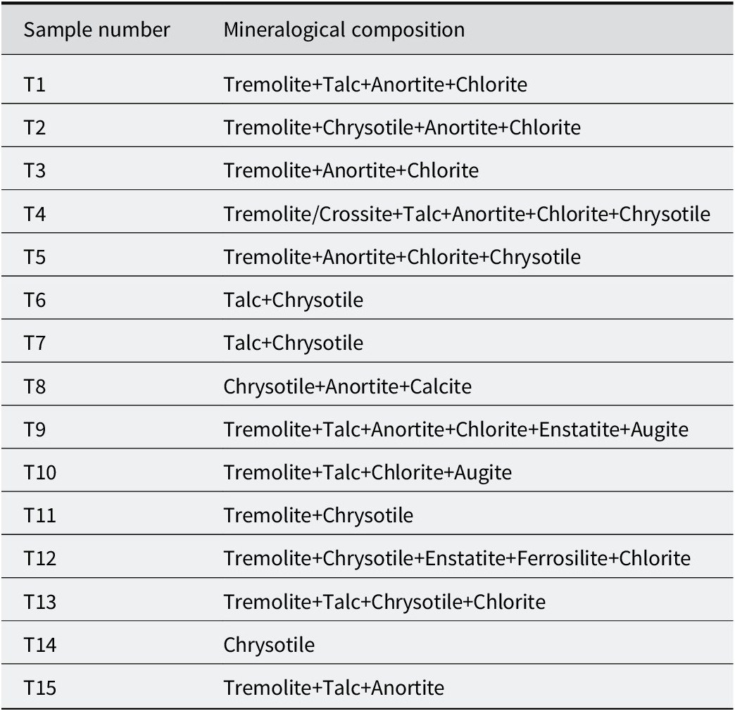
To determine the mineral types and compositions, whole-rock and clay-fraction mineralogical analyses were carried out using X-ray diffraction (XRD) with a Rigaku D/MAX 2200 PC model diffractometer at the Department of Geological Engineering, Hacettepe University, Ankara. The scanning speed was set to 1°2θ per min, and measurements were performed in the range of 2 to 50°2θ. The XRD equipment was equipped with a graphite monochromator, Cu tube (CuKα = 1.5186 Å), Ni filter, 40 kV voltage, and 18 mA current. The goniometer speed was set to 2°/min, sensitivity to 4x10², and chart speed to 2 cm/min. Based on the XRD patterns, the mineral types and their approximate percentages were determined (Table 1).
Mineralogical compositions (XRD) of some representative samples

Table 1. Long description
From the top row, sample T1 contains tremolite, talc, anortite, and chlorite. T2 has tremolite, chrysotile, anortit, and klorit. T3 includes tremolit, anortite, and chlorite. T4 lists tremolite or crossite, talc, anortite, chlorite, and chrysotile. T5 has tremolite, anortite, chlorite, and chrysotile. T6 and T7 both contain talc and chrysotile. T8 includes chrysotile, anortit, and calcite. T9 lists tremolite, talc, anortit, chlorite, enstatit, and ojit. T10 contains tremolite, talc, chlorite, and augite. T11 has tremolite and chrysotile. T12 includes tremolite, chrysotile, enstatite, ferrosilite, and chlorite. T13 lists tremolite, talc, chrysotile, and chlorite. T14 contains only chrysotile. T15 has tremolite, talc, and anortite. Variations in spelling are present for anortite/anortit, chlorite/klorit, tremolite/tremolit, and enstatite/enstatit. The minerals most frequently appearing are tremolite, talc, chlorite, chrysotile, and anortite, with occasional presence of crossite, calcite, augite, ferrosilite, ojit, and enstatite.
Differential thermal analysis–thermogravimetric analysis (DTA–TGA) was used to determine mineral phases by evaluating endothermic and exothermic reaction temperatures and corresponding weight losses. These analyses were performed at Çukurova University using a Mettler Toledo device under the conditions of 25–1200°C, 20.00 K min–1 heating rate, and 40.0 mL min–1 N₂ gas flow.
To identify serpentine polymorphs and tremolite, vibrational spectroscopies (FTIR and Raman) were used. FTIR analyses were performed at the Bayburt University Research Center using a Perkin Elmer instrument in the range of 400–4000 cm-1. The asbestos samples were mixed with KBr and pressed as pellets. Raman analyses were carried out using a Renishaw InVia Qontor model device at the Central Research Laboratory of Çukurova University, in the range 100–4000 cm-1.
Major oxide and trace element compositions of selected samples, based on mineralogical analysis results, were determined using a Panalytical Axios Max WD-XRF instrument at Aksaray University. For chemical analyses, 6 g of powdered samples were mixed thoroughly with 1 g of micro-powder wax. The mixture of wax and sample was then pressed into pellets using a die attachment under a pressure of 14 kN. The chemical compositions of the pressed pellets were analyzed using an X-ray fluorescence (XRF) spectrometer. Loss on ignition (LOI) values of the samples were determined by placing them in a muffle furnace at 900°C for 6 h.
The morphological features and dimensions of fibrous minerals showing asbestos forms were examined using a scanning electron microscope (SEM; FEI Quanta FEG 250 model) at Aksaray University. Semi-quantitative chemical compositions and Mg:Si ratios were obtained through energy-dispersive X-ray spectroscopy (EDX). Furthermore, the size and shape of fibrous minerals were analyzed using a transmission electron microscope (TEM; FEI Talos F200s TEM 200 kV model) at Bayburt University Research Center. In addition to imaging, TEM was used to characterize tremolite and chrysotile crystals and to determine their Mg:Si ratios based on EDX data.
At the final stage of manuscript preparation, an AI language model (ChatGPT, OpenAI) was used solely for language editing and proofreading. All scientific content and conclusions are the authors’ own.
Results
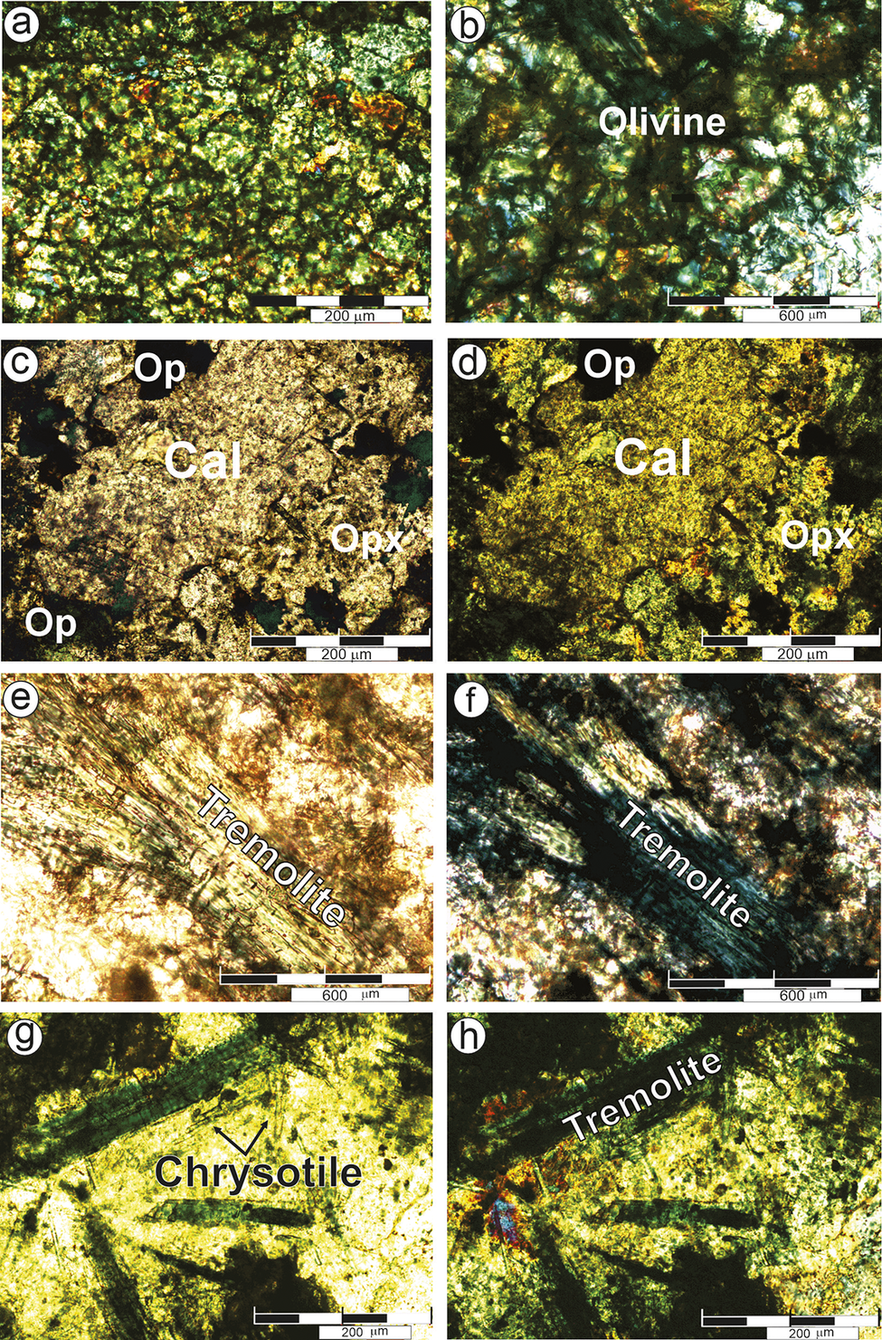
Holocrystalline textures were generally observed in samples taken from gabbro and ultrabasic rocks, and the mineral paragenesis was determined to consist of olivine, pyroxene, amphibole, chlorite, and opaque minerals. In diabase samples, partially porphyritic textures and minor flow structures in the matrix were also detected (Fig. 3a,b). Due to intense alteration and carbonatization, idiomorphic crystals of amphiboles and pyroxenes were only rarely preserved, and their primary petrographic characteristics were commonly obscured (Fig. 3c,d). Chloritization, epidotization, and widespread carbonation in the matrix were observed in olivine and pyroxene crystals, while chloritization and oxidation developed in amphibole and biotite crystals (Fig. 3c,d). The typical lattice structure of olivine alteration was found to be more prevalent (Fig. 3d). Some of the fibers appeared as numerous fine fibrils, probably representing chrysotile, while others were extremely thin and needle-like, displaying the optical properties characteristic of tremolite (Fig. 3e,f). Tremolite fibers were generally thin, straight, and needle-like, with diameters of ~0.2 μm, and commonly observed as rod-like shapes that were transversely fractured. Fibrous minerals, identified as tremolite and possibly chrysotile, occur as slender rods and fine fibrous aggregates, typically arranged in parallel bundles (Fig. 3g,h). In larger tremolite crystals, the edges were observed to be parallel to each other, with an aspect ratio of >3:1 and showing characteristic prismatic cleavage. However, such crystals do not conform to the definition of asbestos fibers (Perkins and Harvey, Reference Perkins and Harvey1993). Quartz was found to be quite rare in the samples examined. In some samples, prismatic biotite crystals exhibiting strong pleochroism and reddish-brown coloration, with single-direction cleavage, as well as amphibole crystals displaying biaxial cleavage, were oxidized along their edges and cleavage planes.
Optical microscopy images of the samples examined: (a,b) crossed nicol images of altered olivine crystals; (c,d) carbonation of orthopyroxenes: c, under plane-polarized light and d, reflected light; (e,f) sub-oriented to oriented long-fiber bundle of tremolite may be formed from olivine and/or enstatite crystals viewed under plane-polarized and reflected light; (g,h) sub-oriented to oriented long-fiber bundle of tremolite and small chrysotile may be formed from olivine and/or enstatite crystals viewed under plane-polarized and reflected light. Cal = calcite, Opx = orthopyroxene, Op = opaque minerals (mineral-name abbreviations after Whitney and Evans, Reference Whitney and Evans2010).

Figure 3. Long description
Panel a shows a crossed-polarized view of altered olivine crystals with granular texture. Panel b, also crossed-polarized, highlights olivine labeled at the center right. Panel c, under plane-polarized light, displays calcite labeled Cal in the center, orthopyroxene labeled Opx at the lower right, and opaque minerals labeled Op at the upper left and lower left. Panel d, under reflected light, repeats the spatial arrangement of Cal, Opx, and Op. Panel e, under plane-polarized light, shows a sub-oriented to oriented long fiber bundle labeled Tremolite diagonally across the field. Panel f, under reflected light, shows the same orientation of Tremolite fibers. Panel g, under plane-polarized light, displays a long fiber bundle labeled Chrysotile at the upper left, with fibers radiating toward the center. Panel h, under reflected light, shows a similar orientation with Tremolite labeled at the center right. All panels include scale bars, with 200 micrometers for panels a, c, d, g, h and 600 micrometers for panels b, e, f. Mineral abbreviations are Cal for calcite, Opx for orthopyroxene, and Op for opaque minerals.
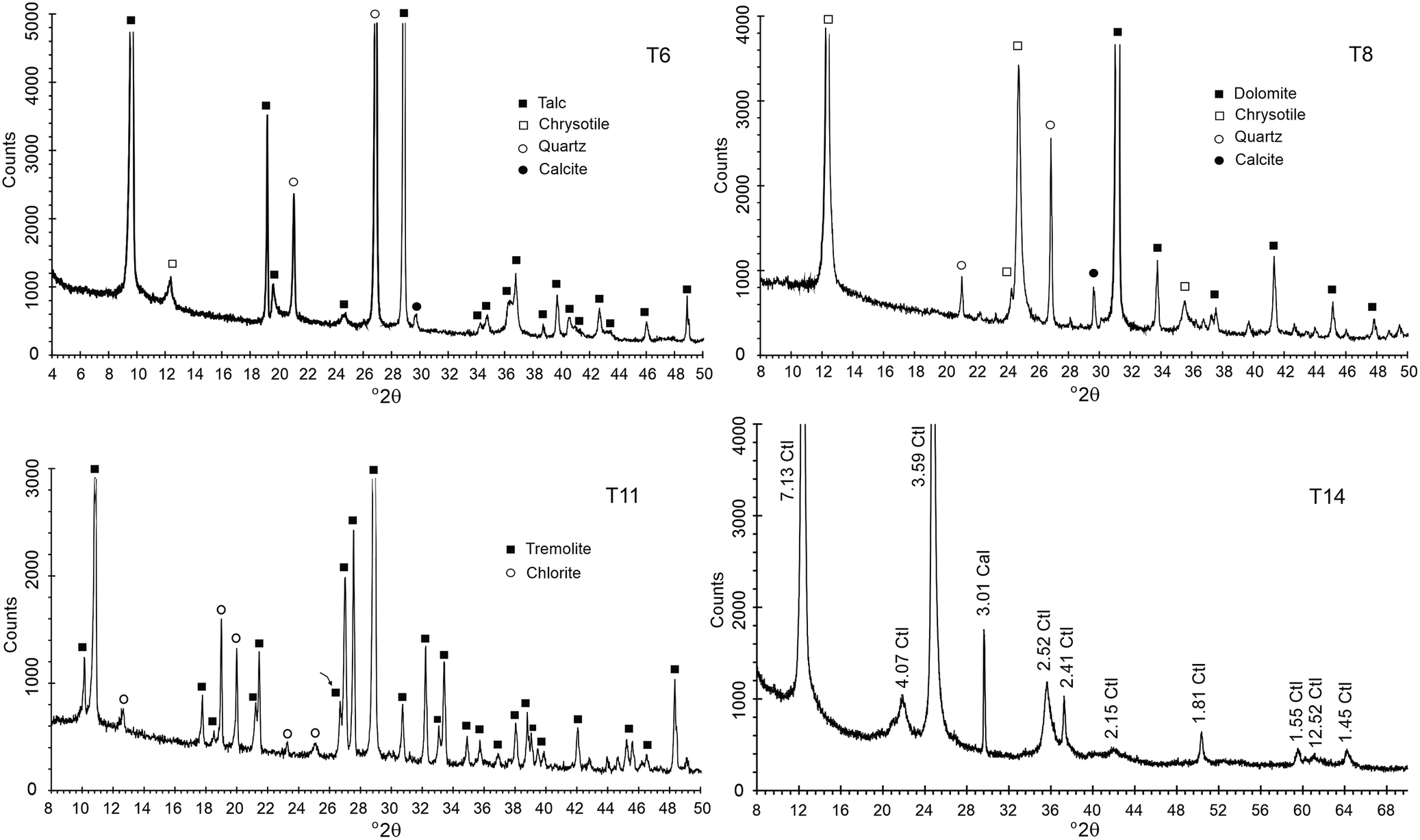
The XRD analysis of samples from regions exhibiting intense alteration revealed the presence of chrysotile, tremolite, and talc as dominant phases, alongside minor amounts of chlorite, calcite, quartz, anorthite, enstatite, and ferrosilite. Many of these minerals were detected in nearly pure phases (Table 1). In the XRD patterns, characteristic diffraction peaks of chrysotile were defined at the d-spacing 7.31, 4.53, 3.65, 2.44, 1.82, and 1.53 Å, while tremolite showed sharp and intense peaks at 9.02, 8.41, 3.38, 3,27, 3.12, 2.94, and 2.71 Å (Fig. 4). The most intense XRD peaks of talc were seen at 9.46, 4.32, 4.55, 3.12, 2.34, and 1.52 Å. Among the serpentine group minerals, the most commonly observed phases were antigorite, lizardite, and chrysotile, all of which exhibit a strong and significant basal reflection near ~7.30 Å. This reflection does not overlap with the kaolinite peak at 7.14 Å, which appears at a different 2θ position. Furthermore, the 002 reflection of serpentine at ~3.65 Å differs in angle from the 3.57 Å 002 reflection of kaolinite (Moore and Reynolds, Reference Moore and Reynolds1997), eliminating any ambiguity in distinguishing these two minerals. Additional specific differences in peak positions and relative intensities between these 1:1 or TO-structure minerals include the 020 and 060 reflections of serpentines, which occur at ~4.60 Å and 1.55 Å, respectively, whereas the dioctahedral kaolinite shows these reflections at 4.47 Å and 1.49 Å (Moore and Reynolds, Reference Moore and Reynolds1997). These distinctions are critical criteria for differentiating the serpentine group minerals from kaolinite.
XRD patterns of the chrysotile, tremolite, and talc-rich asbestos samples. Ctl = chrysotile (abbreviation after Whitney and Evans, Reference Whitney and Evans2010).

Figure 4. Long description
The layout consists of four panels: T6 (top left), T8 (top right), T11 (bottom left), and T14 (bottom right). Each panel displays a line graph with x-axis labeled 2 theta and y-axis labeled counts. In T6, peaks are marked for talc (solid square), chrysotile (open square), quartz (open circle), and calcite (solid circle), with major peaks at approximately 9, 12, 19, 24, 28, 31, 36, and 48 on the x-axis. T8 shows peaks for dolomite (solid square), chrysotile (open square), quartz (open circle), and calcite (solid circle), with prominent peaks at about 12, 19, 24, 28, 31, 36, and 48. T11 highlights tremolite (solid square) and chlorite (open circle), with peaks at roughly 10, 12, 16, 18, 22, 24, 28, 32, 36, and 48. T14 features labeled peaks for chrysotile (Ctl) and calcite (Cal), with specific 2 theta values annotated: 7.13 Ctl, 4.07 Ctl, 3.59 Ctl, 3.01 Cal, 2.52 Ctl, 2.41 Ctl, 2.15 Ctl, 1.81 Ctl, 1.55 Ctl, 1.225 Ctl, and 1.45 Ctl. Each panel’s legend identifies the mineral symbols used. The data show distinct diffraction patterns and mineral compositions for each sample.
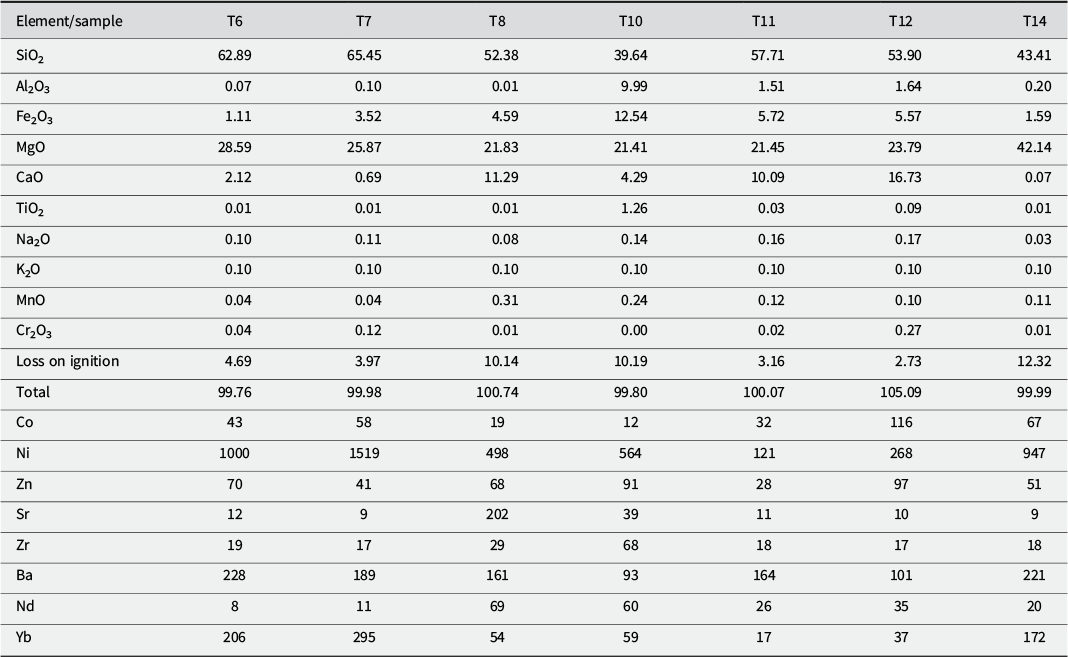
According to the XRD results and chemical compositions presented in Tables 1 and 2, samples T6 and T7 are talc-rich, T11 and T12 are tremolite-rich, and T14 is chrysotile-rich. The elemental compositions of these samples are consistent with the expected mineralogical content. For instance, the chrysotile-rich sample T14 contains 43.41% SiO₂, 42.14% MgO, and 1.59% Fe₂O₃, which closely match the theoretical values for chrysotile (43.55% SiO₂, 33.65% MgO, and ~13% H₂O) (Bolewski and Manecki, Reference Bolewski and Manecki1993). The slightly smaller SiO₂ and MgO contents and the presence of Fe₂O₃ are in line with the presence of Fe-bearing chrysotile detected by TEM-EDX.
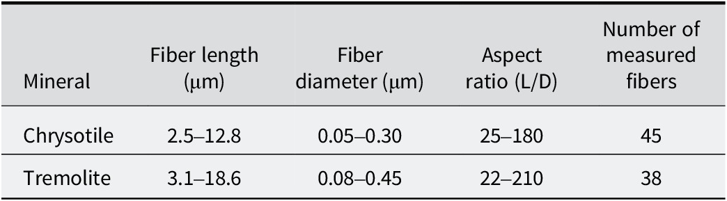
Measured fiber length, diameter, and aspect ratio of chrysotile and tremolite determined from TEM analyses

Table 2. Long description
From left to right, the columns are labeled Mineral, Fiber length in micrometers, Fiber diameter in micrometers, Aspect ratio (L over D), and Number of measured fibers. The first row lists chrysotile with fiber length ranging from 2.5 to 12.8 micrometers, diameter from 0.05 to 0.30 micrometers, aspect ratio from 25 to 180, and 45 measured fibers. The second row lists tremolite with fiber length ranging from 3.1 to 18.6 micrometers, diameter from 0.08 to 0.45 micrometers, aspect ratio from 22 to 210, and 38 measured fibers. All fibers meet the Perkins and Harvey E P A criteria for asbestiform habit, with aspect ratios greater than or equal to 20 to 1 and diameters less than 0.5 micrometers.
All measured fibers satisfy the Perkins and Harvey (Reference Perkins and Harvey1993) (EPA/600/R-93/116) criteria for asbestiform habit, with aspect ratios ≥20:1 and diameters <0.5 μm.
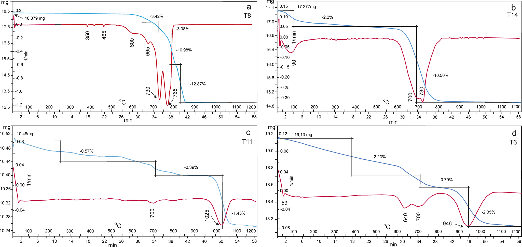
In the DTA analysis of the chrysotile-rich sample, endothermic peaks were observed at 90, 600, and 730°C, respectively, corresponding to dehydroxylation and decomposition–recrystallization reactions, with a weight loss of 12.70% (Fig. 5a,b). In the chrysotile-rich samples (T8, T14), endothermic peaks were recorded in the temperature range of 600–730°C, indicating the dehydration of chrysotile and its transformation into a metachrysotile phase. These observations are in good agreement with previous studies, which report this transformation to occur between ~500 and 750°C (Kusiorowski et al., Reference Kusiorowski, Zaremba, Piotrowski and Adamek2012). In the tremolite-rich sample (T11), a weight loss of 2.39% was observed between 700 and 1025°C, corresponding to an endothermic reaction associated with dehydroxylation due to the release of structural water, as confirmed by DTA–TG analysis (Fig. 5c) (MacKenzie, Reference Mackenzie1957).
DTA-TG curves of talc, chrysotile, and tremolite-rich asbestos samples.

Figure 5. Long description
Panel a, labeled T8, has x-axes for temperature in degrees Celsius and time in minutes, y-axes for mass in milligrams and rate in milligrams per minute. The blue T G curve starts at 18.379 mg and shows three main mass losses: minus 3.42 percent at 790 degrees Celsius, minus 3.08 percent at 865 degrees Celsius, and minus 10.98 percent at 765 degrees Celsius, totaling minus 12.87 percent. The red D T A curve shows endothermic and exothermic events at these points. Panel b, labeled T14, starts at 17.277 mg, with a blue T G curve showing a minus 2.2 percent loss at 90 degrees Celsius and a major loss of minus 10.50 percent at 730 degrees Celsius, ending at 700 degrees Celsius. The red D T A curve mirrors these events. Panel c, labeled T11, starts at 10.486 mg, with the blue T G curve showing a minus 0.57 percent loss at 700 degrees Celsius, minus 0.39 percent at 1025 degrees Celsius, and a total of minus 1.43 percent. The red D T A curve shows a corresponding thermal event. Panel d, labeled T6, starts at 19.13 mg, with the blue T G curve showing a minus 2.23 percent loss at 640 degrees Celsius, minus 0.79 percent at 946 degrees Celsius, and minus 2.35 percent at 1025 degrees Celsius. The red D T A curve shows thermal events at these points. All panels have dual y-axes for mass and rate, and dual x-axes for temperature and time, with annotated mass losses and thermal events.
The talc-rich sample (T6) exhibited an endothermic reaction between 700 and 950°C associated with weight loss (Fig. 5d). It also showed that a weak endothermic reaction at 50°C was due to the loss of absorbed water. In the TG curves of talc, a significant weight loss occurred over the temperature range 600 to 950°C with a broad endothermic DSC signal related to the dehydroxylation of talc and the total weight loss was ~5.37%, consistent for the most part with previous results of dehydroxylation of talc from 800 to 1000°C (Liu et al., Reference Liu, Liu and Hu2014). The thermogravimetric analysis (TGA) results revealed that the LOI value of the talc-rich sample was ~5.37%, which is greater than both the theoretical structural water content of pure talc (4.8%) and the experimental value of 4.3% reported by Liu et al. (Reference Liu, Liu and Hu2014). This elevated LOI is interpreted to result from the presence of a minor amount of chrysotile, as confirmed by complementary mineralogical analyses including FTIR, XRD, Raman spectroscopy, SEM-EDX, and TEM-EDX. Chrysotile, being a fibrous serpentine mineral with a higher hydroxyl content – in sample T8 is 12.7% – contributes additional mass loss through dehydroxylation, typically occurring between 600 and 730°C, and thus increases the overall LOI of the sample. In contrast, the lower LOI reported by Liu et al. (Reference Liu, Liu and Hu2014) compared with the theoretical value may reflect the presence of non-hydrous accessory minerals in their sample, which dilute the talc content and decrease the total dehydroxylation. These variations in LOI emphasize the importance of detailed mineralogical characterization when interpreting thermogravimetric behavior in natural samples.
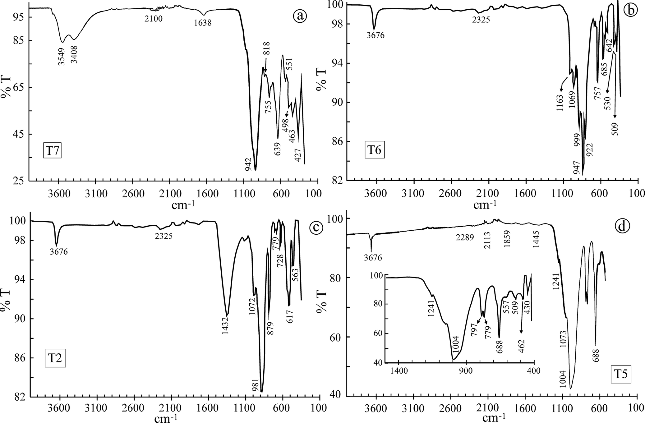
The chemical compositions of talc, chrysotile, and tremolite, the properties of which were investigated here, are characterized by the presence of similar cations (Mg, Si, occasionally Fe, and partially Ca in tremolite) and anions (O and OH). As a result, the absorption bands observed in both FTIR and Raman spectra arise from comparable cation–anion vibrational modes. However, minor variations in band positions occur due to differences in the crystal structure and bonding environments of the individual minerals. The FTIR bands of the investigated asbestos minerals (chrysotile, tremolite) and talc were evaluated in four distinct spectral regions (Fig. 6a–d). Prominent talc absorption bands were observed at 3678 cm-1, corresponding to the stretching vibration of Mg–OH; at ~3549–3408 cm-1, associated with the OH-stretching vibration of adsorbed water; at ~1638 cm-1, attributed to the OH-bending vibration of adsorbed water; at 1069 cm-1, corresponding to the Si–O–Si stretching vibration; at 671 cm-1, related to the Si–O–Mg stretching vibration; at around 462 cm-1, assigned to the Si–O–Si bending vibration; and at 428 cm-1, corresponding to the Si–O stretching vibration (Fig. 6a) (Liu et al., Reference Liu, Liu and Hu2014).
FTIR spectra of talc, chrysotile, and tremolite-rich asbestos samples.

Figure 6. Long description
Top left panel labeled T7 shows talc with broad peaks at 3549 and 3408, smaller peaks at 2100 and 1638, and sharp peaks at 942, 818, 755, 693, 633, 551, 498, 463, and 427. Top right panel labeled T6 shows chrysotile with major peaks at 3676 and 2325, and sharp peaks at 1163, 1065, 999, 947, 922, 757, 685, 642, 530, and 509. Bottom left panel labeled T2 shows tremolite-rich asbestos with peaks at 3676, 2325, 1432, 1072, 981, 879, 728, 779, 653, and 617. Bottom right panel labeled T5 shows a complex spectrum with peaks at 3676, 2289, 2113, 1859, 1445, 1241, 1073, 1004, 688, 579, 509, 430, and 462. An inset in the bottom right panel magnifies the region from 1400 to 400 wavenumbers, highlighting peaks at 1241, 1004, 797, 779, 688, 579, 509, 462, and 430. Each panel displays unique peak positions and intensities, distinguishing the mineralogical composition of the samples.
Chrysotile and tremolite exhibited moderately sharp O–H stretching absorption bands at approximately 3675–3677 cm-1, which are indicative of structurally bound hydroxyl groups within the silicate layers. In contrast, talc showed a broader band centered around 3549–3408 cm-1, consistent with its trioctahedral phyllosilicate structure; a band at 3700–3500 cm-1 region, where sharp O–H stretching bands typical for hydroxyl-bearing phyllosilicates occur; a band in the 1200–800 cm-1 region, which shows six to eight intense to medium bands mainly attributed to lattice Si–O–Si and O–Si antisymmetric stretching vibrations; a band in the 800–600 cm-1 range, containing four to six medium to very weak peaks corresponding to Si–O–Si symmetric stretching or chain deformation modes; and the region below 650 cm-1, where three to four weak and very weak bands are typically attributed to a combination of T–O and M–O bending modes and OH librations. Chrysotile presented prominent absorption bands at 1072, 1030, 981, 879, 778, 619, and 585 cm-1; tremolite at 1165, 1072, 1003, 981, 879, 790, 728, 618, and 564 cm-1; and talc at 942, 818, 639, 552, and 498 cm-1 (Fig. 6). These bands correspond to various lattice vibrations, including Si–O–Si asymmetric and symmetric stretching, T–O and M–O bending modes, and OH librations, thereby confirming the presence and structural characteristics of the asbestiform and related phyllosilicate minerals in the analyzed specimens (Ristic et al., Reference Ristic, Czako-Nagy, Music and Vertes2011).
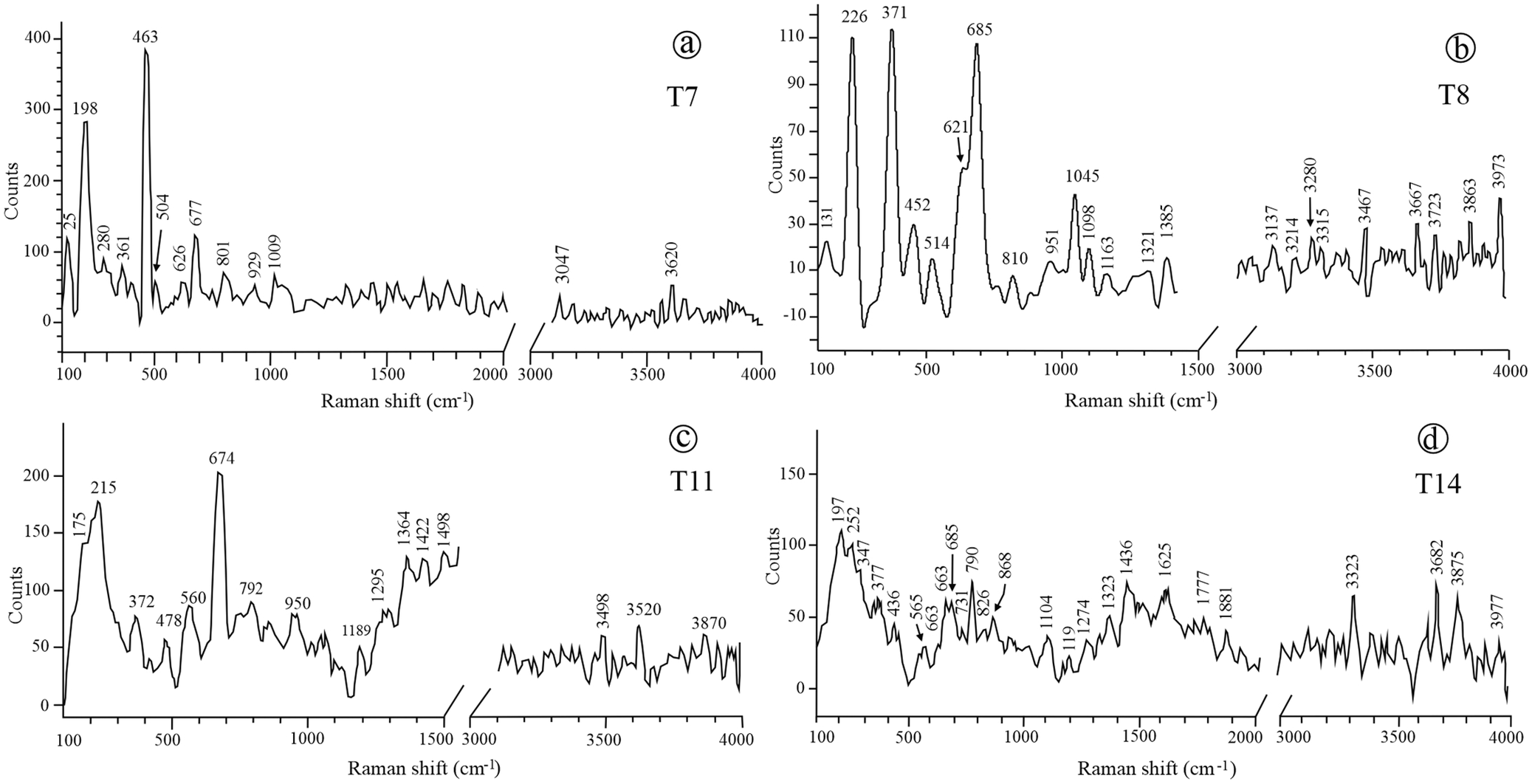
In chrysotile, tremolite, and talc, Raman bands with similar positions were detected between 3660 and 3700 cm-1, corresponding to O–H stretching modes, with low intensity observed across all three minerals. In addition, moderate to weak stretching bands appeared as broad features in the 950–1100 cm-1 region. Strong bands attributed to Si–O–Si bending modes were observed at ~685 cm-1 in chrysotile, 674 cm-1 in tremolite, and 667 cm-1 in talc, reflecting similarities and subtle differences in their silicate frameworks (Fig. 7a,d).
Raman spectra of talc, chrysotile, and tremolite-rich asbestos samples.

Figure 7. Long description
Top left panel labeled T7 shows high intensity peaks at 198, 250, 301, 381, and 463, with additional peaks at 504, 626, 677, 801, 920, and 1009, and lower intensity features near 3047 and 3620. Top right panel labeled T8 displays major peaks at 226, 371, and 685, with smaller peaks at 131, 452, 514, 621, 810, 851, 1045, 1008, 1163, 1385, and a series of minor peaks from 3117 to 3973. Bottom left panel labeled T11 has prominent peaks at 175, 215, 372, 478, 560, 674, 792, 950, 1189, 1295, 1364, and 1498, with broad features at 3498, 3520, and 3870. Bottom right panel labeled T14 shows peaks at 197, 243, 347, 453, 477, 563, 635, 685, 790, 868, 1104, 1197, 1233, 1346, 1625, 1777, 1881, and broad features at 3323, 3582, 3756, and 3977. Each panel demonstrates unique spectral signatures, with T7 showing the highest intensity peaks and T8, T11, and T14 exhibiting distinct but lower intensity patterns.
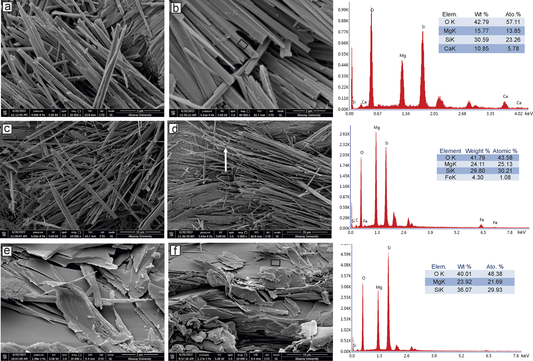
Observations by SEM revealed that the crystals were partially flattened with pointed, splinter-like ends, and bending was particularly evident in typical fibrous forms (Fig. 8a). Compared with tremolite, chrysotile crystals exhibited much smaller diameters (~0.5 μm) but similar lengths (Fig. 8c,d). EDX analyses indicated that these minerals contained primarily Si and Mg, with minor amounts of Fe. Some rod-shaped tremolite crystals were observed to be thinner, with diameters ranging from ~0.3 to 0.5 μm and lengths between 8 and 10 μm. The EDX results also revealed the presence of Ca in addition to Si and Mg. SEM analysis revealed that the talc occurred as irregular, curved, extremely thin platy crystals (Fig. 8e,f). Although the plate thickness could not be measured accurately, the plates exhibited very large aspect ratios. The plates of talc were highly flexible, often showing bending or twisting, and breakage along their edges. EDX analysis indicated MgO and SiO2 contents close to the theoretical composition of talc.
SEM images: (a,b) parallel to subparallel fiber bundles and EDX of tremolite; (c,d) fibrous, parallel to subparallel fiber bundles and EDX of chrysotile; (e,f) platy, curved, extremely thin crystals and EDX of talc.

Figure 8. Long description
Top row, panels a and b, show parallel to subparallel fiber bundles of tremolite at increasing magnification, with panel b highlighting a region analyzed by E D X. The rightmost panel displays the E D X spectrum for tremolite, with peaks for O K, Mg K, Si K, and Ca K, and a table listing weight percent and atomic percent: O K 42.79, 57.11; Mg K 15.77, 13.85; Si K 30.59, 23.26; Ca K 10.85, 5.78. Middle row, panels c and d, show fibrous, parallel to subparallel bundles of chrysotile, with panel d indicating the E D X analysis region by a white arrow. The rightmost panel presents the E D X spectrum for chrysotile, with peaks for O K, Mg K, Si K, and Fe K, and a table: O K 41.79, 43.58; Mg K 24.11, 25.13; Si K 29.80, 30.21; Fe K 4.30, 1.08. Bottom row, panels e and f, display platy, curved, extremely thin crystals of talc at different magnifications. The rightmost panel shows the E D X spectrum for talc, with peaks for O K, Mg K, and Si K, and a table: O K 40.01, 48.38; Mg K 23.92, 21.69; Si K 36.07, 29.93. Each mineral is represented by its characteristic morphology and elemental composition.
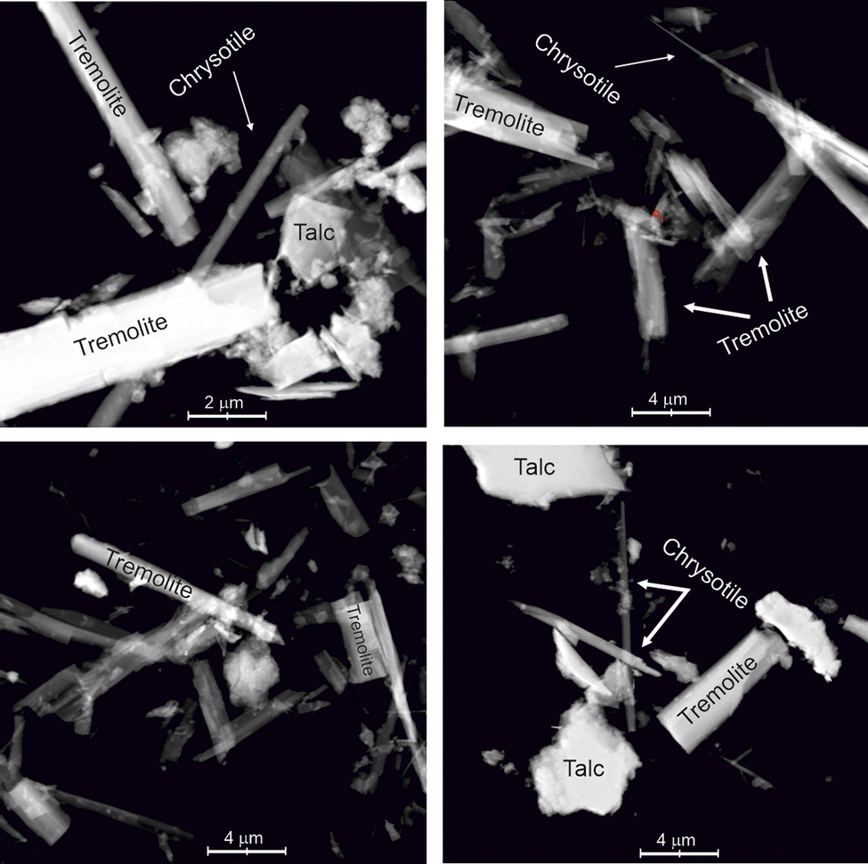
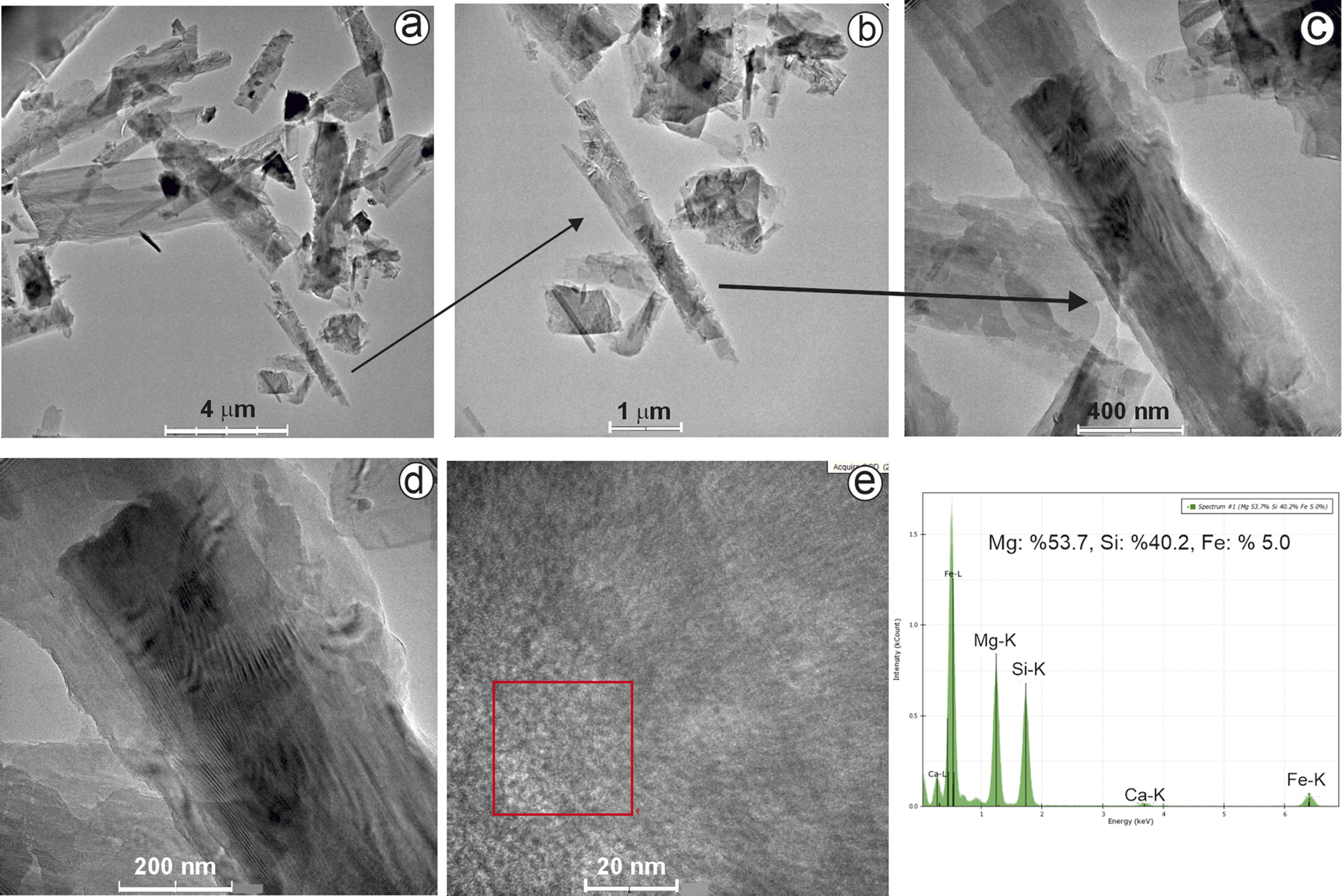
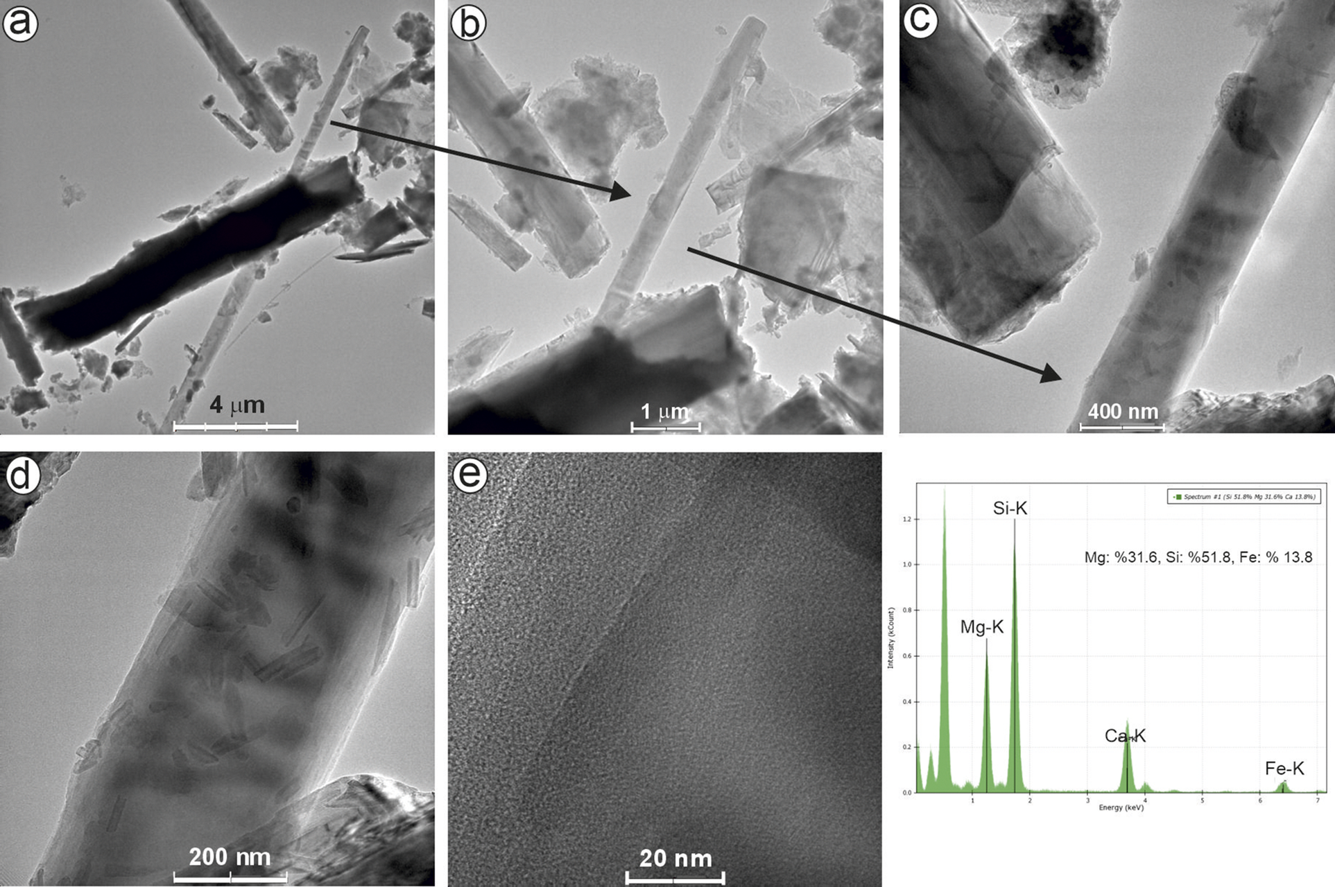
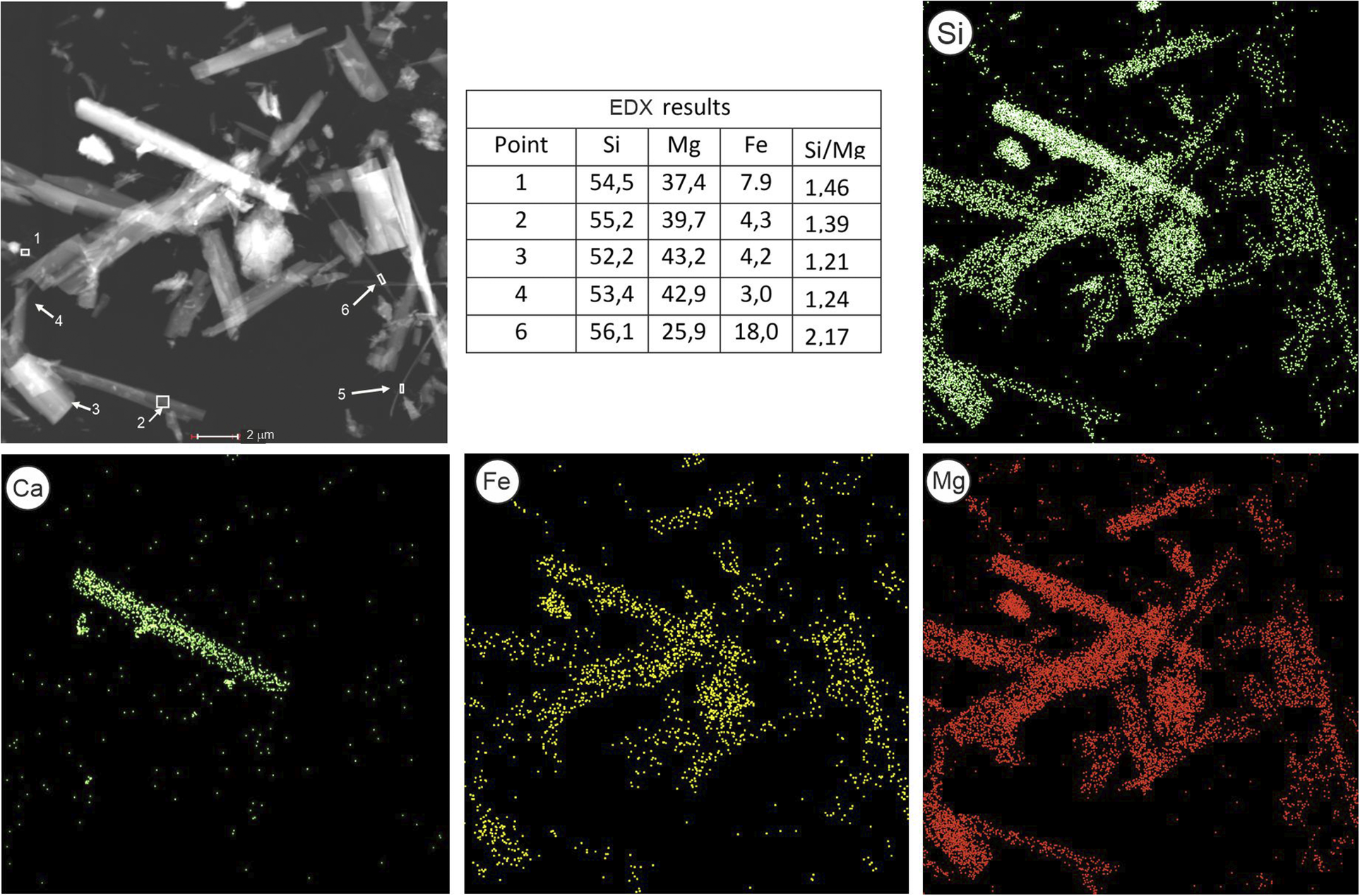
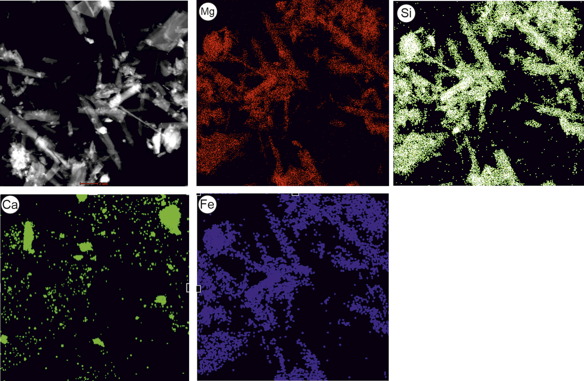
In the three samples analyzed by TEM-EDX (T8, T12, T14), typical needle-like and prismatic morphologies were observed (Figs 9–14). The prismatic crystals ranged in length from 2 to 12 μm, while the needle-like crystals ranged from 1 to 10 μm in length. Their diameters were ~0.5–1.0 μm and 0.1–0.2 μm, respectively. Some crystals overlapped or were observed on top of each other, which made it difficult to precisely determine the size of all crystals (Fig. 9). In each sample, selected areas of prominent crystals were magnified and subjected to EDX analysis (Figs 10–12). According to EDX results, the Mg/Si ratios in samples T8, T12, and T14 were found to be 1.54, 0.70, and 1.40, respectively. The theoretical Mg/Si ratios in the structural formulae of the chrysotile, talc, and tremolite are 3:2, 3:4, and 5:8, respectively. Based on these theoretical ratios, EDX analyses from the points or areas (Figs 10–12) revealed that five analyses in T8 and one in T12 correspond to chrysotile, one in T8 and three in T14 correspond to tremolite, and two analyses in T14 correspond to talc. Considering the structural formulae and element ratios of chrysotile, talc, and tremolite, the EDX results indicate that the analyzed area in T8 (1.54) corresponds to chrysotile, the area in T12 (0.72) corresponds to tremolite, and one analysis in T12 (1.50) and one area in T14 (1.40) are close to chrysotile. Elemental mapping was conducted to identify talc, chrysotile, and tremolite minerals. The distribution of key elements such as Mg, Si, Ca, and Fe provided valuable insights into the compositional zoning of the fibrous and platy crystals. The presence of Ca and Fe within the crystal structures enabled the distinction of tremolite, talc, and chrysotile. Crystals with Ca and high Fe (>5%) contents were interpreted as tremolite, while those with high Mg and low Ca were attributed to chrysotile and talc, and low Fe and no Ca were interprated as chrysotile. The spatial distribution of these elements facilitated the differentiation of crystals exhibiting morphologies characteristic of tremolite and/or chrysotile (Figs 13 and 14).
TEM images showing morphologically similar crystals of talc, chrysotile, and tremolite. The minerals identified are labeled directly on the images.

Figure 9. Long description
Top-left panel shows tremolite as elongated bright rods, talc as irregular clusters, and chrysotile as a fibrous structure, with labels directly on each mineral. The scale bar reads 2 micrometers. Top-right panel displays chrysotile and multiple tremolite crystals, all labeled, with a 4 micrometer scale bar. Bottom-left panel features several tremolite rods labeled at different orientations, with a 4 micrometer scale bar. Bottom-right panel presents talc as bright masses, tremolite as a rod, and chrysotile as fibrous crystals, all labeled, with a 4 micrometer scale bar. Labels are placed directly on or adjacent to the corresponding mineral morphologies in each panel.
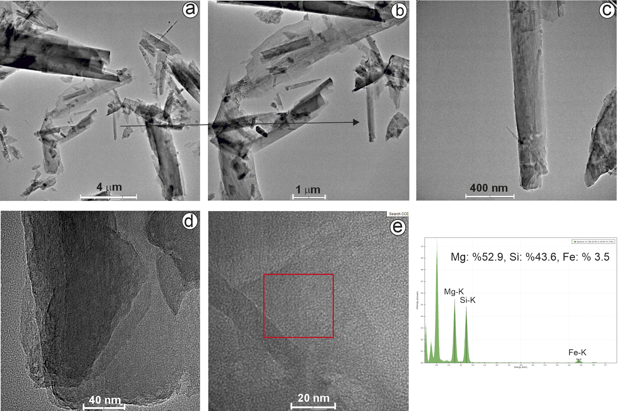
(a) TEM image showing the chrysotile fiber indicated by the arrow. (b–e) TEM images at progressively higher magnifications of the same chrysotile fiber. (f) Corresponding EDX spectrum confirming the chemical composition of the chrysotile analyzed.

Figure 10. Long description
Panel a at top-left shows a low-magnification transmission electron microscopy view of multiple elongated fibers, with an arrow indicating a specific chrysotile fiber. Panel b, to the right, zooms in on the same fiber, with the arrow continuing to highlight its position. Panel c, top-right, presents a further magnified view of the single fiber, revealing its cylindrical structure. Panel d, bottom-left, displays a high-magnification close-up of the fiber wall, showing layered internal morphology. Panel e, bottom-center, offers an even higher magnification of the fiber wall, with a red rectangle marking the region analyzed for composition. Panel f, bottom-right, presents an energy-dispersive X-ray spectrum with three labeled peaks: Mg-K, Si-K, and Fe-K. The spectrum quantifies the elemental composition as magnesium 52.9 percent, silicon 43.6 percent, and iron 3.5 percent.
(a) TEM image showing a chrysotile fiber indicated by the arrow. (b–e) TEM images at progressively higher magnifications of the same chrysotile fiber. (f) Corresponding EDX spectrum confirming the chemical composition of the chrysotile analyzed.

Figure 11. Long description
Panel a at top left shows a low-magnification transmission electron microscopy photo of multiple chrysotile fibers with an arrow pointing to one fiber. Panel b, right of a, presents a closer view of the selected fiber. Panel c, right of b, further magnifies the same fiber, revealing more internal structure. Panel d, bottom left, shows a higher magnification of the fiber, highlighting parallel striations. Panel e, right of d, zooms in on a small region marked by a red square, displaying fine lattice detail. Panel f, at bottom right, is an E D X spectrum with labeled peaks for Mg-K, Si-K, Ca-K, and Fe-K, and annotated elemental percentages: Mg 53.7 percent, Si 40.2 percent, Fe 5.0 percent.
(a) TEM image showing the tremolite crystal indicated by the arrow. (b–e) TEM images at progressively higher magnifications of the same tremolite crystal, displaying predominantly platy–lamellar morphology with locally rod-like features. (f) Corresponding EDX spectrum confirming the chemical composition of the analyzed tremolite.

Figure 12. Long description
Panel a at top-left shows a low-magnification T E M micrograph with an arrow pointing to a prominent elongated tremolite crystal among smaller fragments. Panel b, to the right, zooms in on the same crystal, with an arrow indicating the region of interest. Panel c, further right, presents a higher magnification of the crystal, revealing its platy-lamellar morphology and some rod-like features. Panel d, bottom-left, shows a closer view of the crystal, highlighting internal lamellar structures. Panel e, bottom-center, displays a high-resolution T E M micrograph of the crystal’s lattice fringes. Panel f, bottom-right, presents an E D X spectrum with labeled peaks for Si-K, Mg-K, Ca-K, and Fe-K, and text indicating Mg 31.6 percent, Si 51.8 percent, Fe 13.8 percent. All scale bars are white and labeled: 4 micrometers, 1 micrometer, 400 nanometers, 200 nanometers, and 20 nanometers, respectively.
TEM image of the asbestos-bearing sample showing the locations of six EDX spot analyses (numbered), together with the corresponding EDX results table. Elemental maps illustrating the areal distribution of Si, Ca, Mg, and Fe are also presented. Mineral assignments are primarily based on TEM morphology and supported by EDX data.

Figure 13. Long description
Top left panel is a T E M micrograph of fibrous particles with six numbered arrows marking E D X analysis spots. Center top panel is a table labeled E D X results with columns: Point, Si, Mg, Fe, Si/Mg. Data for points 1 to 6 are: 1: Si 54.5, Mg 37.4, Fe 7.9, Si/Mg 1.46; 2: Si 55.2, Mg 39.7, Fe 4.3, Si/Mg 1.39; 3: Si 52.2, Mg 43.2, Fe 4.2, Si/Mg 1.21; 4: Si 53.4, Mg 42.9, Fe 3.0, Si/Mg 1.24; 6: Si 56.1, Mg 25.9, Fe 18.0, Si/Mg 2.17. Top right panel is the Si map, showing bright signals outlining the fibrous structures. Bottom left is the Ca map, with a single bright region on one fiber. Bottom center is the Fe map, with scattered bright spots, especially on some fibers. Bottom right is the Mg map, with diffuse bright signals overlapping the fibrous network. Mineral assignments are inferred from the T E M morphology and E D X data.
TEM image of the asbestos-bearing sample and corresponding elemental maps showing the areal distribution of Mg, Si, Ca, and Fe. The elemental maps indicate localized Ca enrichment and widespread Mg–Si–Fe signals within the asbestos minerals. Mineral assignments are primarily based on TEM morphology and supported by EDX data.

Figure 14. Long description
The top left panel is a transmission electron micrograph showing elongated, fibrous particles with variable brightness and contrast, indicating mineral morphology. The top center panel is labeled M g and displays a red areal distribution, with magnesium signals matching the fibrous structures seen in the micrograph. The top right panel is labeled S i and shows a green distribution, also following the fibrous pattern. The bottom left panel is labeled C a and presents a green map with calcium signals concentrated in discrete, localized clusters, mostly at the periphery of the fibrous regions. The bottom center panel is labeled F e and shows a blue distribution, with iron signals broadly overlapping the fibrous structures. The bottom right panel is blank. The spatial correspondence between the M g, S i, and F e maps and the fibrous morphology indicates these elements are widespread within the asbestos minerals, while C a is enriched in specific zones. Mineral identification is based on the observed morphology and supported by E D X data.
The spatial distribution of these elements allowed for the differentiation of individual mineral phases, particularly in fibrous aggregates where morphological characteristics alone were insufficient for accurate identification. The integration of elemental mapping with high-resolution imaging supported a more robust mineralogical interpretation, enabling distinction between morphologically similar but compositionally distinct phases such as chrysotile and tremolite.
Discussion
The mineralogical assemblages identified in the highly altered ultramafic zones reveal a complex alteration history shaped by multiple superimposed processes such as serpentinization, carbonation, hydration, and silicification. The dominant alteration products include chrysotile, tremolite, talc, calcite, and chlorite, typically occurring as nearly pure phases indicative of advanced alteration. Minor to moderate amounts of magnesite, quartz, and calcite reflect metasomatic carbonate and silica enrichment. Trace levels of plagioclase group minerals (e.g. anorthite) and pyroxenes (e.g. enstatite, ferrosilite, and augite) suggest relict phases or incomplete alteration of the ultramafic protolith (Table 1).The morphological characteristics of asbestos minerals were evaluated in accordance with Perkins and Harvey (Reference Perkins and Harvey1993), who defined diagnostic criteria including high aspect ratios (typically >20:1), extremely thin fibrils (<0.5 μm in diameter), and splayed or curved fibril bundles. Quantitative measurements of fibre length, diameter, and aspect ratio derived from TEM observations are presented in Table 2. The results confirm that the chrysotile and tremolite fibers analyzed meet the dimensional criteria proposed by Perkins and Harvey (Reference Perkins and Harvey1993) for asbestiform minerals.These features were confirmed in the studied samples through TEM imaging. Chrysotile fibers generally display tubular structures with lighter cores, whereas tremolite fibers exhibited longitudinal, prismatic, or needle-like morphologies. Both minerals were found within respirable size range and exhibited high aspect ratios, characteristics that are commonly associated with increased toxicological concern for asbestos minerals (Strohmeier et al., Reference Strohmeier, Huntington, Bunker, Sanchez, Allison and Lee2010). Measured fiber dimensions obtained from TEM analyses (Table 2) demonstrate that both chrysotile and tremolite fibers meet the Perkins and Harvey (Reference Perkins and Harvey1993) criteria for asbestiform habit, with aspect ratios exceeding 20:1 and diameters consistently <0.5 μm.
Thermal analysis (DTA–TGA) provided further insights into mineral composition and the thermal behavior of the samples studied. The absence of a distinct exothermic peak at ~830°C, commonly associated with chrysotile decomposition in talc-bearing systems, suggests that chrysotile occurs either in low abundance or in a structurally altered form within the investigated assemblages. Instead, progressive dehydroxylation of hydrous silicates was observed. Endothermic reactions between 550 and 650°C are attributed to the release of structural hydroxyl groups from chrysotile, talc, and tremolite, whereas higher-temperature endothermic events occurring between ~900 and 1100°C probably reflect the final stages of dehydroxylation and structural breakdown of these minerals. This thermal behavior is consistent with previous studies reporting broad and overlapping dehydroxylation intervals for fibrous silicates (Paolini et al., Reference Paolini, Tomassetti, Segreto, Borin, Liotta, Torre and Petracchini2019) and aligns well with recent findings by Bloise (Reference Bloise2023), who demonstrated that tremolite exhibits a wide thermal breakdown range and that fibrous varieties tend to decompose at lower temperatures than prismatic crystals. The observed DTA–TG patterns therefore support the mineralogical and morphological interpretations derived from complementary analytical techniques and confirm the presence of thermally reactive fibrous silicates within the studied samples.
Spectroscopic analyses (FTIR and Raman) confirmed the presence of hydroxyl-bearing phyllosilicates. Diagnostic absorption bands appeared in two key regions: 3700–3500 cm-1 (O–H stretching) and 1200–500 cm-1 (Si–O lattice vibrations). Chrysotile and tremolite exhibited sharp O–H bands at ~3675–3677 cm-1, while talc showed broader bands around 3455 cm-1. Raman spectra revealed comparable patterns, with O–H stretching vibrations between 3660 and 3700 cm-1 and prominent Si–O–Si bending bands near 688, 639, and 617 cm-1. These spectral features were consistent with literature data (Anbalagan et al., Reference Anbalagan, Sivakumar, Prabakaran and Gunasekaran2010; Liu et al., Reference Liu, Liu and Hu2014; Reynard et al., Reference Reynard, Bezacier and Caracas2015; Della Ventura et al., Reference Della Ventura, Vigliaturo, Gieré, Pollastri, Gualtieri and Iezzi2018; Zholobenko et al., Reference Zholobenko, Rutten, Zholobenko and Holmes2021), reinforcing the value of using FTIR and Raman in this complementary way for asbestos identification.
Elemental mapping via SEM–EDX and TEM–EDX supported the spectroscopic and petrographic results. Tremolite was identified by high Ca, Mg, Si, and Fe contents, whereas Fe-bearing chrysotile contained Mg, Si, and Fe but lacked Ca. Talc was characterized by Mg and Si, without notable Fe or Ca. Notably, chrysotile fibers contained 1–5 wt.% Fe, aligning with carcinogenic forms reported in previous studies (Janik and Wrona, Reference Janik, Wrona, Worsfold, Townshend and Poole2019; Paolini et al., Reference Paolini, Tomassetti, Segreto, Borin, Liotta, Torre and Petracchini2019). TEM–EDX provided more precise elemental quantification, confirming the substitution of Fe for Mg in the octahedral sheets of chrysotile.
The presence of iron in chrysotile is of particular concern due to its role in enhancing surface reactivity and toxicity. Fe2+/Fe3+ substitution promotes the generation of reactive oxygen species (ROS) through Fenton-type reactions, increasing the mineral’s pathogenic potential (Fubini and Fenoglio, Reference Fubini and Fenoglio2007; Gualtieri, Reference Gualtieri2021). Although chrysotile is often considered less hazardous than amphibole-group asbestos, Fe enrichment significantly influences its bio-persistence and genotoxicity (Jiang et al., Reference Jiang, Chen, Chen, Feng, Jin, Zhong and Lou2022; Walter et al., Reference Walter, Schenkeveld, Geroldinger, Gille and Kraemer2023). The observed Fe-bearing chrysotile with asbestiform morphology and respirable dimensions in the current study reinforces the potential health hazard associated with these minerals.
The chemical compositions of selected samples (T6–T14), determined by XRF spectrometry, support the mineralogical identifications based on XRD, SEM/TEM, and spectroscopic methods (Table 3). The talc-rich samples (T6 and T7) show high SiO₂ (62.89% and 65.45%) and MgO (28.59% and 25.87%) contents, which are close to the theoretical composition of talc (SiO₂: 63.5%, MgO: 31.7%). Slightly lower MgO values may reflect minor Fe substitution (Fe₂O₃: 1.11–3.52%) or the presence of subordinate phases. The LOI values (4.69 and 3.97%) are also consistent with the expected structural water content in talc, and the absence of significant CaO (2.12 and 0.69%) further supports the lack of amphibole phases.
Major oxide (wt.%) and selected trace element (ppm) compositions of representative samples determined by XRF spectrometry

Table 3. Long description
Beginning at the top, the leftmost column lists elements and oxides: Si O sub 2, Al sub 2 O sub 3, Fe sub 2 O sub 3, Mg O, Ca O, Ti O sub 2, Na sub 2 O, K sub 2 O, Mn O, Cr sub 2 O sub 3, loss of ignition, total, Co, Ni, Zn, Sr, Zr, Ba, Nd, Yb. Each row displays values for samples T6, T7, T8, T10, T11, T12, and T14, moving left to right. Si O sub 2 ranges from 39.64 in T10 to 65.45 in T7. Mg O is highest in T14 at 42.14 and lowest in T8 at 21.83. Al sub 2 O sub 3 peaks in T10 at 9.99. Fe sub 2 O sub 3 is highest in T10 at 12.54. Ca O is highest in T12 at 16.73. Ti O sub 2 is low overall, peaking at 1.26 in T10. Na sub 2 O and K sub 2 O are consistently low, with K sub 2 O at 0.10 for all samples. Mn O and Cr sub 2 O sub 3 are minor, with Cr sub 2 O sub 3 highest in T12 at 0.27. Loss of ignition is highest in T14 at 12.32. Total values are near 100 except T12 at 105.09. Among trace elements, Ni is highest in T7 at 1519 and lowest in T11 at 121. Co peaks in T12 at 116. Zn is highest in T12 at 97. Sr is highest in T8 at 202. Zr peaks in T10 at 68. Ba is highest in T6 at 228 and lowest in T10 at 93. Nd is highest in T8 at 69. Yb is highest in T7 at 295 and lowest in T11 at 17. The table reveals significant variation in oxide and trace element concentrations across samples, with Mg O and Si O sub 2 as dominant oxides and Ni as the most variable trace element.
Minor deviations observed in the chemical composition of the chrysotile-rich sample (T14), particularly the slightly reduced SiO₂ and MgO values and the presence of 1.59% Fe₂O₃, support the TEM-EDX results indicating Fe substitution in the chrysotile structure. This is significant because Fe-bearing chrysotile is known to exibit greater oxidative reactivity and is potentially more carcinogenic. The consistency between bulk chemical data and microanalytical observations strengthens the reliability of mineral determination and supports interpretations of associated health risks based on mineral chemistry.
From a public health perspective, the identification of asbestiform chrysotile and tremolite in residential and agricultural zones raises significant concern. Analysis by TEM revealed that many fibers fall within respirable size ranges and possess morphologies associated with carcinogenic potential. These findings are consistent with prior reports of elevated mesothelioma incidence in nearby villages (Canda et al., Reference Canda, Bircan, Akın, Kabadayı and Kurt1988; Canda, Reference Canda1989; Canda et al., Reference Canda, Giray, Topal and Görücü2001), although the specific mineralogical sources had not been characterized thoroughly in the past. The results of the persent study demonstrate that serpentinization, carbonation, and weathering of ophiolitic rocks have produced fibrous chrysotile, tremolite, and talc, confirmed through XRD, FTIR, Raman, DTA–TGA, SEM–EDX, and TEM–EDX analyses.
Beyond their mineralogical significance, the identification of asbestiform chrysotile and tremolite within altered ophiolitic rocks has broader implications for environmental awareness and regional land-use considerations. In areas where naturally occurring asbestos is present, routine activities such as agriculture, construction, and the use of local materials may lead to episodic disturbance of asbestos-bearing rocks and soils. Although the present study does not assess exposure levels or health outcomes, the documented occurrence of respirable asbestiform minerals highlights the importance of incorporating geological information into environmental monitoring programs and land-use planning strategies. Such integration may support informed decision-making aimed at minimizing potential exposure risks in regions characterized by naturally occurring fibrous minerals.
Conclusions
The present study provides integrated mineralogical, chemical, and spectroscopic evidence for the occurrence and characterization of asbestos minerals in altered ultramafic rocks. Detailed analyses revealed that chrysotile and tremolite occur as fibrous minerals within variably altered ultramafic lithologies in the study area. Imaging by SEM and TEM confirmed that many of the fibers exhibit acicular to tubular morphologies, with diameters ranging from 0.1 to 0.5 μm and lengths up to 10–12 μm, placing them within the respirable range. The TEM–EDX analyses show that these fibers are compositionally consistent with chrysotile and tremolite, dominated by Mg and Si, with minor Fe substitution, particularly in chrysotile. Calcium was detected exclusively in tremolite-bearing crystals. Elemental mapping further confirmed the spatial association of Fe-bearing chrysotile and Ca-rich tremolite within the investigated samples.
The use of FTIR and Raman spectroscopy validated the presence of chrysotile, tremolite, and talc, each through diagnostic vibrational bands related to O–H stretching and Si–O lattice vibrations. These identifications were supported by DTA-TG analyses, which revealed endothermic reactions associated with dehydroxylation processes between 650 and 780°C for chrysotile and between 700 and 1025°C for tremolite. Talc exhibited characteristics of thermal behavior, with a broad endothermic peak between 600 and 950°C and a total weight loss of ~5.37%, slightly exceeding theoretical values, probably due to a minor chrysotile admixture.
Several villages in the study area are located on or near asbestos-bearing ultramafic rocks, where agricultural, livestock-, and construction-related activities are common. Although the present study does not quantify airborne fiber concentrations or exposure levels, the documented presence of respirable, Fe-bearing asbestiform chrysotile and tremolite indicates a potential environmental health concern under conditions of natural or anthropogenic disturbance. These findings highlight the importance of detailed mineralogical characterization of naturally occurring asbestos and underscore the need for environmental monitoring and informed land-use planning in regions affected by asbestos-bearing geological formations.
Author contribution
Methodology: M.Ç.K., N.K.; Investigation: M.Ç.K., N.K.; Data curation: M.Ç.K., N.K. Writing - original draft preparation: M.Ç.K.; Writing - review and editing: M.Ç.K., N.K.; Supervision: M.Ç.K.; Project administration: M.Ç.K., N.K. Both authors have read and agreed to the published version of the manuscript.
Acknowledgments
At the final stage of manuscript preparation, an AI language model (ChatGPT, OpenAI) was used for language editing and proofreading only. All scientific content and conclusions are the authors’ own.
Data availability statement
Most of the data generated or analyzed during this study are included in this published article. Representative field photographs and SEM/TEM micrographs are presented in the manuscript, while additional supporting data are available from the corresponding author upon reasonable request.
Financial support
This work was supported by the Scientific Research Projects Support Program of Konya Technical University (211007013).
Formun Üstü
Competing interests
The authors declare that they have no competing interests.
Ethics approval
This study did not involve human participants or animals, and therefore ethics approval was not required.