Introduction
The soft-shelled turtle, Pelodiseus sinensis, widely distributed in China, is the most common terrapin. The eggs of this species of turtle are large and lecithotrophic, which makes them a perfect food source because of their high nutritional value. Over-harvesting of these Pelodiseus sinensis eggs has helped to diminish the abundance of this species of turtle in China (Yang et al., Reference Yang, Niu and Sun1999). Artificial breeding programmes were implemented so that this turtle's eggs could still be used as a food source, while natural populations were protected. However, the nutritional value of harvested eggs is significantly less than that of those from wild populations (Chen et al., Reference Chen, Hua and Chen1998). Thus, in order to pursue both economic and pharmacological values, reproduction studies are necessary to find ways to keep the production and nutritional value of Pelodiseus sinensis eggs at levels acceptable to the communities that require them (Yang et al., Reference Yang, Niu and Sun1999).
Recently, it has been found that male and female Pelodiseus sinensis possess unique features associated with their reproductive system, i.e. sperm storage structures (Han et al., Reference Han, Zhang, Hei and Chen2007, Reference Han, Zhang, Li, Bao, Hei and Chen2008; Zhang et al., Reference Zhang, Han, Qi, Liu and Chen2008). Thus, there might be some corresponding structures in the ovaries of female turtles.
Complete histological details of oogenesis and vitellogenesis in Pelodiseus sinensis are rarely available. The question of whether the vitellogenesis pathway is intrinsic, extrinsic or hybrid is controversial. It was reported that follicular cells could transport nutritive substances from the parent digestive system, via microvilli, to the primary oocyte, which is the source of yolk formation (Chen et al., Reference Chen, Hua and Chen1998). The aim of the present study was, therefore, to identify and to describe the histological changes that occur during oogenesis in Pelodiseus sinensis and to compare them with the findings of previous studies in reptiles (Andreuccetti, Reference Andreuccetti1992; Cai et al., Reference Cai, Li and Chen1999, Reference Cai, Liu and Wang2004; Huang et al., Reference Huang, Pan and Jiang1999; Mari & Guillette, Reference Mari and Guillette2000; Calderón et al., Reference Calderón, De Pérez and Ramírez Pinilla2004; Gómez & Ramírez-Pinilla, Reference Gómez and Ramírez-Pinilla2004; Hernández-Franyutti et al., Reference Hernández-Franyutti, Uribe Aranzábal and Guillette2005) and other vertebrates (Stephen & Goldberg, Reference Goldberg1968; Guraya, Reference Guraya1989; Callebaut et al., Reference Callebaut, Van Nassauw and Harrison1997). In addition, it attempts to show that both intrinsic and extrinsic pathways contribute to vitellogenesis in Pelodiseus sinensis.
Materials and Methods
Animals
All protocols were approved by the Chinese Committee for Animal Use for Research and Education. Mature (3- to 4-year-old) Pelodiseus sinensis females were purchased from an aquafarm in Nanjing, south China, from January to December 2007; five turtles were obtained each month, for a total of 60 turtles. The turtles were anaesthetized with an intraperitoneal injection (20 mg/kg) of sodium pentobarbital (ANPRO Pharmaceuticals) and by cervical dislocation.
Light microscopy
Ovaries were surgically removed under sterile conditions and immediately fixed in Bouin's fluid. After fixation, the tissue blocks were washed in 75% alcohol, dehydrated in a graded ethyl alcohol series (85%, 95% I, 95% II and 100% I, 100% II), cleared in xylene and embedded in paraffin. Tissues were serially sectioned at 5μm thickness on a rotary microtome and stained with hematoxylin and eosin (H–E), periodic acid–Schiff procedure (PAS). For histological descriptions, the oogenesis process was divided into 10 stages (I–X) based on the morphological stages described by Mari et al. (Reference Mari and Guillette2000), one more stage was added to the vitellogenesis phase. Measurements of ovarian structures (n = 20 of each) were made with either a vernier caliper or an ocular micrometer. Photomicrographs were taken with a Spot camera (Typhoon 8600 Phosphorimager, Amersham Biosciences) and digitally enhanced with Adobe Photoshop software (Adobe Systems), to elucidate the histological morphology of the ovaries.
Transmission electron microscopy
Ten oocytes of various sizes were divided into two groups that corresponded to Stages IV and X, as measured by light microscopy results. They were cut into small blocks, immersed in 2.5% glutaraldehyde fixative in phosphate-buffered saline (PBS) (4°C, pH 7.4, 0.1 M) for an additional hour and washed in the same buffer overnight. The tissues were post-fixed with a mixture of 1% osmium tetroxide and 1.25% potassium ferrocyanide for 1.5–2 h, washed in the same buffer, dehydrated in increasing concentrations of ethyl alcohol, infiltrated with a propylene oxide/Araldite mixture and embedded in Araldite. Semi-thin sections of oolemma were stained with toluidine blue for light microscopic examination and five thin sections for electron microscopy were mounted on Formvar-coated grids and stained with uranyl acetate and lead citrate. Ultrastructure of the oocytes was examined with a JEM-1200EX electron microscope.
Results
Light microscopy
General ovarian structure
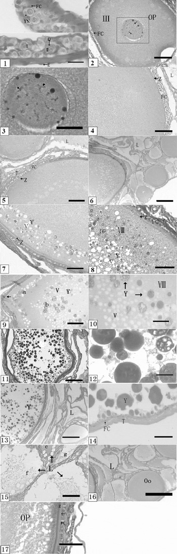
The ovary of Pelodiseus sinensis was seen to be an irregular, elongated cone-shaped structure, lying in the abdominal cavity and connected to the dorsal body wall by the mesovarium. The wall of the ovary was surrounded externally by a simple squamous epithelium. The epithelial cells had a mean height of 1.24μm (Fig. 1-1). Subjacent to the ovarian epithelium was a stroma formed of connective tissue, which enclosed ovarian follicles at various stages of development (Fig. 1-1), including atretic follicles.
Oogonia and early folliculogenesis. (1) Stages I and II of previtellogenesis (I–V) in Pelodiseus sinensis. oogonia (Oo), a yolk nucleus (YN) adjoined to the oocyte nucleus, a follicular cell (FC) surrounds the oocytes, the nucleus contains early diplotene chromosomes with chiasmata (Q), the ovary is covered by a single-layered cuboidal epithelium (E); 10 × 40 H and E. (2) Early previtellogenic oocytes in Stage III have eccentric nuclei containing lampbrush chromosomes (arrowhead) and several nucleoli. ← Indicates the follicular epithelium (FC) completely surrounding the oocyte and consisting of squamous cells. 10 × 10 H and E. (3) Detail of (2), showing the lampbrush chromosomes (arrowhead); 10 × 40 H and E. (4) Stage IV – the ooplasm has a finely granular homogeneous and finely granular dispersed appearance. Small and large lacunae (L) surround the follicle. The follicular epithelium is formed of a single layer of cuboidal cells (FC). The thin ZP (Z) and the theca (T) surround the oocyte; 10 × 20 H and E. (5) Oocyte periphery in Stage V shows the cuboidal follicular cells (FC). The theca (T) contains abundant collagen fibres; 10 × 20 H and E. (6) Stage VI in vitellogenesis (VI–X) of Pelodiseus sinensis – the peripheral ooplasm appears vacuolated (V), the central ooplasm is homogeneous (OP); a cuboidal follicular epithelium (FC), theca (T) and lacunae (L) surround the oocyte; 10 × 4 H and E. (7) Oocytes in Stage VII – the ooplasm has abundant vacuoles (V), most of them containing dense yolk platelets (Y). Both regions of the ZP – the zona radiata (r) and the hyaline band (h) are well defined. The theca (T) is outside the follicular cells; 10 × 20 H and E. (8) Oocytes in Stage VIII – most ooplasm contain abundant vacuoles and dense yolk platelets. Three different regions are observed based on the size of vacuoles and yolk platelets (Y): (i) peripheral small yolk platelets; (ii) larger yolk platelets; and (iii) small, central yolk platelets. In the ZP (Z) two components – the zona radiata (r) and the hyaline band (h) – are distinguished and there are a cuboidal follicular epithelium (FC) and a fibrous theca (T); 10 × 20 H and E. (9) Another morphological variation of the oocyte in stage VIII, showing two layers (a, b) of vacuoles (V) and the yolk platelets (Y) within it and the ZP (Z); 10 × 10 H and E. (10) Magnification of (9), showing that the yolk platelets (Y) exhibit varied sizes, with many of them within the vacuoles (V); 10 × 40 H and E. (11) Oocytes in Stage IX – central yolk platelets (Y) exhibit a shape similar to those of peripheral yolk platelets; 10 × 4 H and E. (12) Magnification of (11), showing that the yolk platelet (Y) is much larger than the one in Stage VIII; 10 × 40 H and E. (13) Oocytes in Stage X – peripheral yolk platelets (Y) attain maximum size and contain abundant yolk spheres of varied sizes and staining affinities. Surrounding the follicle are parallel lacunae (L) and bundles of smooth muscle; 10 × 4 H and E. (14) Oocytes in Stage X – the thick theca (T) has attained its maximum thickness and surrounds the follicle; the yolk platelet (Y) is polygonal; and the follicular cell (FC) consists of squamous cells; 10 × 40 H and E. (15) Lacunae surround various types of follicles in the ovary of Pelodiseus sinensis and exhibit differing morphologies, depending on their contents: hyaline substance with fine granules (g), homogeneous hyaline substance (c), substance with vacuoles and fine fibres (f); 10 × 40 H and E. (16) Detail of (6) – the lacunae surround previtellogenic follicles, vitellogenic follicles and atretic follicles; 10 × 4 H and E. (17) Vitellogenesis oocyte that consists of two layers – an inner striated layer, the zona radiata and an outer homogeneous layer or hyaline band – that exhibit an intense PAS+ reaction (f); 10 × 20 PAS.

The ovary consisted of two histological regions: a cortex and a medulla. The cortex contained germinal elements (follicles in various stages of development, corpora lutea and atretic follicles), which was surrounded by the stroma. The medulla was formed from a stroma with large blood vessels from which the cortical vascular supply was obtained. This stroma consisted of connective tissue that contained collagen fibres, adipocytes, blood and lymphatic vessels, as well as bundles of smooth muscle fibres. Numerous large lacunae distributed throughout the stroma were situated in both the cortex and the medulla (Fig. 1-4–6, -13, -15, -16). These lacunae surrounded follicles at all stages of development: previtellogenic (Fig. 1-4, -5), vitellogenic (Fig. 1-6), postovulatory and atretic. The lacunae were irregular in shape and contained an amorphous substance that exhibited light or deep acidophilic staining. The amorphous substance within each of the lacunae contained fine granules, fibres or gelatinous substances (Fig. 1-15). The germinal beds were distributed within the ovarian cortex as elongated patches of cells (Fig. 1-1) that were thicker than the rest of the ovarian wall and comprised oogonia, early oocytes and epithelial somatic cells. The oogonia were distributed within the medulla; they had the smallest diameter and were spherical or ovoid in shape. The nucleus was round and the ooplasm was relatively homogeneous and lightly stained.
Follicular development and vitellogenesis
Oogenesis consisted of previtellogenesis and vitellogenesis, as determined by the morphological characteristics of the yolk platelets. The oogenesis process was further divided into 10 consecutive phases on the basis of the structures of follicular cells, zona pellucida (ZP) and some other related features.
Previtellogenesis. Stage I: In Stage I, oocyte growth was initiated (Fig. 1-1) and the oocyte diameter was approximately 7.48 ± 3.46μm. The ooplasm was homogeneous and stained lightly, similarly to the oogonia, but the yolk nucleus was a small, basophilic, ovoid structure, adjacent to the nuclear membrane (Fig. 1-1). There was a single nucleolus that stained intensely. The oocyte was incompletely surrounded by a small number of follicular cells that had a squamous appearance (Fig. 1-1).
Stage II: Oocytes were spherical or oval in shape and had a mean diameter of 10.33 ± 4.36μm. The ooplasm was lightly stained and showed fine fibres and granules (Fig. 1-1). The central nucleus was spherical and contained some filiform lampbrush chromosomes, which indicate the diplotene stage of meiosis I. The yolk nucleus was near the oocyte nucleus (Fig. 1-1). At this stage the oocyte was not yet completely surrounded by squamous follicular cells.
Stage III: The oocytes were approximately 86.22μm in diameter. The ooplasm was basophilic and contained fine granules (Fig. 1-2). The yolk nucleus was less dense than in the previous stages. The oocyte nucleus was eccentric and contained obvious lampbrush chromosomes and numerous nucleoli (Fig. 1-2, -3). The follicular epithelium, which was composed of a monolayer of squamous cells that completely surrounded the oocyte, was now termed the granulosa (Fig. 1-2). A thin layer of fibroblasts lying outside the granulosa had begun to form the theca layers (Fig. 1-2).
Stage IV: The oocytes were 118.60 ± 26.04μm in diameter. The morphology of the nucleus and ooplasm was similar to that seen in the previous stages, but no yolk nucleus was observed. The ZP, situated between the oocyte and follicular cells, was observed as a thin layer (1.69 ± 0.22μm in thickness) (Fig. 1-4). The granulosa consisted of a monolayer of cuboidal cells (11.92 ± 2.50μm) that contained centrally located, spherical nuclei (Fig. 1-4).
Stage V: The oocytes at this stage had a mean diameter of 195.56 ± 46.22μm (Fig. 1-5). The morphological features of the ooplasm and granulosa exhibited no apparent differences compared with the preceding stage, but the ZP was significantly thicker, at 3.15 ± 0.92μm. It consisted of two layers: an inner striated layer (the zona radiate) and an outer homogeneous or hyaline band that exhibited an intense PAS+ reaction (Fig. 1-17). The granulosa remained a single layer of cuboidal cells, but the theca continued to grow in thickness (Fig. 1-5).
Vitellogenesis. Stage VI: At the beginning of vitellogenesis the oocytes were 275.24 ± 37.61μm in diameter. The peripheral ooplasm contained fine granules and vacuoles were observed adjacent to this zone, toward the centre of the oocyte (Fig. 1-6). Numbers of vacuoles gradually increased toward the centre of the oocyte and, in parallel, vacuoles at the periphery of the oocyte were surrounded by fine granules (Fig. 1-6), whereas the central ooplasm remained homogeneous at this stage (Fig. 1-6). The granulosa consisted of cuboidal cells and the theca was well developed, with small sinuses (Fig. 1-6).
Stage VII: Continued growth of the oocyte was evident at this stage (diameter approximately 375.34μm) and the peripheral ooplasm was densely populated with fine acidophilic granules (Fig. 1-7). The peripheral ooplasm was densely populated with fine, acidophilic granules. The vacuoles were greater in size than at the preceding stage, with some reaching 11.38 ± 2.74μm in diameter. Toward the centre of the oocyte, some vacuoles in the ooplasm were observed containing one or more large (7.84 ± 1.13μm), dense, acidophilic yolk platelets (Fig. 1-7). The granulosa consisted of a single cuboidal layer. The ZP (4.33 ± 1.65μm in thickness) was formed of two layers: the zona radiata (r) and the hyaline band (h) (Fig. 1-7). Similarly to the previous vitellogenic stages, the theca had sinuses that appeared as large lacunae spread among the collagen fibres that were arranged parallel to the oocyte surface.
Stage VIII: Oocytes in Stage VIII had a greater diameter (623.79 ± 83.98μm) than that in Stage VII (Fig. 1-8). The oocyte showed a regional distribution of yolk platelet features: in region 1, at the periphery, the yolk platelets were small (1.15 ± 0.66μm); in the intermediately located region 2 they become larger (3.11 ± 1.23μm); and in region 3, near the nucleus, they had reverted to a small size (0.83 ± 0.45μm) (Fig. 1-8). Each yolk platelet consisted of several yolk globules (Fig. 1-10). Also, some of the stage VIII oocytes showed a strange feature, characterized by formation of two ring-shaped areas by the vacuole and the inter yolk granules (Fig. 1-9; a, b). The granulosa consisted of a single layer of cuboidal cells and the theca contained large blood vessels, abundant collagen fibres and flattened lacunae.
Stage IX: The oocyte diameter averaged 870.78 ±50.32μm. Large yolk platelets filled most of the ooplasm; they attained a diameter of 12.60 ± 2.33μm and were spherical or polyhedral in shape (Fig. 1-12). Toward the centre of the ooplasm, the platelets contained yolk spheres larger than those observed at the periphery (Fig. 1-11). The follicular epithelium again consisted of squamous cells. The ZP and theca were unchanged in morphology from the previous stages.
Stage X: In Stage X the preovulatory oocytes had increased almost 100-fold in diameter compared with those in Stage I; the maximum diameter observed was 1.63 cm. The large yolk platelets continued to increase in size, to approximately 23.13μm (Fig. 1-4). These platelets contained numerous hyaline or dense yolk spheres of various sizes (Fig. 1-13, -14). Similarly to the previous stages, the central platelets contained yolk spheres larger (20.75 ± 5.36μm) than those at the periphery (Fig. 1-13, -14). The hyaline band of the ZP was well defined. The granulosa were squamous in appearance. The theca increased dramatically in thickness, to 39.94 ± 7.93μm and exhibited extensive layering, with a network of fibroblasts, collagen fibres, lacunae and smooth muscle cells (Fig. 1-13, -14).
Electron microscopy
Transmission electron microscopy was used to observe the follicular ultrastructure during vitellogenesis of Pelodiseus sinensis. To aid clearer description two stages were designated according to the size of the follicle, as below.
The early stage of vitellogenesis
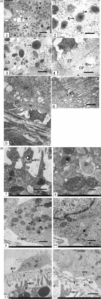
In the early stage of vitellogenesis, there were large numbers of mitochondria (Mt) dispersed under the oolemma, correspondent condition that corresponded to Stage VI as observed via optical microscopy, in which yolk granules gathered first under the oolemma (Fig. 1-6). Some of the Mt were observed gradually transforming into yolk granules: firstly, the cristae layer of the Mt disappeared, then the Mt degenerated and intumesced into vesicular a concentric-circle double-membrane structure (Fig. 2-1); secondly, the double membrane merged and an electron-dense dot appeared in the centre of the Mt and yolk materials were deposited around this dot; thirdly, the Mt were completely filled with yolk materials and transformed to yolk granules (Fig. 2-2, -3); in addition and finally, comparatively large lipid droplets, rough endoplasmic reticula (RERs) and ribosomes were observed (Fig. 2-3).
(a) The early stage of vitellogenesis. (1) Transformation from Mt to yolk materials (1–5): first, the vesicular concentric-circle double-membrane structure ( ), the double membrane then consolidates to a single membrane, an electron-dense dot appears in the centre of Mt and yolk materials are deposited around this dot into Mt (
 ); ×6000. (2) More and more yolk materials are deposited into the Mt (); ×30 000. (3) The Mt is completely filled with yolk materials (indicated by arrowhead), large lipid droplets (LD) and ribosomes (Ri); ×15 000. (4) The follicular cell shows an irregular shape and has a big central nucleus. Spaces between follicular cells (FC) are quite large; and there are ooplasm (OP) and a zona pellucida (ZP); ×4000. (5) The zona pellucida (ZP) is formed tightly and uniformly by the crossed digitations from the oocyte and follicular cell (FC). The microvilli on the oocyte surface are much more regular than those on the follicular cell. The follicular cell is covered by a theca consisting of fibroblast and smooth muscle. There is ooplasm (OP); ×2000. (6) Magnification of (5) There are deep, coated pits (CP) between the microvilli of the oocyte; ×15 000. (b) The advanced stage of vitellogenesis. (7) Microvilli (MVO) from the oocyte become more prominent, irregular in shape and acquire emerging branches; yolk materials are found in it (□); ×10 000. (8) Magnification of (7) – there is a large number of yolk granules in the ooplasm and some of the Mt are still transforming to yolk granules. One or two electron-dense dots (ED) emerge during that transformation period; ×50 000. (9) there are also a great number of Mt () transformed to ‘yellow yolk’ in follicular cells. There are two electron-dense dots (ED), transect of the microvilli of the follicular cell (MVF); ×20 000. (10) Showing follicular cell nucleus (Nu); abundant Golgiosomes (asterisk); ×20 000. (11) The ZP is thicker because of the growth of the microvilli. The shows microvilli of the follicular cell (MVF) and of the oocytes (MVO), laterally dispersed ropes/threads (R) and yolk in the ooplasm (Y); ×5000. (12) Magnification of (11), showing microvilli of the follicular cell (MVF) and the oocytes (MVO), ropes/threads (R) and rough endoplasmic reticulum (RER); ×10 000.

The follicular cell exhibited an irregular shape and a big central nucleus and its cytoplasm contained a large number of free ribosomes and comparatively fewer Mt, ERs and lysosomes, whereas other organelles were seldom observed. Spaces between follicular cells were quite large.
The ZP, whose average thickness was 4.35 ± 1.81μm, was formed tightly and regularly by the crossed digitations (microvilli) from the oocyte and the follicular cell (Fig. 2-4). The microvilli on the oocyte surface were much more regular than those on the follicular cells; moreover, there were deep, coated pits between the digitations (Fig. 2-6).
The follicular cell was covered by theca consisting of fibroblasts and smooth muscle (Fig. 2-5).
The advanced stage of vitellogenesis
In the advanced stage of vitellogenesis, there were myriad yolk granules in the ooplasm and some of the Mts were still being transformed into yolk granules; meanwhile, two electron-dense dots emerged during the transformation period, which was not observed in the earlier stage (Fig. 2-8). Oocyte microvilli became more prominent, with irregular shapes and emerging branches contained yolk materials (Fig. 2-7).
The same phenomenon of transformation of great numbers of Mt into ‘yellow yolk’ precursors was observed in follicular cells also. We also found one or two electron-dense dots emerging during that period (Fig. 2-8, -9). Notably, the presence of organelles inside the follicular cell was obvious: it contained abundant SERs and apparently expanded Golgiosomes (Fig. 2-10, -12).
The ZP was thicker, at 7.47 ± 2.27μm, because of growth of the microvilli. The microvilli of the follicular cell exhibited little growth compared with that of the oocyte, which was growing rapidly and branching conspicuously. This feature made the structure of the ZP more complicated, because the microvilli emerging from the oocyte and the follicular cell were irregular and the spaces between them were much wider; moreover some ropes (or named threads) were also seen, laterally distributed in the ZP. This kind of material was located in the microvilli, adjacent to the follicular cells (Fig. 2-11, -12).
Discussion
Structural histology of the ovary
The ovary of Pelodiseus sinensis was surrounded by a simple squamous epithelium and was divided into two distinct regions: a cortex and a medulla. The germinal beds of the ovary were distributed in the dorsal side of the cortex and contained oogonia that proliferated by mitotic divisions and the medulla, which had bird's nest-like structure, was amongst the follicles during the previtellogenesis stage. In lizards (Mabuya, Sceloporus), there were one or two germinal beds, depending on the species (Callebaut et al., Reference Callebaut, Van Nassauw and Harrison1997; Gómez & Ramírez-Pinilla, Reference Gómez and Ramírez-Pinilla2004). The ovary of the crocodile was surrounded by a simple cuboidal epithelium and possessed larger germinal beds than Pelodiseus sinensis, in terms of area (Mari & Guillette, Reference Mari and Guillette2000; Calderón et al., Reference Calderón, De Pérez and Ramírez Pinilla2004).
Only one nucleolus could be observed during the early stages of oogenesis in Pelodiseus sinensis; it changed to polynucleoli in the advanced stages of previtellogenesis. This condition, termed peripheral nucleoli, was consistent with what had been observed in other reptiles and birds (Guraya, Reference Guraya1989). The position of the nucleoli in traditional reptiles and birds might aid in the transport of nucleoprotein and ribosome from the nucleus to the ooplasm (Saxena, Reference Saxena1979; Mari & Guillette, Reference Mari and Guillette2000; Hernández-Franyutti et al., Reference Hernández-Franyutti, Uribe Aranzábal and Guillette2005).
Early oocytes of Pelodiseus sinensis in the prelampbrush-chromosome stage had a basophilic juxtanuclear Balbiani's vitelline body, similar to those of the other reptiles and birds (Hernández-Franyutti et al., Reference Hernández-Franyutti, Uribe Aranzábal and Guillette2005). This structure contained mitochondria, SERs, Golgisomes, lysosomes and lipids, which were dispersed throughout the cytoplasm while the oocyte was growing. This body acted as the initial centre of increased metabolic activity and could be related to intensive transportation and accumulation of substances that were necessary for the growth and development of telolecithal eggs (Tammaro et al., Reference Tammaro, Andreuccetti, Filosa, Indolfi, Prisco and Motta1998; Mari & Guillette, Reference Mari and Guillette2000).
Yolk platelet structure
The morphology of yolk platelets showed gradual changes throughout the vitellogenic process. Initially, yolk platelets surrounded by vesicles were seen. As vitellogenesis advanced, the vesicles contained numerous yolk spheres, with slowly growing platelets and these spheres continued to increase in size and number within the vacuoles. In birds, three main types of yolk are successively deposited during early, intermediate and late stages of yolk formation. Primordial yolk is deposited first and its formation begins with the appearance of clear vacuoles in the ooplasm, in which yolk is deposited. The yolk appears as round, dense spheres, similar to those in Stage VII of oogenesis in Pelodiseus sinensis. The intermediate yolk in birds was described as vacuoles containing several small yolk spheres, similar to the observations found in Stage VIII of our present study (Guraya, Reference Guraya1989). Late yolk formation in birds is associated with vacuoles that contain numerous yolk spheres of varied sizes; this stage is characterized by polyhedral yolk units, similar to those seen in Stage IX of oogenesis in Pelodiseus sinensis. We observed yolk platelets in preovulatory oocytes in Stage X and the maximum size of the vacuoles and the spherical yolk increased to 23.13. Only a thin layer of small yolk platelets separated the large vitelline platelets from the ZP. The morphology of yolk platelets of Pelodiseus sinensis was spherical or polyhedral, whereas in birds and alligators, respectively, only spherical and polyhedral platelets were observed (Guraya, Reference Guraya1989; Mari & Guillette, Reference Mari and Guillette2000; Calderón et al., Reference Calderón, De Pérez and Ramírez Pinilla2004).
Construction features and functions of the follicular cell
Histological features
The oocyte of Pelodiseus sinensis was surrounded by a granulosa formed of a single layer of follicular cells. This epithelium exhibited simple squamous morphology during the early stages of oogenesis (I–III), which changed to cuboidal during the middle and late stages (IV–VIII) and finally back to squamous (IX and X). Thus, the morphology of the granulosa changed only slightly during oogenesis, as described previously for turtles (Rahil & Narbaitz, Reference Rahil and Narbaitz1973). The granulosa cells of turtles and crocodilians resembled those of birds more closely than those of snakes and lizards (Taddei & Andreuccetti, Reference Tadei and Andreuccetti1990), which exhibited polymorphic and stratified follicular epithelia that contained three types of cells during previtellogenesis and vitellogenesis (Andreuccetti, Reference Andreuccetti1992; Cai et al., Reference Cai, Li and Chen1999; Huang et al., Reference Huang, Pan and Jiang1999; Gómez & Ramirez-Pinilla, Reference Gómez and Ramírez-Pinilla2004). Turtles were phylogenetically related to birds and crocodilians as both were archosaurs (Guraya, Reference Guraya1989). However, although they shared some similar structures, they still differed in some respects, which is not surprising in light of the millions of the years it had taken to separate these phylogenetic relatives.
Relationship between follicular cells and the oocyte
Structures and functions of follicular cells changed dramatically during oogenesis: in the early stage there were few cellular organelles connected with synthesis and secretion and the digitations between the oocyte and the follicular cell were regular and thin. Thus it is most probable that the follicular cell did not itself synthesize the ‘yellow yolk’ precursors but transported some macromolecular substances to the oocyte via the ZP (Matsumoto et al., Reference Matsumoto, Yamano, Kitamura and Hara2008; Mou et al., Reference Mou, Xu and Yang2008); in the advanced stage, there were well developed Golgiosomes, tubiform RERs and Mt and lipid droplets in follicular cells. All of these characteristics are shared with the steroidogenesis cells of the vertebrates (Zhang et al., Reference Zhang, Yang and Jin2004). These characteristics also endowed follicular cells with their secretory function. The follicular cell fabricated the vitellogenic materials and transferred them to the ‘yellow yolk’ precursor, after which the accumulation of yolk in the oocyte started (Robin et al., 1990). When the spawning period began many immature oocytes would turn into atretic follicles; meanwhile, the follicular cell became a phagocyte through vacuolization, invaded the ooplasm and digested the yolk materials. The lysosome and phagocytic vacuole that were observed in the follicular cell could justify the phagocytosis. To sum up, the follicular cell not only contributed to the oogenesis, it also reabsorbed the immature or over-matures oocytes. Deeper understanding of the development and function of the follicular cell would provide a significant contribution to advanced research on oocytes (Zhang, Reference Zhang2001; Mou et al., Reference Mou, Xu and Yang2008; Reston et al., Reference Reston, Lekha, Mathew, Oommen and Mohammed2008).
Relationship between follicular cells and the ropes (threads)
In the advanced stage of vitellogenesis, the ropes (threads) were secreted by follicular cells, for the following reasons: first, the ropes (threads) were laterally distributed in ZP, located near to the follicular cell rather than the oocyte; and second, the anabolism of the follicular cell was very productive at that stage. It was supposed that the ropes (threads) were involved in the transportation of yolk materials, but its exact role in vitellogenesis and some other processes needs further investigation.
The ZP and the pathway of vitellogenesis
As in other species, the oocyte was surrounded by a well defined ZP that was comprised of a striated inner layer (the zona radiata) and a homogeneous outer layer (the hyaline band) (Callebaut et al., Reference Callebaut, Van Nassauw and Harrison1997; Mari & Guillette, Reference Mari and Guillette2000). During folliculogenesis the ZP surrounding the oocytes of Pelodiseus sinensis progressively increased in thickness. In particular, the ZP grew in size, which was indicative of the development of abundant microvilli associated with cellular transportation between follicular cells and the oocyte during the short period of vitellogenesis (Rahil & Narbaitz, Reference Rahil and Narbaitz1973; Saxena, Reference Saxena1979). The special structure formed by the digitations from the oocyte and follicular cells plays a significant role in transportation of nutrient substances in most animals (Karen et al., Reference Karen, Archie and Anne1985). These histological results supported the extrinsic pathway of vitellogenesis in turtles (Chen et al., Reference Chen, Hua and Chen1998), whereas the intrinsic pathway needed to be proved by electron microscopy.
The electron microscopy results confirmed the existence of the intrinsic pathway and at the same time reinforced the idea of the extrinsic one. Some of the Mt were gradually transformed into yolk granules, which demonstrated the existence of the intrinsic pathway of vitellogenesis in oocytes during the early stage; from the structure of ZP, digitations could dramatically enlarge the interfacial area between the oocyte and the follicular cell and thereby contribute to the transportation and accumulation of nutritive materials as a basis for the structure and to supply its contents. Likewise, the oocyte absorbed ‘yellow yolk’ precursors by forming pinocytic vesicles, as indicated by the existence of the coated pits. All the above findings had been confirmed in fishes and amphibians (Mordechai et al., Reference Mordechai, Voiker, Shoshana and Helene1984; Robin et al., 1990; Reston et al., Reference Reston, Parameswaran, Lekha, Oommen and Mohammad2007). However, the ‘yellow yolk’ precursors, Golgiosomes and RERs were seldom seen in follicular cell during the early stage, but the comparatively large spaces between follicular cells might enable the transportation of nutritive substances into the oocyte, via the blood circulation. The oocyte would then be able to produce yolk materials by itself (Perry et al., Reference Perry, Gilbert and Evans1978). In light of the above results and of the presence of rich nutritive substances in the oocyte, it was supposed that the intrinsic pathway of vitellogenesis, rather than the extrinsic one, played a primary role in the early stage.
In the advanced stage of vitellogenesis, the oocyte was nearly mature when filled with yolk platelets. Furthermore, transformations from Mt to yolk granules still continued, via the intrinsic pathway. Follicular cells had changed dramatically when compared with what seen in the previous stage: first, many electron-dense materials (‘yellow yolk’ precursors) were deposited in it; and second, the presence of Golgiosomes, SERs and other organelles involved in the synthesis and secretion function proved that this function was intensely active in the follicular cell, in order to prepare for the efficient transportation of yolk materials. Furthermore, the yolk materials in the oocyte microvilli might originate in follicular cells, which, if true, would clearly demonstrate that the oocyte absorbed yolk materials from follicular cells during this period (Perry et al., Reference Perry, Gilbert and Evans1978). The above observations suggest that both the intrinsic and the extrinsic pathways contribute to the vitellogenesis in the advanced stage; in comparison with the previous stage, the extrinsic pathway was distinctly enhanced.
The lacunae system
The lacunae system that was observed in all the investigated ovaries contained vascularized chordae and large lacunae that might be important during follicular development and that might participate in ovulation (Callebaut, Reference Callebaut1988).
These numerous large lacunae were seen in both cortex and medulla. They were also present among the stroma and around follicles in all stages of development, including the previtellogenesis, vitellogenesis and follicular atresia stages. As animals age, the medulla also increases in size, because of expansion of the lacunae. These lacunae in Pelodiseus sinensis were larger and more abundant than their counterparts in birds (Guraya, Reference Guraya1989; Callebaut et al., Reference Callebaut, Van Nassauw and Harrison1997). The lacunae of alligators were larger and more numerous and dispersed more widely during previtellogenesis, vitellogenesis, the luteal phase and atresia (Mari & Guillette, Reference Mari and Guillette2000). Although some lizards displayed large, well developed vascular sinuses around follicles, no real lacunae system was found in other reptiles, such as most other Squamates and Sphenodons (Callebaut et al., Reference Callebaut, Van Nassauw and Harrison1997; Gómez & Ramírez-Pinilla, Reference Gómez and Ramírez-Pinilla2004).
The lacunae system in Pelodiseus sinensis contained three substances: particle-like, fibroid and colloid substances. In contrast, the lacunae of crocodilians contained tiny particles, fibres and vacuoles. Also, there were many blood vessels around the Pelodiseus sinensis lacunae systems – a situation identical to that reported in birds and alligators (Guraya, Reference Guraya1989; Mari & Guillette, Reference Mari and Guillette2000). It was inferred that the lacunae system had some effect on the nutrient supply, by promoting vascularization rather than simply holding follicular cells in place in the structure (Chen et al., Reference Chen, Hua and Chen1998).
Conclusion
Oocyte growth in Pelodiseus sinensis was due not only to the uptake of exogenous molecules, but also to the activity of the oocyte itself and of the granulosa cells. The oocyte synthesized large amounts of yolk platelets and later, as the oocyte grew, the cytoplasm of granulosa cells progressively gave rise to numerous islets of yolk platelets. These were also evident in the oocyte cortex, which suggests that they might flow from granulosa cells to the oocyte. The contribution of granulosa cells seemed to become more relevant during vitellogenesis (Marina et al., Reference Marina, Maurizio, Loredana, Ermelinda and Piero2002).
In light of comparisons of ovarian structure among Pelodiseus sinensis, other reptile species and birds, it could be concluded that the general structures of ovaries, yolk platelets, follicular cells, ZP and lacunae systems in turtles were more similar to those in Archosaurians than to those in birds. Moreover, these structures in Pelodiseus sinensis were largely different from those in lizards and snakes (Tammaro et al., Reference Tammaro, Andreuccetti, Filosa, Indolfi, Prisco and Motta1998; Huang et al., Reference Huang, Pan and Jiang1999). The characteristics described indicated that turtles, crocodiles and birds showed similarities in their functional reproductive morphology (Hedges & Poling, Reference Hedges and Poling1999).
According to traditional views, among present reptiles, diapsida Crocodilians have the closest genetic relationships to birds; diapsida Rhynchocephalia have the closest genetic relationship to Squamata; and the anapsid Terrapin lies at the bottom of the phylogenetic tree (Hedges & Poling, Reference Hedges and Poling1999). According to Hedges & Poling (Reference Hedges and Poling1999), crocodiles had a closer genetic relationship with the terrapin. However, Zhou (Reference Zhou2001) claimed that squamata should be seated at the bottom of the reptile phylogenetic tree, according to his research, which focused on the sequence analysis of mtDNA and 22 varieties of gene. Furthermore, other researchers supported the view that the terrapin should be regarded as a sister group to crocodiles and avians, rather than as the bottom species in the reptilian phylogenetic tree (Rieppel, Reference Rieppel1996; Rogers, Reference Rogers1999; Iwabe et al., Reference Iwabe, Hara, Kumazawa, Shibamoto, Saito, Miyata and Katoh2005). There is a lack of morphological evidence in the cited studies, but our present findings could support their views by providing appropriate morphological evidence that further demonstrates that the terrapin, crocodile and avians belong to the same branch in phylogenetic tree.
Acknowledgements
We would like to thank Dr Mark Pines of the Volcani Center Research Institute in Rehovot, Israel, for linguistic corrections.
This research was funded by grants (nos. 30671513 and 30871833) from the National Science Foundation of China.