INTRODUCTION
Correct protein folding depends on the cis–trans isomerization of X-Pro bonds, where X is any other amino acid (Brandts et al. Reference Brandts, Halvorson and Brennan1975). Uniquely among naturally occurring amino acids, peptidyl-prolyl bonds have a relatively low difference in free energy between the cis- and trans-conformations. X-Pro bonds spontaneously adopt their intended conformations only extremely slowly, and this effectively limits the rate of folding of some proteins (Fischer and Schmid, Reference Fischer and Schmid1990). Catalysis of cis–trans isomerization can be mediated by four classes of peptidyl-prolyl cis–trans isomerase (PPIase) (Galat, Reference Galat2003): cyclophilins (CYPs), FK506-binding proteins (FKBPs), pin1/parvulins and trigger factors. Almost all organisms characterized possess at least one protein from one of these families of PPIases (Galat, Reference Galat2003). There is no significant sequence homology between the four groups, but they do exhibit some overlap in sequence specificity for X-Pro bonds in peptide substrates. Their active sites also have different architectures and bind to small molecules with totally dissimilar structures (Galat, Reference Galat2003). Immunophilins (CYPs and FKBPs) are grouped together because of their similar roles in the action of the immunosuppressive peptide cyclosporin A (CsA, for which CYPs are the major receptors) and the immunosuppressive macrolactones FK506 and rapamycin (both of whose major receptors are FKBPs). The immunosuppressive actions of CsA, FK506 and rapamycin are mediated by drug–immunophilin complexes. CsA–CYP and FK506–FKBP target the phosphoprotein phosphatase calcineurin (PPP3) and rapamycin–FKBP complex inhibits the protein kinase mTOR (mammalian [or mechanistic] target of rapamycin) (Ho et al. Reference Ho, Clipstone, Timmermann, Northrop, Graef, Fiorentino, Nourse and Crabtree1996). CsA, FK506 and rapamycin are used clinically as immunosuppressants to prevent rejection of transplanted organs. Non-immunosuppressive derivatives of CsA and FK506 have antimalarial activity similar to or better than the parent compounds (Bell et al. Reference Bell, Wernli and Franklin1994; Monaghan et al. Reference Monaghan, Fardis, Revill and Bell2005). The antimalarial activity of these non-immunosuppressive derivatives suggests that a target or targets exists in the parasite that is distinct from calcineurin.
Are immunophilins required for survival in biological systems? – The answer depends on the species studied. Caenorhabditis elegans possesses a number of immunophilin isoforms, many of which have been well-characterized. Some of these immunophilins are essential; RNAi experiments have shown some associated phenotypes such as embryonic lethality (Kamath et al. Reference Kamath, Fraser, Dong, Poulin, Durbin, Gotta, Kanapin, Le Bot, Moreno, Sohrmann, Welchman, Zipperlen and Ahringer2003). In bacteria, Escherichia coli PPIase SurA is dispensable for growth in culture but required for biogenesis of the pilus that is required for urinary tract invasion (Justice et al. Reference Justice, Hunstad, Harper, Duguay, Pinkner, Bann, Frieden, Silhavy and Hultgren2005), and mutants of Bacillus subtilis with both of the organism's PPIases deleted had much reduced growth under near-starvation conditions (Gothel et al. Reference Gothel, Scholz, Schmid and Marahiel1998). ESS1 is a pin1/parvulin of Saccharomyces cerevisiae that is known to be essential (Hanes et al. Reference Hanes, Shank and Bostian1989) but it appears that immunophilins are not required for growth of S. cerevisiae under the usual culture conditions (Dolinski et al. Reference Dolinski, Muir, Cardenas and Heitman1997). To summarize, with some exceptions immunophilins are only required in response to certain stress conditions or environmental cues. Immunophilins have a number of known roles in disease, including several viral infections and neurodegenerative diseases, and immunophilin ligands are actively being pursued as novel treatments (Kang et al. Reference Kang, Ye, Dhe-Paganon and Yoon2008; Galat and Bua, Reference Galat and Bua2010; Frausto et al. Reference Frausto, Lee and Tang2013).
Plasmodium falciparum, the most prevalent and deadly malaria parasite, possesses 13 immunophilin or related genes, encoding 11 CYP or CYP-like proteins, an FKBP and an FKBP-like protein (Bell et al. Reference Bell, Monaghan and Page2006; Krucken et al. Reference Krucken, Greif and von Samson-Himmelstjerna2009; Marin-Menendez and Bell, Reference Marin-Menendez and Bell2011). Of these 13 proteins only three, PfCYP19A, PfCYP19B and PfFKBP35, are known to retain the activities characteristic of most immunophilins, i.e. PPIase activity and ability to bind immunosuppressive ligands. All the three are also capable of acting as molecular chaperones on model substrates in vitro, a feature common to many immunophilins (Monaghan and Bell, Reference Monaghan and Bell2005; Marin-Menendez et al. Reference Marin-Menendez, Monaghan and Bell2012). The identities of substrates in the parasite are however unknown. PfCYP19A and PfCYP19B appear to be the most abundant of the blood-stage P. falciparum CYPs (making up ~1·2 and ~0·5% of total cellular protein, respectively) and are located predominantly in the cytosol (Gavigan et al. Reference Gavigan, Kiely, Hirtzlin and Bell2003). Additionally, they are the only two proteins that are pulled down from extracts of these stages by cyclosporin-coupled affinity columns (Gavigan et al. Reference Gavigan, Kiely, Hirtzlin and Bell2003). PfCYP19B has also been detected at the surface of infected erythrocytes (Wu and Craig, Reference Wu and Craig2006). PfFKBP35 (Braun et al. Reference Braun, Barglow, Lin, Akompong, Briesewitz, Ray, Haldar and Wandless2003) is the only FKBP in P. falciparum. It contains an FK506-binding domain (FKBD) linked to a tetratricopeptide repeat-containing domain (Kumar et al. Reference Kumar, Adams, Musiyenko, Shulyayeva and Barik2005; Monaghan and Bell, Reference Monaghan and Bell2005) and was the only parasite protein detected on affinity columns containing the ethyl FK506 analogue ascomycin (Kumar et al. Reference Kumar, Adams, Musiyenko, Shulyayeva and Barik2005). During the ring stage, PfFKBP35 is predominantly cytosolic, but as the parasites mature into trophozoites and schizonts, most of it moves to the nucleus (Kumar et al. Reference Kumar, Adams, Musiyenko, Shulyayeva and Barik2005).
In this study, we set out to identify the interacting protein partners of the three major P. falciparum immunophilins PfCYP19A, PfCYP19B and PfFKBP35, with a view to elucidate their functions. No previous studies have looked specifically at the protein–protein interactions of immunophilins, though whole proteome yeast two-hybrid (Y2H) analysis identified one CYP–protein interaction, namely that between PfCYP19A and the product of the gene PF3D7_0604500, a conserved Plasmodium protein of unknown function (LaCount et al. Reference LaCount, Vignali, Chettier, Phansalkar, Bell, Hesselberth, Schoenfeld, Ota, Sahasrabudhe, Kurschner, Fields and Hughes2005). Other studies have shown interactions in vitro between PfFKBP35 and heat shock protein 90 (Hsp90) (Kumar et al. Reference Kumar, Adams, Musiyenko, Shulyayeva and Barik2005) and between PfFKBP35 and calcineurin (Kumar et al. Reference Kumar, Adams, Musiyenko, Shulyayeva and Barik2005; Monaghan et al. Reference Monaghan, Fardis, Revill and Bell2005) but in neither case is there evidence that the interaction occurs in intact cells. Therefore, before the present study, almost nothing was known about the immunophilin interactome in P. falciparum. We have identified a large cohort of putative interacting partners for the three immunophilins by two separate methods, co-immunoprecipitation (co-IP) and Y2H, with significant overlap of interacting partners between all three. Follow-up investigation by a number of different methods revealed a specific interaction between PfCYP19B and Hsp70, a potential role for PfFKBP35 in regulating histone methylation and a potential role for CYPs in chaperoning the rhoptry-associated protein 1 (RAP1) to its destination. These data suggest key roles for immunophilins in protein transport and quality control, gene regulation and host cell invasion and may give clues as to the mechanisms of antimalarial action of immunophilin ligands.
MATERIALS AND METHODS
Chemicals and reagents
All chemicals and reagents used in this study were purchased from Sigma Aldrich Ireland Ltd. unless otherwise stated. All general chemicals were of analytical grade. All reagents used during electrophoresis were of electrophoresis grade. All chemicals used for cell culture were cell culture tested. [MeVal]4-cyclosporin ([MeVal]4-Cs) was a gift from Sandoz AG, Basle, and BC556 from Biotica, Cambridge, UK. AntiRAP11–14 antibody was a kind gift from Prof G. Pluschke, Swiss Tropical and Public Health Institute, Basle.
Culture, harvesting and lysis of parasites
Plasmodium falciparum line 3D7 was cultured in human erythrocytes as previously described (Fennell et al. Reference Fennell, Naughton, Dempsey and Bell2006). Free parasites were generated from parasite cultures with high parasitaemia by standard methods (Zuckerman et al. Reference Zuckerman, Spira and Hamburger1967). Free parasites were lysed by incubation with parasite lysis buffer (phosphate-buffered saline [PBS] containing 10% w/v glycerol, 1× complete mini protease inhibitor [Roche Diagnostics, Mannheim, Germany] and 0·5% [v/v] Triton X-100) on ice for 30 min with agitation every 5 min to enhance lysis. The lysate was clarified by centrifugation at 18 000 g for 10 min at 4 °C, and the supernatant was carefully removed to a fresh microcentrifuge tube, leaving behind the unwanted cellular debris. This process was repeated twice more to ensure removal of cellular debris and insoluble material.
Generation of anti-immunophilin antibodies
Escherichia coli strains previously generated in our laboratory harbouring plasmids pMAL-PfFKBD-His6 (Monaghan et al. Reference Monaghan, Fardis, Revill and Bell2005) and pET22b-PfCYP19A (Marin-Menendez et al. Reference Marin-Menendez, Monaghan and Bell2012) were grown and the proteins encoded by these plasmids were overproduced and purified as described (Monaghan et al. Reference Monaghan, Fardis, Revill and Bell2005; Marin-Menendez and Bell, Reference Marin-Menendez and Bell2011). These proteins were used as antigens for generation of custom polyclonal antibodies by CovalAb (St John's Innovation Centre, Cowley Road, Cambridge, UK). Briefly, immunization was performed on two female New Zealand white rabbits for each protein by the following method: day 0, rabbits were bled (4–5 mL) to harvest pre-immune serum which was stored at −20 °C, 1 mL injection consisting of 0·5 mL antigen (between 0·5 and 1 mg mL−1) and 0·5 mL incomplete Freund's adjuvant was administered. Injections were repeated on days 14, 28 and 42. Test bleeds were performed on day 39 (4–5 mL) and day 53 (10–15 mL) with storage of the sera at 4 °C, with final bleed performed on day 67. Antibodies were purified on a protein-A column by standard methods (Phizicky and Fields, Reference Phizicky and Fields1995).
Co-immunoprecipitation (co-IP)
A preparation of 9·62 × 108 parasites was harvested as described in section ‘Culture, harvesting and lysis of parasites’. Parasites prepared in this manner formed the ‘bait and prey’ fraction for use in co-IP. Co-IP was performed using the Pierce co-IP Kit (Product #26149) according to the manufacturer's instructions with the following modifications. Columns were prepared using 200 µL of 50% (v/v) resin slurry and approximately 500 µg of the relevant antibody. During co-IP, all wash steps were increased to 400 µL, the 500 µL of ‘bait and prey’ prepared as above were diluted in 400 µL of IP lysis/wash buffer and mixed with the prepared column resin suspended in 200 µL of IP lysis/wash buffer. This mixture was incubated with gentle shaking at 4 °C in a 1·5 mL microcentrifuge tube. The procedure was then completed as per the manufacturer's instructions. Concentration of the eluted Co-IPs was performed using 0·5 mL Amicon Ultra 10 kDa centrifugal filter units (Millipore), in a benchtop centrifuge at 14 000 g for 25 min at 4 °C. This was followed by a buffer exchange (by re-diluting the concentrated eluate in desired buffer and centrifuging at 14 000 g for 25 min at 4 °C in the same centrifugal filter unit, repeated four times) to reduce background staining in subsequent electrophoretic analysis. The concentrated immunoprecipitates were analysed by sodium dodecyl sulphate – 10% polyacrylamide gel electrophoresis (SDS-PAGE), with component solutions filtered through a 0·2 µM filter to ensure removal of contaminating particles such as keratin, and bands corresponding to immunoprecipitating partners were cut out with clean scalpels and analysed by liquid chromatography/mass spectrometry (LC/MS) at the University College Dublin Conway Institute MS facility on either a Thermo Fisher Q-exactive LC/MS or a Thermo Fisher Orbitrap LC/MS. Details of LC/MS methodology and database searching are given in Supplementary Methods 1. Two control columns were also prepared, one using 100 µL pre-immune serum from the same rabbit in which the anti-immunophilin serum was produced, the second using Pierce control agarose resin (cross-linked 4% [v/v] beaded agarose) and the co-IP procedure was repeated as above and analysed by SDS-15% PAGE in the same manner.
Y2H screening
A pLexA-N bait construct containing the FKBD of PfFKBP35 was generated from pMal-FKBP-His6 (Monaghan et al. Reference Monaghan, Fardis, Revill and Bell2005) as follows. Primers PfFKBP35fw and PfFKBDrev (5′–GACGAATTCATGACTACCGAACAAG–3′ and 5′–GTCCTGCAGTCATCTAAAGCTTAATAATTC–3′, respectively) were used to amplify the coding sequence for the FKBD of PfFKBP35 with an EcoRI site and a PstI site at the 5′ and 3′ ends, respectively, to facilitate subsequent cloning into the pLexA-N expression vector. Polymerase chain reaction (PCR) was performed using ~100 ng of pMal-PfFKBP35-His6 template, 0·3 µ m primers and 1X KAPA HiFi HotStart® ReadyMix (KAPA Biosystems) in a Techne TC-3000 thermocycler (95 °C for 5 min; followed by 35 cycles of 98 °C for 20 s, 65 °C for 15 s, 72 °C for 30 s; followed by 72 °C for 5 min).
pLexA-N and the PCR-amplified FKBD coding sequence purified from agarose gel slices were digested with EcoRI and PstI (Roche). Briefly, 3 µL reactions were set up in microcentrifuge tubes containing 0·02–1 µg DNA, 10 units each of PstI and EcoRI, 3 µL of 10X buffer ‘H’ (Roche), 0·3 µL of 100× (10 mg mL−1) bovine serum albumin (BSA) and 20·7 µL of deionized water. Tubes were incubated at 37 °C for 3 h in a water bath. Ligation of pLexA-N-PfFKBP35 and pLexA-N-FKBD was performed using a total of ~100 ng DNA in 1:1 and 1:3 ratios of vector:insert with one unit of T4 DNA ligase (Roche) and the reaction incubated overnight at 4 °C. The ligation mixture was transformed by the heat-shock method (Maniatis and Sambrook, Reference Maniatis and Sambrook1982) into competent E. coli XL1-Blue cells and plated onto L-agar supplemented with 100 µg tetracycline mL−1. Resulting colonies were screened for presence of the desired constructs by restriction digestion using EcoRI and PstI endonucleases and agarose gel electrophoresis. Y2H screening was performed commercially by Dualsystems Biotech AG, Zurich, Switzerland. Details of the methodologies involved can be found in Supplementary Methods 2.
Histone purification and far-western blotting
Histones were harvested by the method of Longhurst and Holder (Reference Longhurst and Holder1997). Far-western blotting was performed essentially by the method of Wu et al. (Reference Wu, Li and Chen2007). Briefly, after SDS-PAGE, and transfer to polyvinylidenedifluoride (PVDF) membrane, the membrane was incubated with 1 µg of the protein probe mL−1 in 5% (v/v) skimmed milk in Tris-buffered saline (50 mm Tris-HCl, 150 mm NaCl, pH 7·5) with gentle shaking overnight at 4 °C. Western blotting was then continued from primary antibody step by standard methods.
Thermal melt and stability shift assay
The protein being assessed (1 µ m) was prepared in a final volume of 50 µL into 0·2 mL thin-walled PCR tubes (VWR, Dublin, Ireland) with one of 11 buffers. The buffers were as follows: Buffer 1: 100 mm 4-(2-hydroxyethyl)-1-piperazineethanesulphonic acid (HEPES), 150 mm NaCl, pH 7·5. Buffer 2: 100 mm potassium phosphate, pH 7·0. Buffer 3: 100 mm sodium phosphate, pH 7·5. Buffer 4: 100 mm sodium citrate, pH 5·5. Buffer 5: PBS. Buffers 6–11 consisted of 100 mm NaCl and 50 mm HEPES at pH values 6·2, 6·6, 7·0, 7·4, 7·8 and 8·2, respectively.
The fluorescent dye used in this assay was SYPRO® Orange (Invitro-gen™ Molecular Probes™). Triplicates of each sample were heated from 30 to 80 °C at a rate of 2 °C min−1. Fluorescence readings were taken for each sample at 0·2 °C increments at 470 nm excitation wavelength and 585 nm emission wavelength in a Rotor Gene-3000 thermal cycler (Corbett Research, Sydney, Australia). The melting temperature (T m) was determined by obtaining the first derivative of the curve and identifying the curve's maximal point.
Immunofluorescence microscopy
Eight-well multitest immunofluorescence microscopy slides (Thermo Scientific) were pre-treated with 0·1% (w/v) poly-l-lysine overnight at room temperature in a humid chamber. They were then washed five times for 10 min with wash medium (RPMI 1640 supplemented with 25 mm HEPES, 0·18% w/v sodium bicarbonate, 50 µg hypoxanthine mL−1, 0·16% w/v glucose). Infected erythrocytes from cultures of P. falciparum at about 10% parasitaemia or treated for 14–16 h overnight with relevant inhibitors were washed two times in wash medium at room temperature. Twenty µL of 4% (w/v) paraformaldehyde/0·1% (v/v) Triton X-100 were pipetted into each window of the slide and 30 µL of cells (resuspended in wash medium) were added. Wells were washed five times for 10 min with PBS and blocked with 30 µL 5% (v/v) normal goat serum for 30 min at room temperature. Immunostaining was started by incubating the cells with 30 µL of the relevant antibody (0·2 mg PfRAP11–14 mL−1, or a 1:40 dilution of PfCYP19B serum) for 1 h at room temperature. After five washes with 5% (v/v) goat serum, 30 µL of a 1:500 dilution of the relevant secondary antibody (donkey antimouse conjugated Alexafluor®-488, donkey antirabbit conjugated Alexafluor®-546 [Invitrogen], or goat antimouse conjugated fluorescein isothiocyanate [DakoCytomation]) were pipetted onto each window and incubated for 1 h at room temperature. Afterwards slides were washed five times for 10 min each with PBS, incubated for 2 min with 0·2 µg 4′,6-diamidino-2-phenylindole (DAPI) mL−1, and washed again three times for 10 min with PBS. Slides were mounted with 2 µL per window Prolong Gold antifade reagent (Bio-Sciences, Dun Laoghaire, Ireland) and covered with a coverslip. The coverslip was sealed to the slide using a clear nail varnish and left to set overnight. Antibody binding and DNA staining were assessed by confocal fluorescence microscopy (on an Olympus FV1200 Biological Laser Scanning Confocal Microscope).
RESULTS
Identification of interacting partners by co-IP
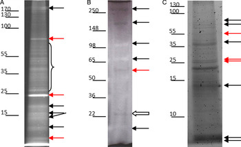
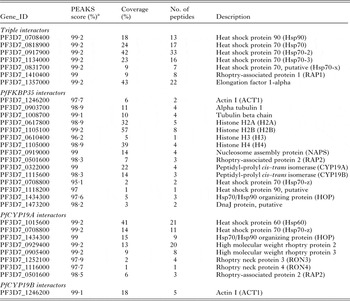
Co-IPs of PfCY19A, PfCYP19B and PfFKBP35 were analysed by SDS-PAGE (Fig. 1). Bands of interest were excised from the gels and analysis by MS revealed 161, 11 and 113 high-confidence (PEAKS analysis score >95%)Footnote 1 protein identifications for PfCYP19A, PfCYP19B and PfFKBP35, respectively. A number of putative interactions of interest have been highlighted in Table 1. A full list of the putative interactions is available in online Supplementary Table S1. The difference between the numbers of high-confidence protein identifications may be largely due to the lower sensitivity of the Thermo Fisher Orbitrap LC/MS used for PfCYP19B.
Sypro Ruby® stained SDS-4–20% polyacrylamide gel electrophoretograms showing concentrated co-IP eluates from anti-immunophilin (A) PfCYP19A, (B) PfCYP19B, (C) PfFKBP35 columns. Molecular weight marker positions are indicated to the left of each image: numbers indicate mass in kDa. Co-IP eluates were concentrated using a 9 kDa cut-off protein concentrator: 100% of the fraction was loaded, containing ~80–100 ng of protein. Concentration was performed at 5000 g for 30 min. Red arrow = co-precipitating protein also present in pre-immune serum control. White arrow = PfCyp19B. Black arrows = putative interacting partners. Bracket = ~25 to ~70 kDa section excised as a whole from gel. Abbreviations: SDS, sodium dodecyl sulphate; Co-IP, Co-immunoprecipitation.

Examples of putative immunophilin–protein interactions identified by co-IP

a See footnote in text.
Identification of interacting partners of PfFKBP35 by Y2H
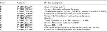
Y2H screening ultimately revealed 11 putative interacting partners for the FKBD of PfFKBP35 (Table 2). Three of these proteins were identified twice in the screen (class B) and the remainder were found only once (class C). Of particular note in view of the co-IP results reported above was the identification of the histone subunits H2B and CenH3 (an H3 variant) by this method.
Putative PfFKBD–protein interactions identified through Y2H screening

a ‘Class B’ interactors were identified two times (out of three possible) and represent highly likely interactors with the bait; ‘Class C’ interactors were found only once in the screen.
Interaction between PfCYP19B and PfHsp70
In order to confirm that PfCYP19B interacts with PfHsp70, we attempted to pull down the CYP using co-IP of the Hsp. Antibodies to Hsp70 from other organisms are readily available. We sourced a polyclonal antibody which had been generated against a recombinant full length Hsp70 from Homo sapiens to increase the possibility of cross-reactivity, due to the high level of sequence similarity between Hsp70 s, and confirmed that it was able to detect a band of apparent molecular mass 70 kDa on a western blot of a crude parasite lysate (online Supplementary Figure S1).
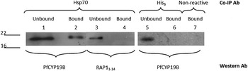
After confirming cross-reactivity of the HsHsp70 antibody, we used it to generate a co-IP column. The co-IP eluate from this column contained PfCYP19B when analysed by western blotting with an antiPfCYP19B antibody (Fig. 2, lanes 1 and 2). We can conclude that this ability to pull down PfCYP19B is specific to the antiHsp70 column since neither co-IPs performed using a column made with an irrelevant antibody (Fig. 2, lanes 3 and 4) nor a non-reactive column that is unable to bind antibody (Fig. 2, lane 5) were able to pull down PfCYP19B. Similarly, the co-IP eluate from the antiHsp70 column was negative for an irrelevant protein (Fig. 2, lanes 6 and 7). Taken together, these results lead to the conclusion that PfCYP19B specifically interacts with PfHsp70, at least under the conditions used for co-IP.
Co-IP and western blot investigation of PfCYP19B–Hsp70 interaction. Co-IP was performed using antiHsp70 to pull down PfCYP19B by its affinity for Hsp70 (lanes 1 and 2). Controls comprising an Hsp70 co-IP western blot probed with an antibody to an irrelevant protein (lanes 3 and 4), an irrelevant antibody column (antiHis6: lanes 5 and 6) and a non-reactive column (lane 7) are also shown. In the cases of the unbound fraction approximately 20 µL of a 400 µL fraction were loaded, and in the cases of the bound fractions the eluate was concentrated and a volume equalling the total fraction was loaded onto the gel. Abbreviations: Co-IP, Co-immunoprecipitation; Hsp70, heat shock protein 70.

Interaction between PfFKBP35 and histones
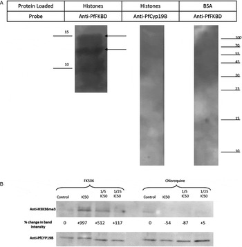
As mentioned above, a putative interaction between PfFKBP35 and histones was identified by both co-IP and Y2H in the initial screening. To investigate this interaction, we first employed the method of Longhurst and Holder (Reference Longhurst and Holder1997) to purify histones from P. falciparum cultures and demonstrated an interaction between recombinant PfFKBP35 and 2–3 P. falciparum histones by far-western blotting (Fig. 3A). This interaction was specifically between PfFKBP35 and histones because loading with no histones (BSA lane) did not reveal any bands. In the same way, PfCYP19B (as an unrelated protein control) did not bind histones since no PfCYP19B was detected in the corresponding lane. Additionally, we investigated whether the action of FK506 had an effect on the methylation of the lysine residue at position 36 of histone H3 (H3K36). Previously, H3K36 methylation was shown to be controlled by the PPIase action of the yeast FKBP Fpr4p. This regulation is governed by cis–trans isomerization of the prolines P30 and P38 on histone H3 by Fpr4p (Nelson et al. Reference Nelson, Santos-Rosa and Kouzarides2006). In this experiment, we incubated parasites in culture with FK506 or chloroquine for 14 h. As shown (Fig. 3B), there was an increase in H3K36 methylation with increasing concentrations of FK506, which is known to inhibit the PPIase activity of PfFKBP35. The standard antimalarial drug chloroquine, whose primary action is to disrupt haemozoin formation, had a much less significant effect; bands for H3K36me3 disappeared at high chloroquine concentration possibly due to the toxicity of the drug.
(A) Far-western blotting analysis of P. falciparum histone interactions. Thirty micrograms of P. falciparum histones or 10 µg of BSA were separated by SDS-15% PAGE and transferred to PVDF membrane, which was probed with 1 µg µL−1 recombinant PfFKBD-His6 or 1 µg µL−1 recombinant PfCYP19B-His6, extensively washed and the interaction was detected by standard western blot using an antibody for PfFKBD-His6 or PfCYP19B as appropriate. Arrows (A and B: ~15·5 and ~13·1 kDa) indicate bands corresponding to the apparent masses of histones H3 and H2B. Numbers and lines to the left and right indicate the positions and sizes of molecular mass markers in kDa. (B) Representative western blot of extracts from triplicate experiments on parasites incubated with decreasing concentrations of either FK506 or chloroquine for ~14 h. Parasites were lysed by incubation on ice with Triton X-100 for 30 min and the clarified lysate was separated by SDS-12·5% PAGE. The blot was probed with either antiH3K36me3 (Abcam®, ab9050) or antiPfCYP19B as a loading control. Numbers underneath the antiH3K36me3 panel indicate the band intensity relative to the control lane as estimated by densitometry. Abbreviations: BSA, bovine serum albumin; SDS PAGE, sodium dodecyl sulphate polyacrylamide gel electrophoresis; PVDF, polyvinylidenedifluoride.

Interaction between CYP and RAP1
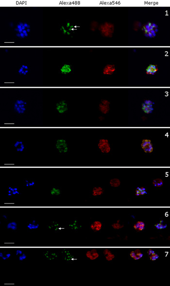
Immuno-staining schizonts for RAP1 under normal conditions resulted in the bi-punctate staining characteristic of rhoptry proteins, while PfCYP19B was located in the cytoplasm as expected (Fig. 4, 1). After 14–16 h of incubation with CsA at 5× IC50 the characteristic staining of PfRAP1 was disrupted and RAP1 and PfCYP19B co-located (Fig. 4, 2). The disruption was still evident at the IC50 (Fig. 4, 3) and somewhat evident at 0·2× IC50 and 0·04× IC50, while the characteristic bi-punctate staining for RAP1 was restored after reduction below this concentration (data not shown). At none of the concentrations tested was there evidence of an effect on the location of PfCYP19B.
Confocal immunofluorescence microscopic images of P. falciparum schizonts treated with: (1) vehicle only control, (2) 5× IC50 CsA, (3) IC50 CsA, (4) 5× IC50 BC556, (5) 5× IC50 [MeVal]4-Cs, (6) 5× IC50 chloroquine and (7) 5× IC50 artemisinin. Schizonts were stained with DAPI (nuclear stain), Alexafluor-488 (PfRAP1) and Alexafluor-546 (PfCYP19B). White scale bars indicate 5 µ m. White arrows show characteristic bi-punctate rhoptry staining. Abbreviations: CsA, cyclosporin A; DAPI, diamidino-2-phenylindole.

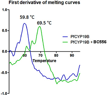
In order to exclude the possibility that this effect was due to calcineurin inhibition by the ligand–CYP complex, we tested the effects of the non-calcineurin binding immunophilin ligands [MeVal]4-Cs (Bell et al. Reference Bell, Wernli and Franklin1994) and BC556 (Fischer et al. Reference Fischer, Gallay and Hopkins2010) on RAP1. These ligands were both able to disrupt RAP1 location in schizonts at similar relative concentrations to CsA, i.e. 5× IC50 (Fig. 4, 4 and 5) and IC50 (not shown). Also tested were the classic antimalarial drugs chloroquine and artemisinin, which had no effect on RAP1 location after 14–16 h at 5× IC50 (Fig. 4, 6 and 7). Taken together these results suggested that the disruption of proper RAP1 location was not associated in a non-specific way with parasite damage or growth inhibition but was likely mediated by interference with the action of one or more CYPs. It is known that BC556 binds to other CYPs (Fischer et al. Reference Fischer, Gallay and Hopkins2010) but in order to confirm ligand binding to Plasmodium CYP, we utilized the thermal stability shift assay. Briefly, an increase in the peak of the first derivative of the melting curve indicates binding of a ligand to a protein. BC556 was able to bind to recombinant PfCYP19B (Fig. 5).
Binding of BC556 to PfCYP19B assessed by thermal stability shift assay. Binding was indicated by an increase in the peak of the first derivative of the melting curves of a protein in the presence of a ligand. T m values are given above the peaks.

DISCUSSION
This study has investigated the protein–protein interactome of the major immunophilins PfCYP19A, PfCYP19B and PfFKBP35 of P. falciparum with a view to understand better the cellular functions of these immunophilins. Two methodologies, co-IP and Y2H, were used. Co-IP specifically and reproducibly pulled down a number of protein bands and identified a large cohort of putative immunophilin–protein interactions. Below we have highlighted a number of these putative interactions that may be important to the biology of the parasite, for which there are similar data from other organisms, or for which we have confirmation of the interaction from a second experimental source.
Specifically, we believe all three immunophilins to interact with large portions of P. falciparum's heat shock machinery: all co-IPs pulled down Hsp90 and four Hsp70 isoforms (Hsp70, Hsp70-2, Hsp70-3 and Hsp70-x). PfFKBP35 pulled down a putative Hsp90 and a putative DnaJ (Hsp40) protein, though with low peptide coverage; DnaJ was also indicated as a putative interaction by our Y2H study. PfCYP19A pulled down Hsp60 with 21 peptides identified by MS covering 41% of the protein. Additionally, both PfCYP19A and PfFKBP35 pulled down another Hsp70 isoform (Hsp70-z) and Hsp70/Hsp90-organizing protein. We believe these putative interactions potentially to be important because of analogous interactions in other organisms such as the steroid receptor complex in humans (Ratajczak et al. Reference Ratajczak, Ward and Minchin2003), as well as the general importance of Hsps for parasite biology (Acharya et al. Reference Acharya, Kumar and Tatu2007).
We subsequently demonstrated that PfCYP19B was specifically pulled down with Hsp70 in co-IP experiments using whole parasite lysate in vitro. These data, along with the known interaction between PfFKBP35 and Hsp90 (Kumar et al. Reference Kumar, Adams, Musiyenko, Shulyayeva and Barik2005), lend confidence to the idea that P. falciparum possesses a chaperone complex similar to the high molecular weight chaperone machinery known to exist in other organisms. This machinery, usually consisting of immunophilins (CYPs and FKBPs), Hsp90 and p23 along with accessory proteins Hsp70, Hsp40, Hip and Hop, appears to be present in most eukaryotes. In P. falciparum such machinery might be involved in chaperoning correct folding and regulating activities of various proteins. In other organisms, different immunophilins are associated with this complex depending on the substrate which is chaperoned, for example FKBP51 and 52 are associated with the complex during steroid receptor assembly, while CYP 40 is associated during oestrogen receptor chaperoning (Galat, Reference Galat2003). This may explain in part why the parasite requires a large repertoire of immunophilins.
We also highlighted the putative interaction of PfFKBP35 with the nucleosome complex of P. falciparum. Our Y2H study indicated a putative interaction between PfFKBP35 and the histones H2B and CenH3. Co-IP with PfFKBP35 also pulled down H2B and H3, along with the other histones H2A and H4 and the nucleosome assembly protein. We believe that the direct interactions may be with H2B and H3 and since these proteins exist as heterodimers of H2A–H2B and H3–H4 they may pull down H2A and H4 by that association. These interactions have a precedent in the literature in that nuclear FKBPs in S. cerevisiae and Schizosaccharomyces pombe were shown to possess histone chaperone activity (Kuzuhara and Horikoshi, Reference Kuzuhara and Horikoshi2004) and the nuclear FKBP Fpr4p in S. cerevisiae regulates methylation of amino acid lysine-36 on histone H3 (Nelson et al. Reference Nelson, Santos-Rosa and Kouzarides2006). We demonstrated by far-western blotting that recombinant PfFKBD bound to purified histones immobilized on PVDF membrane and appeared to bind with higher affinity to bands corresponding to the molecular weights of PfH2B and PfH3. Treatment of parasites with the FKBP ligand FK506 increased H3K36 methylation. In S. cerevisiae, H3K36 methylation is regulated by the PPIase activity of the FKBP Fpr4p, and inhibition of this protein leads to increased H3K36 methylation. It appears that PfFKBP35 via its PPIase activity is also involved in regulation of H3K36 methylation, which in P. falciparum is known to affect expression of var genes encoding clonally variant antigens that are exported to the surface of the parasitized erythrocyte (Jiang et al. Reference Jiang, Mu, Zhang, Ni, Srinivasan, Rayavara, Yang, Turner, Lavstsen, Theander, Peng, Wei, Jing, Wakabayashi, Bansal, Luo, Ribeiro, Scherf, Aravind, Zhu, Zhao and Miller2013).
The co-IP study also indicated putative interactions between all three immunophilins and RAP1, as well as between PfFKBP35 and PfCYP19A and a number of other rhoptry proteins. RAP1 is known to be critical for invasion of erythrocytes by P. falciparum merozoites (Cowman et al. Reference Cowman, Berry and Baum2012). When analysed by immunofluorescent microscopy with antibodies directed against it, RAP1 exhibits a characteristic bi-punctate staining in parasite schizonts, indicative of location in the rhoptry body (Moreno et al. Reference Moreno, Poltl-Frank, Stuber, Matile, Mutz, Weiss and Pluschke2001). We demonstrated that when parasites were grown in the presence of the CYP ligands CsA, [MeVal]4-Cs or BC556, RAP1 lost its bi-punctate pattern and instead appeared in the cytosol of immature merozoites within the schizont. There was no detectable effect of ligand treatment on PfCYP19B location but given that this is an abundant protein its presence in a compartment other than the cytosol cannot be excluded. Short treatments (2 h) of parasite schizonts with the same ligands indicated that they had little effect on merozoite invasion, consistent with the hypothesis that the action of these ligands occurs at some point before arrival of RAP1 at the rhoptry (data not shown).
Among the large number of other putative interacting partners that were identified from the co-IP and Y2H studies, there was a significant representation of proteins involved in protein translation, chaperoning and digestion. From these data, it appears that these major P. falciparum cytosolic immunophilins may be involved in a wide variety of cellular functions in the parasite. Some of these interactions may be analogous to immunophilin–protein interactions in other organisms, like the known role of immunophilins in cytoskeletal architecture, molecular chaperone machinery and nucleosome assembly and modification, while some may represent novel immunophilin–protein interactions specific to P. falciparum and/or critical for its life cycle.
In summary, with the results from our co-IP experiments and our Y2H screen, we have been able to generate an interaction map which provides a body of evidence not only to support predictions of protein–protein interactions inferred from other organisms but also as a starting point for further research. Our own follow-up work confirmed a number of these putative interactions, namely immunophilin interactions with Hsp70 and histones. We were also able to demonstrate a potential novel role for immunophilins in parasite biology, that of chaperoning RAP1. These results may also have relevance for the mechanisms of antimalarial action of cyclosporins, macrolactones and other immunophilin ligands that have shown promise as antimalarial agents (Bell et al. Reference Bell, Monaghan and Page2006; Harikishore et al. Reference Harikishore, Leow, Niang, Rajan, Pasunooti, Preiser, Liu and Yoon2013a , Reference Harikishore, Niang, Rajan, Preiser and Yoon b ).
SUPPLEMENTARY MATERIAL
To view supplementary material for this article, please visit http://dx.doi.org/ 10.1017/S0031182015000803.
ACKNOWLEDGEMENTS
We thank Sandoz AG, Biotica Technology Ltd., and G. Pluschke for kind gifts of reagents and G. McManus (Dept. of Biochemistry) for advice on confocal fluorescence microscopy.
FINANCIAL SUPPORT
This work was supported by grant 09-RFP-BMT2128 from Science Foundation Ireland to A. B.