INTRODUCTION
Unicellular protozoan parasites belonging to the genus Leishmania are the causative agent of a spectrum of human diseases known as leishmaniasis. Over 350 million people worldwide are at risk of contracting the disease which is endemic in 98 countries (Alvar et al. Reference Alvar, Vélez, Bern, Herrero, Desjeux, Cano, Jannin and de Boer2012). Moreover, current long-term conflicts and consequent migration of infected individuals from endemic areas has increased not only the number of reported cases but also the risk of an increase in the number of countries becoming endemic of the disease (Kedzierski, Reference Kedzierski2010; Stamm, Reference Stamm2016). Depending on the species of parasite and host genetic background, disease manifestation ranges from asymptomatic to cutaneous skin ulceration to life threatening infection of liver and spleen, leading to death, if untreated. No vaccine is currently available and effectiveness of existent drugs is limited due to toxicity and emergence of resistant parasites, making research on disease development and host–pathogen interaction a major priority in public health. The transmission of leishmaniasis is initiated when the sandfly injects the parasite infective stage (metacyclic promastigotes) during a blood meal. The total number of promastigotes inoculated is low (100–1000) (Rogers et al. Reference Rogers, IIg, Nikolaev, Ferguson and Bates2004; Kimblin et al. Reference Kimblin, Peters, Debrabant, Secundino, Egen, Lawyer, Fay, Kamhawi and Sacks2008) with approximately 90% killed instantaneously by the host complement system despite the protective action of the sandfly saliva (Dominguez et al. Reference Dominguez, Moreno, Aizpurua and Torano2003; Gomez and Oliveira, Reference Gomez and Oliveira2012). Disease can only develop in those cases where the surviving parasites successfully establish themselves within the definitive host cells, the macrophages.
Survival inside host macrophages depends on the ability of well characterized virulence factors (e.g. GIPLs, GP63, LPG, PPG and KMP-11) to inhibit macrophages anti-microbial activity (Bifeld and Clos, Reference Bifeld and Clos2015). Leishmania inhibits multiple host pathways including lysosomal fusion (Lodge and Descoteaux, Reference Lodge and Descoteaux2005), inflammatory cytokine and chemokine production (Cameron et al. Reference Cameron, McGachy, Anderson, Paul, Coombs, Mottram, Alexander and Plevin2004; Gregory et al. Reference Gregory, Godbout, Contreras, Forget and Olivier2008), and MHC class II expression (De Souza et al. Reference De Souza, Lang, Prina and Hellio1995). These virulent functions provide sufficient time for promastigotes to successfully transform into amastigotes, which establish themselves within the parasitophorous vacuole and start to actively replicate. Eventually intracellular parasites are released and spread to uninfected cells. In mice models, a silent phase, lasting 4–5 weeks was identified whereby the amplification of parasites in the dermis takes place without the formation of a lesion. The development of a lesion coincides with the killing of the parasites (Belkaid et al. Reference Belkaid, Mendez, Lira, Kadambi, Milon and Sacks2000). Although the cellular and molecular mechanism behind Leishmania spread remains largely unknown, the above findings indicate a silent spreading takes place. The prime reason for this slow advancement is the lack of an effective in vitro model to represent this stage of host–parasite interaction. It has been widely assumed that Leishmania could egress via host cell lysis as previously observed for numerous human pathogens such as Shigella flexneri and Listeria monocytogenes (Ashida et al. Reference Ashida, Mimuro, Ogawa, Kobayashi, Sanada, Kim and Sasakawa2011). Nevertheless, multidimensional live cell imaging has shown evidence that spreading of L. (L). amazonensis amastigotes between mice macrophages can also take place via a regulated mechanism resembling apoptosis (Real et al. Reference Real, Florentina, Reis, Ramos-Sanchez, Veras, Goto and Mortara2014). Interestingly the existence of this mechanism is species-specific with no evidence of apoptotic induction detected during L. (V). guyanensis (DaMata et al. Reference DaMata, Mendes, Maciel-Lima, Menezes, Dutra, Sousa and Horta2015). However, no data are available for other Leishmania species or, more importantly, on whether a similar mechanism exists in human macrophages. The choice of host cell to support Leishmania infection makes significant differences to the parasites susceptibility to drugs (Seifert et al. Reference Seifert, Escobar and Croft2010) and it cannot be excluded that it would also impact on the parasites behaviour during spreading. Studies carried out directly on human cells are needed but are complicated by the fact that available models allow study of infection over a relatively short period of times (72 h maximum), which is generally insufficient for spreading of amastigotes to take place (Getti et al. Reference Getti, Cheke and Humber2008).
This study aimed to develop an in vitro model, which allowed the study of long-term Leishmania infection and dissemination in human macrophages. This was successfully achieved by employing a cell-to-cell infection strategy whereby enriched populations of Leishmania aethiopica and Leishmania mexicana infected cells were used to infect uninfected recipient macrophages. The percentage of infected cells in the mixed population doubled within 12 h from co-culture without significantly affecting viable cell number, suggesting infection spreading was taking place. Live microscopy confirmed parasite movement between infected and recipient cells and gave an initial insight into the process of Leishmania extrusion.
MATERIALS AND METHODS
Parasite culture
Previously described promastigotes of Leishmania aethiopica (MHOM/ET/72/L100) and Leishmania mexicana (MNYC/BZ/62/M379) constitutively expressing GFP were used in this study (Patel et al. Reference Patel, Deacon and Getti2014). The reporter gene, GFP, was integrated in the genome, downstream of the promoter of the 18S rRNA. The presence of several copies of the 18S rRNA gene tandemly arrayed in the Leishmania parasite genome is advantageous in that introduction of transgenes into the chromosome has no detrimental effect on parasite survival (Mißlitz et al. Reference Mißlitz, Mottram, Overath and Aebischer2000).
For simplicity, GFP expressing clones were named as: L8G for L. aethiopica and M5G for L. mexicana. The promastigotes were grown in Schneider's Drosophila Medium (Thermo Fischer Scientific, UK) supplemented with 23% Foetal Calf Serum (FCS) (Thermo Fischer Scientific) and 1× Penicillin-Streptomycin-Glutamine (Thermo Fischer Scientific) at neutral pH. The parasites were sub-cultured once a week at a concentration of 1 × 10⁶ mL−1, supplemented with 700 µg mL−1 of G418 (Sigma-Aldrich, UK) and were incubated at 24 °C cooling incubator.
Human cell culture
Human THP-1 monocytes (ATCC, TIB-202) were sub-cultured every 3 days at a concentration of 2·5 × 105 mL−1 in complete RPMI 1640 medium (Thermo Fischer Scientific) and incubated at 37 °C in a humidified 5% CO2 incubator. The medium was made complete following supplementation with 10% FCS and 1× Penicillin-Streptomycin-Glutamine.
Infection of differentiated THP-1 macrophages with GFP expressing Leishmania
Experimental infection with L. aethiopica and L. mexicana was carried out as previously described (Patel et al. Reference Patel, Deacon and Getti2014). Briefly, peanut lectin agglutination was used to isolate the metacyclic promastigotes from stationary phase cultures. This infectious form of the parasite was used to infect differentiated THP-1 macrophages at a ratio of 10 : 1 (parasites to cell ratio). This differentiation was achieved by treating THP-1 monocytes with 1 µ m of retinoic acid (Sigma-Aldrich) for 72 h prior to infection (Ogunkolade et al. Reference Ogunkolade, Colomb-Valet, Monjour, Rhodes-Feuillette, Abita and Froomel1990). The infected cells was seeded on a 24 well culture plate and incubated at 37 °C in a humidified 5% CO2 incubator. The percentage of infection for L. aethiopica and L. mexicana was determined through flow cytometry (BD Bioscience, UK) at 24, 48, 72 and 96 h.
Induction of apoptosis
Apoptotic induction was carried out by treatment with 3 µ m of camptothecin (Sigma-Aldrich) at 0 h after infection. Excess camptothecin from L. mexicana and L. aethiopica infected cells were washed away with 1× PBS after 5 and 24 h after infection, respectively. Following the completion of washing step, the infected cells were re-suspended in complete RPMI medium and incubated at 37 °C in a humidified 5% CO2 incubator for 72 h.
Detection of cellular apoptosis
PE Annexin V assay kit I (BD Biosciences) was used to determine apoptosis. The assay is based on Annexin V protein binding onto the externalized phosphatidyl serine (PS), which is an early marker for apoptotic cells. The kit also contains 7-AAD dye, which intercalates with DNA if the cell membrane is damaged, hence measuring necrosis. Briefly, the harvested cells were centrifuged at 500 g for 10 min and washed twice with ice-cold 1× PBS. After re-suspension in 1× binding buffer at a cell concentration of 1 × 10⁶ mL−1, 100 µL of cells were then stained with 5 µL of PE Annexin V and 5 µL of 7AAD and incubated for 15 min in the dark at room temperature. Finally, 400 µL of 1× binding buffer was added to the stained cells and the percentage of Annexin V positive/7AAD negative (apoptosis) cells were analysed through flow cytometry within 1 h of staining.
Infection of differentiated THP-1 macrophages with Leishmania infected cells
After 72 h incubation of camptothecin treated L. aethiopica and L. mexicana infected cells, the concentration of viable cells was determined via trypan blue stain (Sigma-Aldrich). The infected cells were then centrifuged at 500 g for 10 min and the supernatant removed before co-culturing with a freshly prepared batch of differentiated THP-1 macrophages. Three different infection ratios were tested including 10 : 1, 5 : 1 and 1 : 1 (infected cell per differentiated THP-1 macrophage ratio). The percentage of infection was determined after 0, 12, 24 and 48 h of co-culture through flow cytometry. For this experiment, 72 h incubated non-treated L. aethiopica and L. mexicana infected cells co-cultured with differentiated THP-1 macrophages were taken as a negative control.
Fluorescent microscopy
THP-1 macrophage from 48 and 96 h incubation were centrifuged at 500 g for 5 min before washing twice with 1× PBS. Equal volume (20 µL) of suspended cells was added onto microscopic slides before applying a drop of Prolong gold anti-fade mount with diamidino-2-phenylindole (DAPI) (Thermo Fisher Scientific). The resulting samples were then visualized through a fluorescent microscope (Nikon, UK).
Real time lapse microscopy
A fresh population of differentiated THP-1 macrophages at a concentration of 1 × 10⁵ mL−1 was labelled with 0·5× CellMask™ orange plasma membrane stain (Thermo Fischer Scientific) for 15 min at 37 °C. After washing three times with 1× PBS, the stained cells were re-suspended in a previously described cell imaging buffer solution (Richardson et al. Reference Richardson, Wallom, Ferguson, Deacon, Davies, Powell, Piper and Duncan2008). After seeding in a 6 well culture plate containing 1 × 10⁵ of apoptotic induced L. aethiopica and/or L. mexicana infected cells, the co-culture sample was then mounted on 37 °C heated stage connected to a fluorescent microscope. Using an in built digital camera (Nikon), live images were acquired every 5 min with bright field and TRITC filters for a maximum period of 12 h under 20× objectives (Nikon). After acquisition, images were processed by NIS Elements version 4.2 software, which allowed for the construction of multidimensional time-lapse videos.
Flow cytometry
To determine the percentage of infection, the infected cells were extensively washed with 1× PBS before analysis with flow cytometry. Following an initial gating with Forward scatter (FSC) and Side scatter (SSC) parameters, the THP-1 population was further gated using a histogram plot to separate GFP positive (infected cell) from GFP negative (uninfected cell) population.
Statistical analysis
Data shown are representative of at least three independent experiments performed in three biological replicates. Data represents mean values with statistical error mean (s.e.m.). Statistical analysis was performed using student t test provided by GraphPad Prism (CA, USA) software and P values less than 0·05 (P < 0·05) were considered significant differences between two independent samples.
RESULTS
Infection of THP-1 macrophage with Leishmania promastigotes
Current in vitro models of Leishmania infection in human cells are based on promastigotes infection of macrophage cell lines such as THP-1 (Ogunkolade et al. Reference Ogunkolade, Colomb-Valet, Monjour, Rhodes-Feuillette, Abita and Froomel1990; Getti et al. Reference Getti, Cheke and Humber2008; Hsiao et al. Reference Hsiao, Ueno, Shao, Schroeder, Moore, Donelson and Wilson2011). In order to identify whether such model is suitable to represent parasite spread, differentiated THP-1 macrophages were infected with L. aethiopica (L8G) and L. mexicana (M5G) promastigotes. The percentage of cells harbouring GFP expressing parasites during early (24 and 48 h) and late (72 and 96 h) stages of infection was quantified via flow cytometry.
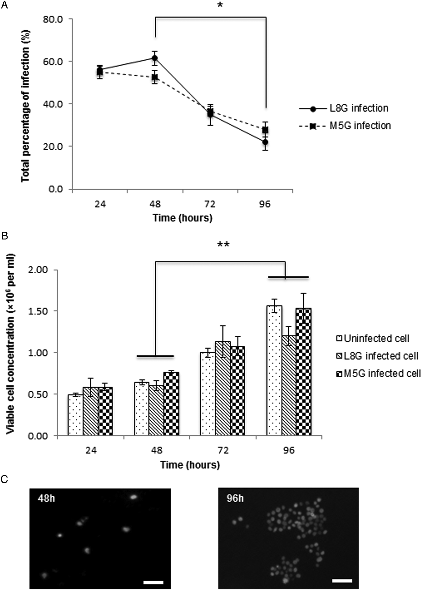
After 24 h, 57·1 ± 0·4 and 57 ± 9·1% of cells were infected with L. aethiopica and L. mexicana, respectively (Fig. 1A). This level of infection for both species was maintained till 48 h of continuous incubation. However, no spreading was observed as, there was a significant decrease (P < 0·05) in the percentage of L. aethiopica and L. mexicana infected cells from 48 to 96 h (Fig. 1A). To clarify the cause of this reduction, cell viability was quantified and showed a 2-fold increase in the concentration of viable cells from 48 to 96 h in both uninfected and infected cells suggesting that the cells had resumed replication (Fig. 1B and C).
Infection of THP-1 macrophage with Leishmania promastigotes. (A) Differentiated THP-1 macrophages were infected with two promastigote species, L. aethiopica and L. mexicana at 10 : 1 ratio (parasites to cell ratio). The percentage of infection was determined through flow cytometry at 24, 48, 72 and 96 h of incubation. Data indicate mean percentage of infection ± s.e.m. from three independent experiments. (B) Uninfected, L. aethiopica infected and L. mexicana infected cells were stained with trypan blue solution at 24, 48, 72 and 96 h of incubation. Data indicate mean concentration of viable cells ± s.e.m. from three independent experiments. (C) Fluorescence microscopic imaging of DAPI positive nuclei of uninfected cells from 48 and 96 h. Bar = 50 µm. L8G, L. aethiopica; M5G, L. mexicana; *P < 0·05 and **P < 0·01.

Effect of apoptotic induction on the rate of Leishmania infection
Induction of host cell apoptosis has been reported to promote cell-to-cell dissemination of L. amazonensis during mice macrophage infection in vitro (Real et al. Reference Real, Florentina, Reis, Ramos-Sanchez, Veras, Goto and Mortara2014). Therefore, we investigated whether apoptotic induction could also support L. aethiopica and L. mexicana spreading within the infection model based on THP-1 macrophage. Camptothecin is a quinoline alkaloid, which binds to topoisomerase I and DNA complex, stabilising it and causing DNA damage which results in apoptosis. The optimal concentration and length of camptothecin treatment was identified for each infecting species (data not shown). Based on optimised conditions, L. aethiopica and L. mexicana infected cells were treated with camptothecin for 24 and 5 h respectively, after infection and the percentage of infected cells was detected over a period of 96 h.
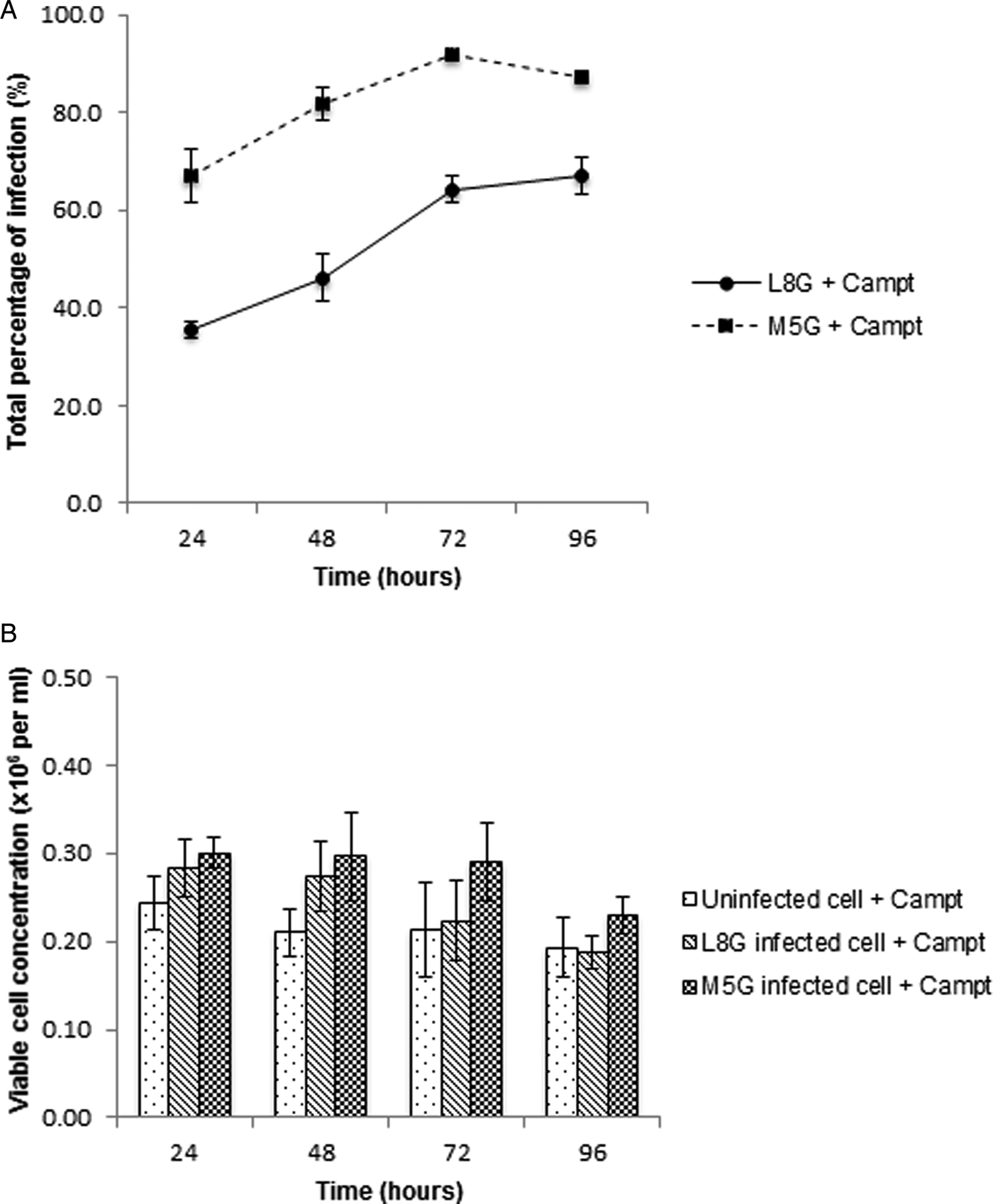
At 24 h, 35·4 ± 2 and 67·1 ± 5·4% of cells were infected by L. aethiopica and L. mexicana, respectively, in the presence of camptothecin treatment (Fig. 2A). Interestingly, the treatment caused a significant increase (P < 0·05) in the percentage of infection for both species, reaching a maximum of 64·1 ± 3% for L. aethiopica and 92 ± 1·2% for L. mexicana within 72 h before plateauing at 96 h (Fig. 2A). This indicated that camptothecin treatment did not affect intracellular parasites viability. The concentration of viable cells was similar from 24 to 96 h infection, but significantly lower than the non-treated samples (Figs 1B and 2B).
Effect of apoptotic induction on the rate of Leishmania infection. (A) Differentiated THP-1 macrophages were infected with L. aethiopica and L. mexicana promastigotes at 10 : 1 ratio (parasite to cell ratio) and treated with Campt to induce apoptosis. Excess Campt was washed away from culture medium at 24 h after L. aethiopica and 5 h after L. mexicana infection. The percentage of infection was determined through flow cytometry at 24, 48, 72 and 96 h of incubation. Data indicate mean percentage of infection ± s.e.m. from three independent experiments. (B) Campt treated uninfected or infected cells by L. aethiopica and L. mexicana were stained with trypan blue solution at 24, 48, 72 and 96 h of incubation. Data indicates mean concentration of viable cells ± s.e.m. from three independent experiments. L8G, L. aethiopica; M5G, L. mexicana; Campt, Camptothecin; *P < 0·05.

Infection of differentiated THP-1 macrophages with L. aethiopica and L. mexicana infected cells
Despite the low concentration of viable cells in camptothecin treated L. aethiopica and L. mexicana-infected samples (compared with non-treated), a heavy proportion of infected cells were obtained at 72 and 96 h. To determine which of the infected culture, 72 or 96 h, would be suitable as a donor population, the long-term effect of camptothecin treatment on THP-1 cells was determined. Annexin V assay showed no significant difference between the percentage of apoptosis of L. aethiopica and L. mexicana infected cells with and without 72 h camptothecin treatment (Supplementary Fig. S1A). In contrast, the treated L. aethiopica and L. mexicana-infected cells displayed a significantly higher percentage of apoptosis than the non-treated culture at 96 h (Supplementary Fig. S1B). Therefore, to remove any experimental bias due to camptothecin treatment, the 72 h infected population (64·1 ± 3 and 92 ± 1·2%) was used as a donor to infect newly differentiated THP-1 macrophages. Different infection ratios were tested (1 : 1, 5 : 1 and 10 : 1 infected to uninfected cell ratio) and the course of infection was detected at 0, 12, 24 and 48 h after co-culture through flow cytometry.
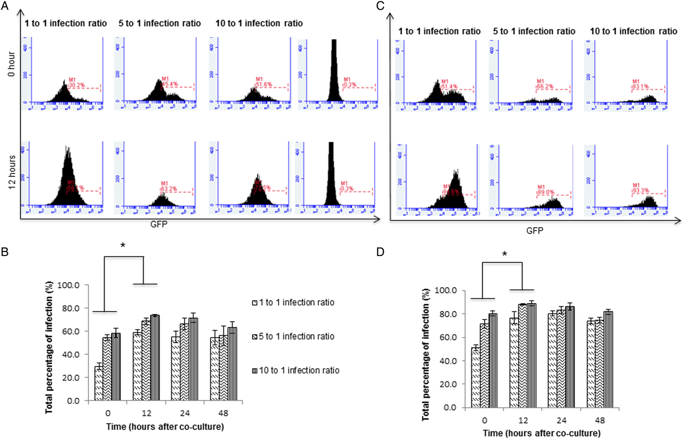
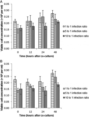
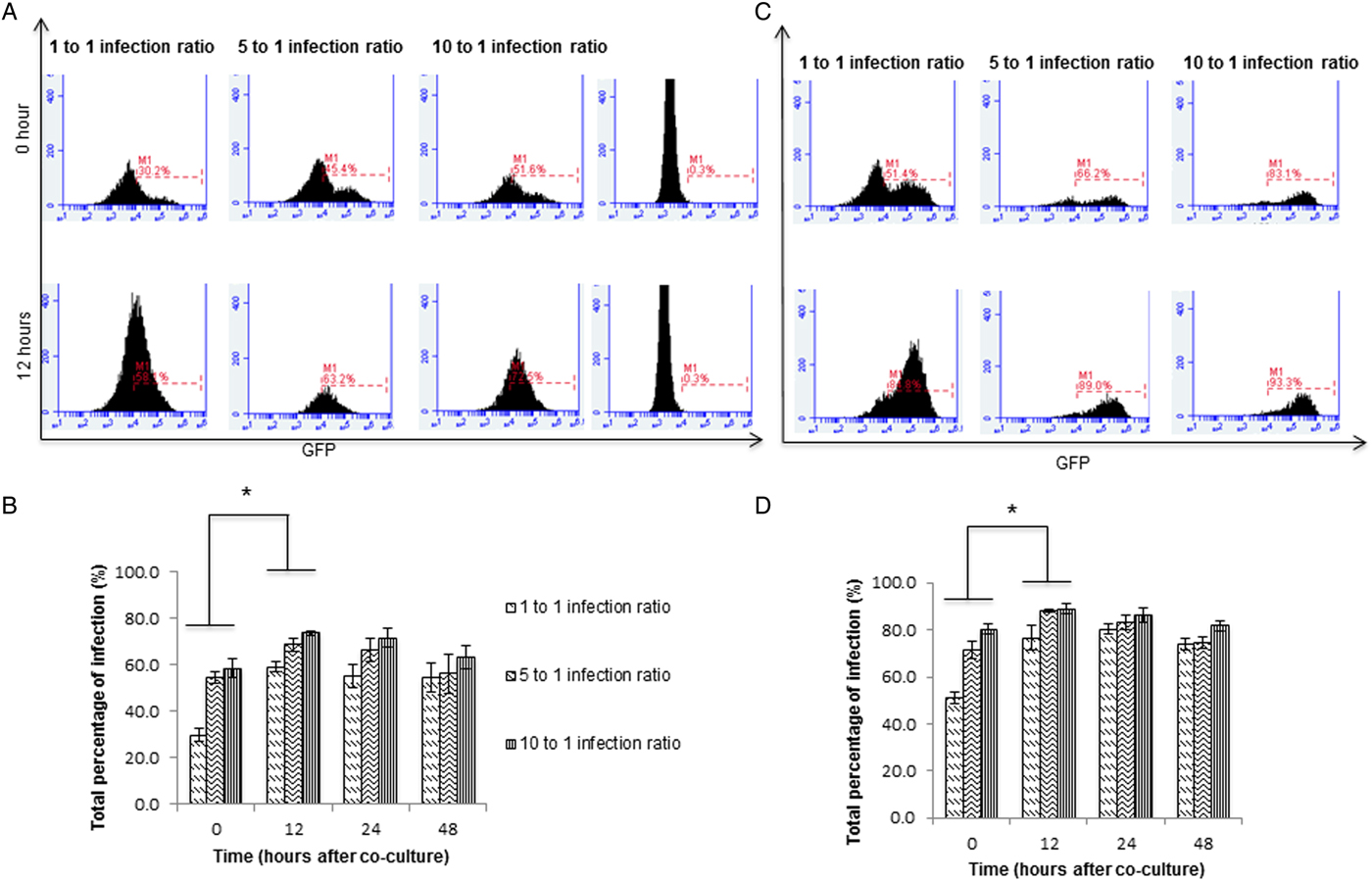
Immediately (0 h) after co-culture between infected and uninfected cells, a dilution effect on the percentage of infection for both L. aethiopica and L. mexicana was detected as expected (Fig. 3A and C). Following 12 h of co-culture, the percentage of L. aethiopica-infected cells almost doubled from 35 ± 2·3 to 60 ± 2·1% for 1 : 1 and significantly increased (P < 0·05) for 5 : 1 (54 ± 2·4 to 69 ± 3%) and 10:1 (58 ± 4·1 to 74 ± 0·4%) infection ratios (Fig. 3B). This was also observed during co-culture with L. mexicana-infected cells; 51 ± 2·3 to 76 ± 5·2% for 1 : 1, 71 ± 3·3 to 88·2 ± 1% for 5 : 1 and 80 ± 2·1 to 90 ± 2·3% for 10 : 1 infection ratios (Fig. 3D). However, such effects were not detected in the negative control, comprising non-treated infected cells co-cultured with uninfected cells. The trypan blue assay showed no significant difference in the concentration of viable cells between 0 and 12 h of co-culture (Fig. 4A and B). This suggested that the infection increase from 0 to 12 h time frame was due to spreading of L. aethiopica and L. mexicana amastigotes from the 72 h donor infected cells to healthy cells and not caused by the reduction of uninfected cells. No further increase in the percentage of L. aethiopica and L. mexicana-infected cells was detected from 12 to 24 h after co-culture (Fig. 3B and D).
Infection of THP-1 macrophages with Leishmania infected cells. (A–D) After 72 h of infection, apoptotic induced L. aethiopica and L. mexicana infected cells were co-cultured with newly differentiated THP-1 macrophage at different infection ratios, 1 : 1, 5 : 1 and 10 : 1 (infected to uninfected cell ratio). (A) Flow cytometry histogram plot representative of L. aethiopica infected cells ‘M1’ at 0 and 12 h after co-culture. (B) Data indicate mean percentage of L. aethiopica infection ± s.e.m. from three independent experiments at 0, 12, 24 and 48 h after co-culture. (C) Flow cytometry histogram plot representative of L. mexicana infected cells ‘M1’ at 0 and 12 h after co-culture. (D) Data indicate mean percentage of L. mexicana infection ± s.e.m. from three independent experiments at 0, 12, 24 and 48 h after co-culture. L8G, L. aethiopica; M5G, L. mexicana; *P < 0·05.
Measurement of viable cell concentration during infection with Leishmania infected cells. L. aethiopica infected co-culture (A) and L. mexicana-infected co-culture (B) cells at different infection ratios were stained with trypan blue solution at 0, 12, 24 and 48 h after co-culture. Data indicate mean concentration of viable cells ± s.e.m. from three independent experiments. L8G, L. aethiopica; M5G, L. mexicana.

Overall, the above results provided quantitative evidence supporting the hypothesis that L. aethiopica and L. mexicana amastigotes spreading takes place from 0 to 12 h after co-culture. This in vitro spreading model provides an initial platform to address which mode of exit is utilized by both species of Leishmania in order to transfer from cell-to-cell.
Live cell imaging of intracellular L. aethiopica and L. mexicana amastigotes spreading
During cell-to-cell infection spreading, intracellular pathogens within the donor cells can trigger either a lytic or a non-lytic release (Friedrich et al. Reference Friedrich, Hagedorn, Soldati-Favre and Soldati2012). To verify whether such a process might occur during L. aethiopica and L. mexicana release, time lapse microscopy was performed.
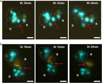
Initially, differentiated THP-1 macrophages (pre-stained with CellMask™ orange plasma membrane stain) were co-cultured with L. aethiopica and/or L. mexicana infected cells and imaged over 12 h. From 8 h, L. aethiopica amastigotes started migrating towards the peripheral membrane of the donor cells, before completely transferring into the already infected recipient stained cells within 10 min of recording (Fig. 5A, Supplementary Video 1). This phenomenon was also visualized in L. mexicana-infected co-culture whereby after 3 h, an amastigote appeared to be extruded into the recipient stained cells (Fig. 5B, Supplementary Video 2). Since donor cell (in both scenarios) remained intact during amastigote transfer, it is evident that Leishmania spread can take place via a non-lytic mode in human cells. It remains unclear whether recipient macrophages selectively engulf L. aethiopica and L. mexicana via recognition of parasite or host associated markers present in the donor cell.
Cell-to-cell spreading of Leishmania amastigotes. Freshly differentiated THP-1 cells acting as recipient uninfected cells (labelled as R), were pre-stained with CellMask™ orange plasma membrane dye. The recipient cells were then co-cultured with L. aethiopica (A) and L. mexicana (B) infected cells, which act as donor cells (labelled as D). The time of acquisition is represented by hours (h) and minutes (min). Image acquisition started at 1 hour after co-culture. Bar = 25 µm.

DISCUSSION
The development of leishmaniasis and associated pathogenesis is dependent on Leishmania amastigotes ability to successfully disseminate to uninfected macrophages. Despite the importance of this virulence mechanism on the outcome of infection, a lack of a reliable infection model representative of parasite–host interaction has led to a major gap in knowledge pertaining to the understanding of Leishmania spreading in human macrophages. Here, we have reported the development of a co-culture-based experiment, which models cell-to-cell dissemination of L. aethiopica and L. mexicana parasites. The spreading of parasites was evaluated by monitoring the total percentage of infection as well as cell viability before and after co-culture. This approach, along with live imaging, not only confirmed spreading but also supported a previous observation that some Leishmania species can exit host cells in a non-lytic process.
During natural Leishmania infection, macrophages are the definitive human cells for parasite growth and replication. To model this host–parasite interaction in vitro, many investigators have used either macrophage-like cell lines or human blood derived macrophages (Ogunkolade et al. Reference Ogunkolade, Colomb-Valet, Monjour, Rhodes-Feuillette, Abita and Froomel1990; Lisi et al. Reference Lisi, Sisto, Acquafredda, Spinelli, Schiavone, Mitolo, Brandonisio and Panaro2005; Ruhland et al. Reference Ruhland, Leal and Kima2007). Since primary blood derived macrophages do not support replication and spreading of amastigotes (Hsiao et al. Reference Hsiao, Ueno, Shao, Schroeder, Moore, Donelson and Wilson2011), cell lines were selected in our in vitro model. The choice of human THP-1 is further supported by the fact that they are non-adherent, which makes them ideal for flow cytometry studies. The use of adherent cell lines requires detachment through trypsin treatment that might damage cell membranes causing subsequent parasite release, leading to inconsistent data (Huang et al. Reference Huang, Hsing, Lai, Chen, Lee, Chan, Lyu, Wu, Lu, Lin, Lin, Lai, Chang, Chou and Chan2010). Direct infection of differentiated THP-1 macrophages with promastigotes was not sufficient to detect spreading as the effect of retinoic acid started to subside after 48 h. At this point, THP-1 replication resumed causing the percentage of infected cells to decrease, eventually making the model unsuitable for further studies on parasitic spreading.
Treatment with an apoptotic inducer immediately after infection was successful in overcoming the above challenge and producing a population containing a high percentage of infected cells. Induction of host cell apoptosis is considered as one of the major routes for the spreading of many virulent pathogens including, Mycobacterium tuberculosis, Chlamydia and adenovirus (Mi et al. Reference Mi, Li, Ni, Steinwaerder and Liber2001; Byrne and Ojcius, Reference Byrne and Ojcius2004; Aguilo et al. Reference Aguilo, Marinova, Martin and Pardo2013). The link between apoptosis of host macrophages and Leishmania infection is complex. Parasites have shown the ability to protect host cells from apoptotic induction at early stages of infection (Moore and Matlashewski, Reference Moore and Matlashewski1994; Akarid et al. Reference Akarid, Arnoult, Micic-Polianski, Sif, Estaquier and Claude Ameisen2004; Lisi et al. Reference Lisi, Sisto, Acquafredda, Spinelli, Schiavone, Mitolo, Brandonisio and Panaro2005; Ruhland et al. Reference Ruhland, Leal and Kima2007; Donovan et al. Reference Donovan, Maciuba, Mahan and McDowell2009) but a link between apoptosis and spreading has also been reported (Getti et al. Reference Getti, Cheke and Humber2008; Real et al. Reference Real, Florentina, Reis, Ramos-Sanchez, Veras, Goto and Mortara2014; DaMata et al. Reference DaMata, Mendes, Maciel-Lima, Menezes, Dutra, Sousa and Horta2015). Based on either set of evidence, apoptotic induction was expected to produce a large proportion of infected macrophages, which it did over a period of 72 and 96 h. Therefore, to mimic a natural form of parasitic dissemination, a high proportion of L. aethiopica and L. mexicana-infected cells from either 72 or 96 h were chosen as the donor population before co-culturing with uninfected freshly differentiated THP-1 macrophages. This form of cell-to-cell infection has been successfully utilized in studies of other infectious agents such as HIV-1 (Groppelli et al. Reference Groppelli, Starling and Jolly2015), Mycobacterium (Hagedorn et al. Reference Hagedorn, Rohde, Russell and Soldati2009) and L. amazonensis (Real et al. Reference Real, Florentina, Reis, Ramos-Sanchez, Veras, Goto and Mortara2014). However, prior to carrying out such an experiment, it was very important to select a suitable time point for co-culture. Specifically, a time during which the effect of camptothecin treatment on infected cells viability had subsided. Careful analysis of early apoptotic markers showed that after 72 h infection the treated population of infected cells expressed a similar percentage of apoptosis to the untreated, infected population. The time point of 72 h was therefore identified as an appropriate point of co-culture.
Results from flow cytometry and trypan blue assay confirmed that the increase in percentage of infection from 1 to 12 h was due to intracellular amastigote dissemination and not caused by a decrease in viable cell number. Moreover, microscopy clearly showed parasites spreading during which both species were filmed exiting the host cell via non-lytic mode. This corresponds with a previous report by Real et al. (Reference Real, Florentina, Reis, Ramos-Sanchez, Veras, Goto and Mortara2014), where the authors observed that L. amazonensis amastigotes were surrounded by a host membrane during their egress from mice macrophages. It cannot be excluded that amastigotes could also cause macrophages to rupture and once released be picked up by neighbouring macrophages as extracellular parasites. Nevertheless, our findings suggest that L. aethiopica and L. mexicana parasites have developed a mechanism whereby they do not need to burst the host cells to spread to uninfected macrophages. Parasites can be slowly released from the host cells without damaging them. Such mechanism would explain the silent phase of amastigote spreading within the host body, when no inflammatory response is detected (Belkaid et al. Reference Belkaid, Mendez, Lira, Kadambi, Milon and Sacks2000). This mechanism in L. aethiopica and L. mexicana could be regulated via apoptotic pathways, or could involve an alternative form of induction of membrane budding and/or actin based extrusion, which needs to be investigated further.
In summary, for the very first time, we have successfully developed an in vitro model representative of Leishmania spreading in human macrophages. This was achieved in two steps. First, a population containing a high percentage of infected cells was obtained via camptothecin treatment of infected cells. This population was then used as a donor to infect a new and healthy population of differentiated THP-1 macrophages. This form of co-culture experiment allowed us to identify the timeframe for the inter-cellular dissemination of L. aethiopica and L. mexicana spreading to uninfected cells. Live microscopy revealed non-lytic release of amastigotes from cell-to-cell and provided confirmation of a strong link with quantitative flow cytometry data on infection spreading. Following the establishment of the above model, the next step will be to functionally characterize virulence factors associated with amastigote spreading and decipher the cellular and molecular mechanism by which Leishmania exit the host cells in non-lytic mode, an issue neglected in the study of these parasites.
SUPPLEMENTARY MATERIAL
The supplementary material for this article can be found at https://doi.org/10.1017/S0031182017001366
ACKNOWLEDGEMENTS
We thank Samantha Ingram, Richard Anthony and Rachael Nice for expert technical assistance and Stuart Connop for proofreading the manuscript. The work was funded by the University of Greenwich VC PhD studentship.