Introduction
Laryngocele is a rare benign lesion of the larynx with an incidence of approximately one in 2.5 million.Reference Stell and Maran1 Laryngocele is a pathologic abnormal dilatation of the laryngeal saccule connected to the laryngeal ventricle. Depending on the location of the laryngocele and its relationship to the thyrohyoid membrane, it can be categorized into three typesReference Stell and Maran1, Reference Verret, DeFatta and Sinard2: (1) the internal type, which is confined to the endolarynx and medial to the thyrohyoid membrane; (2) the external type, which protrudes from the thyrohyoid membrane to the neck and forms a cervical mass; (3) the combined type, which has both internal and external component.
The laryngocele is typically filled with air. When the orifice of the laryngocele saccule is blocked, the air is absorbed, and the gland secretes mucus. In this case, the saccule is filled with fluid and termed a laryngomucocele. In another case, if infection occurs, the saccule will be filled with pus, and the laryngocele is termed a laryngopyocele. Laryngopyocele may result in acute dyspnoea and even require tracheotomy.Reference James, Garry, Corbett and Lang3, Reference Zelenik, Stanikova, Smatanova, Cerny and Kominek4
The aetiopathogenesis of laryngocele remains unclear. To date, it can be categorized into three main theories: congenital factors, increased pressure in the laryngeal lumen, and mechanical obstruction.Reference Verret, DeFatta and Sinard2–Reference Zelenik, Stanikova, Smatanova, Cerny and Kominek4 Some articles report that laryngocele has a certain correlation with laryngeal cancer.Reference Micheau, Luboinski, Lanchi and Cachin5–Reference Close, Merkel, Burns, CW and Schaefer7 However, the reason for this correlation remains unclear and controversial.
Treatment is based on the location and size of the laryngocele, including the external approach and the transoral approach.Reference Zelenik, Stanikova, Smatanova, Cerny and Kominek4 For external and combined types, the external approach is the classic surgical approach.Reference Myssiorek, Madnani and Delacure8 Transoral approach surgery has unique advantages for the internal type, and this approach has also been gradually adopted for the combined type.Reference Purnell, Haught and Turner9, Reference Heuveling and Mahieu10 However, controversy still exists regarding the choice of surgical approach for laryngocele.
Determining the aetiology of laryngocele, achieving an accurate diagnosis, and implementing appropriate management are clinically significant. This report outlines the clinical features and experience of our department in recent years.
Materials and methods
Patients
A retrospective study was accomplished in the Department of Otorhinolaryngology, the Second Affiliated Hospital, School of Medicine, Zhejiang University from June 2014 to June 2024. This study includes 14 patients with different types of laryngoceles. All patients underwent pre-operative laryngoscope. Almost all patients underwent pre-operative computed tomography (CT) or magnetic resonance (MR) imaging to diagnose and assess the extent of the lesion.
Clinical data
The following clinical data were collected and analyzed: sex, age at diagnosis, clinical symptoms, type, side, contents (air/fluid), concomitant disease, surgical technique, post-operative hospitalization and recurrence.
Post-operative treatment and follow-up
Post-operatively, the patients were routinely followed at two weeks, one month, three months and then every six months, with laryngeal assessment via laryngoscope.
Results
Clinical characteristics
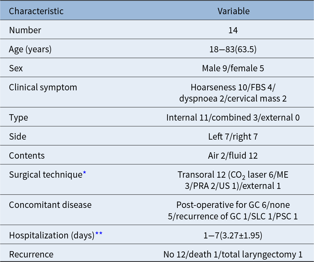
This study included 14 adult patients with 14 laryngoceles (Table 1), and clinical features were analyzed (Table 2). There were nine males and five females. Their ages ranged from 18 to 83 years, with a median age of 63.5 years. Eleven patients (78.6 per cent) had an internal laryngocele, three patients (21.4 per cent) had a combined laryngocele, and there was no external laryngocele. Twelve of them contained fluid and were determined to be laryngomucocele. The clinical symptoms were hoarseness in 10 cases, foreign body sensation in five cases, inspiratory dyspnoea in two cases and cervical mass in two cases.
Enrolled patients

* Open partial laryngectomy five years ago.
CT = computed tomography; F = female; FBS = foreign body sensation; GC = glottic carcinoma; M = male; ME = monopolar electrotome; MRI = magnetic resonance imaging; US = ultrasonic scalpel.
Characteristic of the patients

* A total of 13 patients underwent surgery.
** Of 11 internal laryngocele patients via transoral approach surgery.
GC = glottic carcinoma; SLC = supraglottic laryngeal carcinoma; PRA = plasma radiofrequency ablation; PSC = pyriform sinus carcinoma.
Sub-mucosal smooth mass in the false vocal fold via pre-operative laryngoscope was found in all patients. Pre-operative CT scanning was performed in seven patients, showing a low-density mass. The mass was found connected to the laryngeal ventricle, containing air or fluid. No enhancement was observed on contrast-enhanced CT. Ten patients underwent pre-operative MRI examination. On T1-weighted and T2-weighted MRI, the laryngocele demonstrated a hypointense mass without contrast enhancement, connecting with the laryngeal ventricle. Laryngomucocele contained fluid and showed a hyperintense mass on T2-weighted MRI.
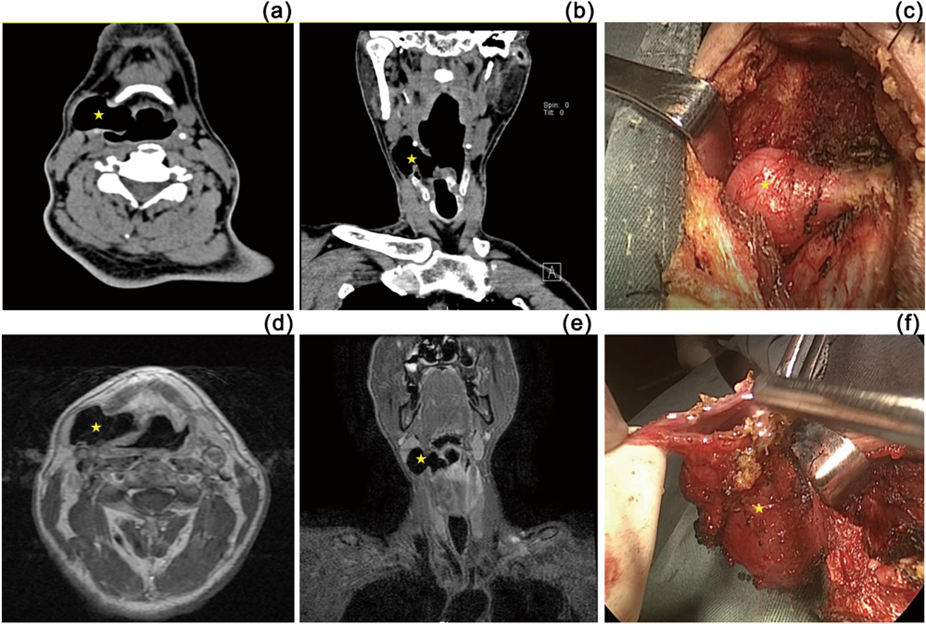
Nine laryngocele patients (n = 9; 64.3 per cent) were accompanied by cancer in larynx. One patient (case 9; Figure 1) was found to have a recurrence of glottic carcinoma after open partial laryngectomy five years ago. Two patients had concomitant malignant tumours in the larynx: one patient of pyriform sinus carcinoma (case 4; Figure 2) and one patient of supraglottic laryngeal cancer (case 5). Six patients had a history of surgery for glottic carcinoma. Notably, the laryngocele detected in these six patients was found on the same side as the previous carcinoma surgery.
Imaging examination and laryngoscopy of a left combined laryngomucocele (yellow pentagram). (a) Axial enhanced CT scan showing a left-sided low-density mass with no enhancement. (b) Coronal enhanced CT scan showing the mass (yellow circle) exceeding the superior border of the thyroid lamina, with both internal and external component. (c) Coronal T2-weighted MRI scan showing a left hyperintense mass, well demarcated from the surrounding tissue. (d) Preoperative laryngoscopy showing a smooth mass (yellow pentagram) on the left false vocal fold and a right glottic neoplasm (bule triangle).

Imaging examination and intraoperative photo of a right combined laryngocele (yellow pentagram). (a) Axial CT scan of right-sided combined laryngocele. (b) Coronal CT scan showing the mass exceeding the superior border of the thyroid lamina, with both internal and external component. (c) Photo showing the exposed external portion of the laryngocele. (d) Axial T1-weighted MRI and (e) coronal T2-weighted MRI showing a hypointense mass, with both internal and external component. (f) Photo showing the resected laryngocele, characterized by a cystic space.

Treatment and follow-up
Within the 14 cases, all 11 patients of internal type underwent transoral approach surgery. Six patients were treated with CO2 laser, three with monopolar electrotome, one with plasma radiofrequency ablation and one with the ultrasonic scalpel. These 11 patients of internal type had no complications after surgery. The hospitalization time of them was 1–7 days, with a mean of 3.27 ± 1.95 days. One patient (case 9, Figure 1) underwent an open partial laryngectomy five years ago. During the current hospitalization, a recurrence of laryngeal cancer was detected, necessitating a total laryngectomy.
Out of the three patients of combined type, one underwent transoral surgery with plasma radiofrequency ablation, one underwent external cervical surgery, and the other refused surgery. The one patient (case 4; Figure 2) underwent external approach resection surgery, and a right combined laryngocele was completely resected. A biopsy of the neoplasm in the left pyriform sinus was also performed during the operation, and the case was diagnosed as left pyriform sinus squamous cell carcinoma (T2N2M0). The hospitalization time for this surgery was 9 days. This patient received post-operative radiotherapy and chemotherapy. No recurrence was identified during the seven-year follow-up period. Another patient (case 5) with a combined laryngocele refused surgery. This patient was admitted to the hospital with a cervical mass, and a laryngeal neoplasm biopsy suggested squamous cell carcinoma, which was diagnosed as supraglottic laryngeal carcinoma (T2N1M0) with laryngocele. The patient refused surgery and died one year after radiotherapy and chemotherapy due to a rupture of neck vasculature.
Histopathologic examination
Post-operative histopathologic examination of the surgical specimen was performed for 13 (13/14) patients. Pathological examination reported a fibrous capsule tissue covered with flat or ciliated columnar epithelium consistent with a cyst. All cases were followed up for more than one year without recurrence.
Discussion
In 1867, VirchowReference Virchow11 was the first to propose the term “laryngocele” and considered it to be caused by the expansion of the laryngeal saccule. The laryngeal saccule is located at the anterior one-third of the top of the laryngeal ventricle, between the vocal fold and the false vocal fold. The upper part of the laryngeal saccule is located between the false vocal fold and the thyroid lamina, extending upwards and outwards to the superior border of the thyroid lamina, situated within the pre-epiglottic space and anterolateral to the para-glottic space.Reference Holinger, Barnes, Smid and Holinger12 A laryngocele is caused by the abnormal expansion of the laryngeal saccule due to various factors.
Laryngocele is more common in males, with a male:female ratio of approximately 5:1.Reference Stell and Maran1 The age range of peak incidence is between the 50s and 60s, and laryngocele is mostly unilateral, with no significant preference for either side.Reference Stell and Maran1, Reference Zelenik, Stanikova, Smatanova, Cerny and Kominek4 Our own series had a male:female ratio of 9:5 and a median age of 63.5 years. All 14 cases were unilateral, with no preference for either side (left 7: right 7). The higher incidence in males in our study may be related to concomitant laryngeal cancer.
Based on the location of the laryngocele and its relationship with the thyrohyoid membrane, laryngocele can be classified into three types: internal type, external type, and combined type. Mobashir et al.Reference Mobashir, Basha, Mohamed, Hassaan and Anany13 considered that laryngocele originated from the laryngeal saccule and believed that a purely external type does not exist, classifying laryngocele only as internal and combined type, with the internal type being more common (63.6 per cent, 7/11). In our study, there were 11 internal laryngoceles, three combined laryngoceles, and no external type, consistent with Mobashir’s opinion.
The etiology of laryngocele remains unclear, and there are several different theories: congenital factors, increased pressure in the laryngeal lumen and mechanical obstruction. (1) Congenital laryngocele can be found in newborns and infants, and it may occur due to congenitally wide laryngeal saccule or congenital weakness of the laryngeal tissue.Reference Verret, DeFatta and Sinard2–Reference Zelenik, Stanikova, Smatanova, Cerny and Kominek4, Reference Holinger, Barnes, Smid and Holinger12, Reference Mobashir, Basha, Mohamed, Hassaan and Anany13 (2) MacfieReference Macfie14 stated that prolonged periods of increased pressure within the laryngeal lumen could result in gradual dilation of the saccule. This situation may occur in individuals with chronic cough and those who practice excessive breath-holding due to their professions, such as glassblowers and trumpet players. (3) It is generally believed that acquired laryngocele is caused by partial or complete obstruction of the laryngeal saccule orifice. This mechanical obstruction is caused by chronic laryngeal inflammation, scars or tumours.Reference Verret, DeFatta and Sinard2, Reference Zelenik, Stanikova, Smatanova, Cerny and Kominek4, Reference Marom, Roth and Cinamon15, Reference Dhaha, Jbali, Dhambri, Mahjoub, Touati and Gritli16 The orifice of the saccule becomes narrowed due to blockage or external pressure, forming a valvular effect restricting the exit of air from the saccule.Reference Dhaha, Jbali, Dhambri, Mahjoub, Touati and Gritli16
The mechanical obstruction theory is supported by the frequent association of laryngocele with laryngeal cancer. Some authors noted that 5 per cent to 45 per cent of laryngocele patients have concomitant cancer in the larynx.Reference Micheau, Luboinski, Lanchi and Cachin5–Reference Close, Merkel, Burns, CW and Schaefer7, Reference Slonimsky, Goldenberg, Hwang, Gagnon and Slonimsky17 Benign laryngeal diseases such as amyloidosis, chondroma and scleroderma can also cause laryngocele.Reference Celin, Johnson, Curtin and Barnes6 There is also literature suggesting that post-operative scar in the larynx can affect the drainage of the laryngeal saccule orifice, leading to a laryngocele.Reference Marom, Roth and Cinamon15, Reference Dhaha, Jbali, Dhambri, Mahjoub, Touati and Gritli16 In our study of 14 patients, nine had concomitant laryngeal disease (64.3 per cent; 9/14 cases), including six patients with post-operative glottic carcinoma, one patient with recurrence of laryngeal carcinoma after partial laryngectomy, one patient with supraglottic laryngeal carcinoma, and one patient with piriform fossa carcinoma invading the supraglottic area. Notably, the laryngocele detected in these six patients who underwent partial laryngectomy nine months to four years prior was found on the same side as the previous laryngeal surgery. It is speculated that the formation of the laryngocele is related to scarring contraction or tumour compression obstructing the saccule orifice, supporting the mechanical obstruction theory. Considering the correlation between laryngocele and laryngeal cancer, imaging examinations are extremely important in patients with laryngocele to exclude possibilities of laryngeal diseases, especially laryngeal carcinoma. Long-term follow-up after partial laryngectomy is critical, especially with laryngoscopy or imaging examinations to detect laryngocele or early recurrence of cancer.
The diagnosis of laryngocele is based on clinical symptoms, laryngoscopy and imaging examination. The clinical symptoms are related to the size, location and whether there is an accompanying infection of the laryngocele. The most common symptom of the internal type is hoarseness, unclear or loss of voice. Laryngoscopy may reveal a smooth mass on one side of the false vocal fold, which can obscure the glottis and partially obstruct it. The external or larger combined laryngocele may present as a cervical mass, which can increase in size during Valsalva maneuvers.Reference Mobashir, Basha, Mohamed, Hassaan and Anany13 In rare cases, patients presented severe dyspnoea due to significant enlargement or infection of the cyst, requiring emergency treatment, even tracheotomy.Reference James, Garry, Corbett and Lang3, Reference Zelenik, Stanikova, Smatanova, Cerny and Kominek4 In our series, clinical symptoms included hoarseness (n = 10), foreign body sensation (n = 4), dyspnoea (n = 2) and cervical mass (n = 2). Laryngoscopies of all 14 patients revealed smooth mass on the false vocal fold. These were consistent with previous literature.Reference Stell and Maran1, Reference Mobashir, Basha, Mohamed, Hassaan and Anany13 Imaging examination is important to show the extension, anatomic relationship with surrounding structures,and content of both internal and external components of laryngocele.Reference Mobashir, Basha, Mohamed, Hassaan and Anany13, Reference Alvi, Weissman, Myssiorek, Narula and Myers18 CT scanning is more accurate in distinguishing the upper edge of the thyroid cartilage and helpful in differentiating laryngocele from saccular cysts of the larynx. MRI is more sensitive than CT in soft tissues, which is particularly important to exclude laryngeal disease, especially laryngeal carcinoma.
The treatment for laryngocele includes regular observation and surgical resection. Most laryngoceles are asymptomatic, for asymptomatic and smaller internal types, regular follow-up is feasible. Despite this, it is indispensable to undergo detailed imaging and other examinations to exclude tumours in the larynx. In our study, one patient (case 9) who had a history of partial laryngectomy for laryngeal cancer five years ago detected a left combined laryngocele with a right glottic neoplasm. The post-operative pathological report of neoplasm revealed spindle squamous cell carcinoma, served as a cautionary example. For external and combined types, especially those accompanied by dyspnoea, surgical treatment is recommended. Surgery can be performed using either an external approach or a transoral approach.
The external approach is suitable for external and combined types with a larger external component. This can be accomplished via a thyrohyoid membrane approach, a lateral thyrotomy approach or a combination of the two.Reference Myssiorek, Madnani and Delacure8 Several modifications of this lateral thyrotomy approach included a resection of the superior segment of the thyroid lamina, a rectangular fold-down thyroplasty, and an inferiorly based thyroid chondroplastic FLAP.Reference El-Naggar, Abdelaziz and Mandour19 These modified lateral thyrotomy approaches have proved to be effective in exposing the paraglottic space and facilitating complete resection. Caution should be taken to protect the superior laryngeal neurovascular bundle during surgery. The external approach has the advantages of easy access to the laryngocele and a low recurrence rate. In our study, one patient (case 4) with a combined laryngocele underwent external approach resection and no recurrence was identified during the seven-year follow-up period.
The transoral approach is suitable for internal types and combined types with a predominant internal component. Techniques include using CO2 laser, monopolar electrotome, plasma radiofrequency ablation and ultrasonic scalpel. This approach offers advantages such as minimal trauma, precision, safety and shorter hospital stays.Reference Zelenik, Stanikova, Smatanova, Cerny and Kominek4, Reference Purnell, Haught and Turner9, Reference Heuveling and Mahieu10 In our study, 12 patients underwent transoral approach surgery. Except for one combined laryngocele patient (case 9; recurrence of laryngeal carcinoma), the other 11 patients with internal laryngoceles had no complications after surgery. The hospitalization time of these 11 patients was 1–7 days, with a mean of 3.27 ± 1.95 days. However, this approach also has limitations, such as limited exposure, post-operative scarring and incomplete excision.Reference Zelenik, Stanikova, Smatanova, Cerny and Kominek4 Heuveling and MahieuReference Heuveling and Mahieu10 reported 20 patients undergoing complete excision of combined laryngoceles using transoral endoscopic CO2 laser surgery, with good outcomes and no intra-operative complications. A total of 75 per cent of patients were discharged on the first post-operative day, with a low recurrence rate (9.1 per cent; 2/22). However, they also proposed that, for patients with a history of significant neck surgery or abscess drainage, the external approach might be more suitable to ensure complete excision. Since 2000, transoral robotic surgery (TORS) has been used to resect laryngocele. TORS offers better exposure, a larger angle, and a broader field of view, but it remains expensive and is not yet widely used.Reference Zelenik, Stanikova, Smatanova, Cerny and Kominek4, Reference Purnell, Haught and Turner9 With technological advancements, TORS holds a promising future.
In this study of 14 cases, our experience indicates that the transoral approach is effective for treating internal laryngocele. Based on the mechanical obstruction theory, the key to the transoral approach is to resect the anterior part of the false vocal fold up to the thyroid cartilage to ensure complete removal of the laryngeal saccule orifice. With this technique, even if part of the cyst wall remains, recurrence can be avoided. During the external approach, adequate exposure of the paraglottic space should be achieved. During the surgery, a simultaneous examination of the laryngeal lumen and hypopharyngeal area should be performed under an endoscope, particularly the base of the false vocal fold, laryngeal ventricle and medial wall of the piriform sinus, to avoid missing any potential tumourous diseases. For those patients with concomitant laryngeal cancer, the laryngocele should be considered a potential invasion area, and the excision range should be expanded accordingly.
• Laryngocele is a rare clinical condition that often presents with nonspecific symptoms, and there remains no consensus on the optimal treatment approach
• Nine laryngocele patients (n = 9, 64.3 per cent) were accompanied by laryngeal cancer, suggesting a strong association between the two conditions
• The laryngocele detected in six out of the nine patients was observed on the same side as the patient’s prior partial laryngectomy. It is speculated that the formation of the laryngocele is related to scarring contraction or tumour compression obstructing the saccule orifice
• The transoral approach is effective for the internal type, and the key is to resect part of the anterior end of the false vocal fold up to the thyroid cartilage
• Besides regular inspections, thorough intra-operative endoscopic examination and post-operative histological examination are critical, especially for those patients with concomitant laryngeal cancer to reduce the potential reoccurrence risk of cancer
Conclusion
In summary, laryngocele is a rare disease, often without typical manifestations, but it can be associated with life-threatening malignancy in the larynx. Laryngoscopy and imaging examination are helpful for diagnosis. Laryngocele can be resected via an external or transoral approach. The transoral approach is effective for the internal type, and the key is to resect part of the anterior end of the false vocal fold. Careful examination before and during surgery is necessary to avoid missed diagnoses or potential tumourous diseases. For those patients with concomitant laryngeal cancer, the laryngocele should be proactively managed and removed, as it is considered a potential invasion area. After a thorough evaluation, the treatment should be tailored based on the size, type and location of the cyst, as well as the patient’s condition.
Acknowledgments
The authors thank all the members of Department of Otorhinolaryngology—Head and Neck Surgery, the Second Affiliated Hospital, School of Medicine, Zhejiang University, for their invaluable help.
Funding statement
The authors declared that this study has received no financial support.
Competing interests
None declared.
Ethical standards
The study was approved by the Ethics Committee of the Second Affiliated Hospital, School of Medicine, Zhejiang University (Approval No. 2024LSYD0871). Informed consent was waived by our Institutional Review Board because of the retrospective nature of our study.