Tetralogy of Fallot is a malformation with concordant atrioventricular connections and is characterised by anterosuperior deviation of the infundibular septum. This results in narrowing or atresia of the pulmonary outflow, malalignment ventricular septal defect, and biventricular origin of the aorta. Hearts with tetralogy of Fallot will most often have some degree of right ventricular hypertrophy.Reference Béland, Franklin and Jacobs1 Tetralogy of Fallot is the most common form of cyanotic CHD, with an incidence of 421 cases per million live births, constituting around 10% of CHD.Reference Hoffman and Kaplan2 Types of branching of the aortic arch have been described for the normal population (i.e., with no intracardiac abnormalities), the commonest being left aortic arch with innominate artery (dividing into right common carotid artery and right subclavian artery), left common carotid artery, and left subclavian artery, which occur in 64.9%.Reference Liechty, Shields and Anson3 The same series mentions the incidence of right aortic arch as 0.1%.Reference Liechty, Shields and Anson3
Variations in aortic arch anatomy associated with tetralogy of Fallot, other than the incidence of right aortic arch, have not been frequently reported. The incidence of right aortic arch in tetralogy of Fallot varies between 13% and 34%.Reference Nadas4–Reference Hastreiter, D’Cruz, Cantez, Namin and Licata7 The other arch variations, such as cervical aortic arch, double aortic arch, aorto pulmonary window, interrupted aortic arch, and coarctation of the aorta, are rarely seen in tetralogy of Fallot patients, although there are scattered case reports and case series in the literature.Reference Patel, Hurwitz and Clauss8–Reference Kothari, Rajani and Shrivastava13 A comprehensive analysis of a large number of tetralogy of Fallot cases is required to arrive at a compendious description of aortic arch anatomy.
In this article, we have described the variations of aortic arch anatomy in tetralogy of Fallot and their clinical implications. Surgically important arch variations are discussed.
Materials and methods
All patients with a diagnosis of tetralogy of Fallot who had cardiac CT or MRI either pre or post procedures between 1 January 2010 and 31 December 2019 at our hospital were included in the study. The inclusion did not depend on which procedures they underwent, which might have been intra-cardiac repair, intra-cardiac repair with right ventricle to pulmonary artery (PA) conduit, systemic to pulmonary artery shunt (Blalock-Taussig shunt, central shunt), isolated right ventricular outflow tract obstruction relief (Brock-type procedures), right ventricular outflow tract stenting, or pulmonary valve implantation post tetralogy repair. Tetralogy of Fallot patients who underwent procedures based only on echocardiograms were excluded from the study, since the arch anatomy could not be reliably or precisely determined.
Using the master registers of the operating room and paediatric catheter laboratory for these 10 years, we identified patients who had tetralogy of Fallot-related procedures. From the electronic medical records, we identified those having a CT or MRI scan anywhere in the course of treatment (pre-procedure, post-procedure, inpatient or outpatient basis). Using radiology consultants’ reports and our own analysis of individual images, we identified the various morphological patterns of aortic arches prevalent in this particular cohort of tetralogy of Fallot patients.
All anatomic variations were assessed, including the presence or absence of coarctation and hypoplastic aortic arch. The prevalence of surgically significant variations in the anatomy was determined, which we defined to be the associations which warrant a change in the standard surgical procedure or lead to additional operative steps.
Ethics clearance
The ethics committee of the Narayana Institute of Cardiac Sciences approved the study (NHH/AEC-CL-2020-494) and waived need for the individual consent.
Result
In the study period, there were 5779 tetralogy of Fallot patients who underwent procedures as outlined above. Out of these, 2684 patients (46%) met the inclusion criteria for the present study and 1627 (60.6%) were males and 1057 (39.4%) were females.
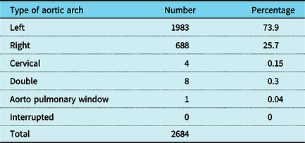
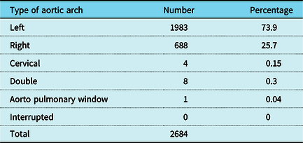
Totally, 1983 patients had left aortic arch (73.9%), 688 patients had right aortic arch (25.7%), four patients had cervical aortic arch (an anomaly in which the ascending aorta arises from the left ventricle and courses into the neck on either side, in a supra-clavicular position) (0.15%) (one with a coarctation), eight patients had double aortic arch (0.3%), and one patient had an aorto pulmonary window (0.04%) (Table 1). None of the patients in the series had interrupted aortic arch. There was no significant gender difference in the type of aortic arch and branching in tetralogy of Fallot (p = 0.304 by Chi-square test). The rarity of combined right and left-sided obstruction was evident, with only one patient with tetralogy of Fallot having a coarctation (0.04%).
Types of aortic arch in tetralogy of Fallot
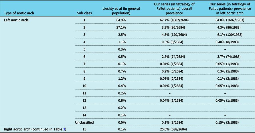
The variation of aortic arch position and its branching pattern is summarised in Table 2, and it is compared with patterns in the general population based on observations of Liechty et al (type 15 of this classification is right aortic arch, and it is described separately).Reference Liechty, Shields and Anson3 As in the general population, the commonest aortic arch pattern in tetralogy of Fallot was Liechty type 1 (innominate artery – left common carotid artery – left subclavian artery) which occurred in 62.7%. In left aortic arch, there were three unclassified arch anatomies. One patient had a separate origin of each arch vessel (right subclavian artery – right common carotid artery – left common carotid artery – left subclavian artery). Two patients had normal branching pattern (Liechty type 1) with a persistent fifth arch (a systemic to systemic connection leading to double barrel aortic arch).
The variations of aortic arch and its branching in tetralogy of Fallot, compared with patterns in the general population based on a publication by Liechty et al (type 15 of this classification is right aortic arch, which is described separately)

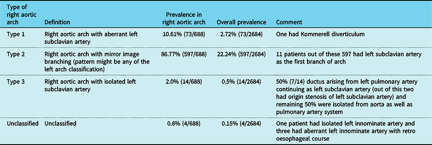
In Table 3, we have classified the right aortic arch branching patterns as per Shuford et al.Reference Shuford, Sybers and Edwards14 Within this group, 10.6% (73/688) had a right aortic arch with aberrant left subclavian artery (overall 2.7%), 86.8% (597/688) had a right aortic arch with mirror image branching with various branching patterns (overall 22.24%), 2.0% (14/688) had right aortic arch with isolated left subclavian artery (overall 0.52%), and 0.6% (4/688) had an unclassified right aortic arch (overall 0.15%). In right aortic arch type 3 (isolated left subclavian artery), our series demonstrated that 50% (7/14) had a ductus arteriosus arising from the left pulmonary artery and continuing as a left subclavian artery (two amongst these had origin stenosis of left subclavian artery). The remaining 50% were isolated from the aorta as well as pulmonary artery system. Out of four patients with unclassified right aortic arches, one patient had an isolated innominate artery, an arch variation that has been described by Mart et al.Reference Mart, Zachary, Kupferschmid and Weber15 The remaining three patients had an aberrant left innominate artery with retro-oesophageal course, which then branched into left common carotid artery and left subclavian artery.
Classification of right aortic arch and its proportion in tetralogy of Fallot

Common carotid trunk is defined as the branching of both common carotid arteries from a single branch of the aorta, also called as bovine trunk, which is a misnomer.Reference Layton, Kallmes, Cloft, Lindell and Cox16 In the general population, the prevalence of common carotid trunk is 29.5%.Reference Liechty, Shields and Anson3 In tetralogy of Fallot patients, the prevalence of common carotid trunk was 3.7% in our series. This anatomical variant has clinical significance (vide infra).
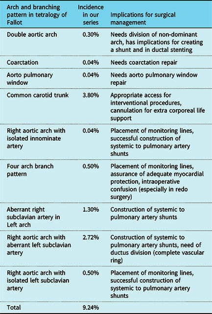
Overall, about 10% of patients had anatomical variations which could have implications for the surgical management (palliative or corrective) of tetralogy of Fallot (Table 4).
Aortic arch anatomy, branching pattern, and implications for surgical and interventional management of tetralogy of Fallot

Discussion
The potential treatment issues and clinical implications of variations in tetralogy of Fallot arch anatomy are as follows:
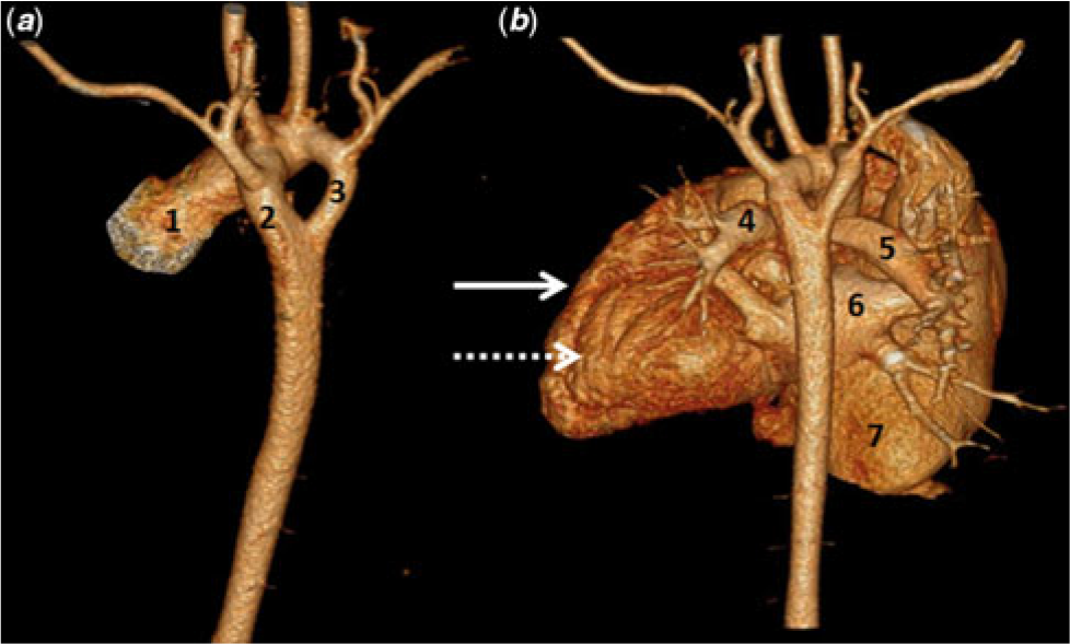
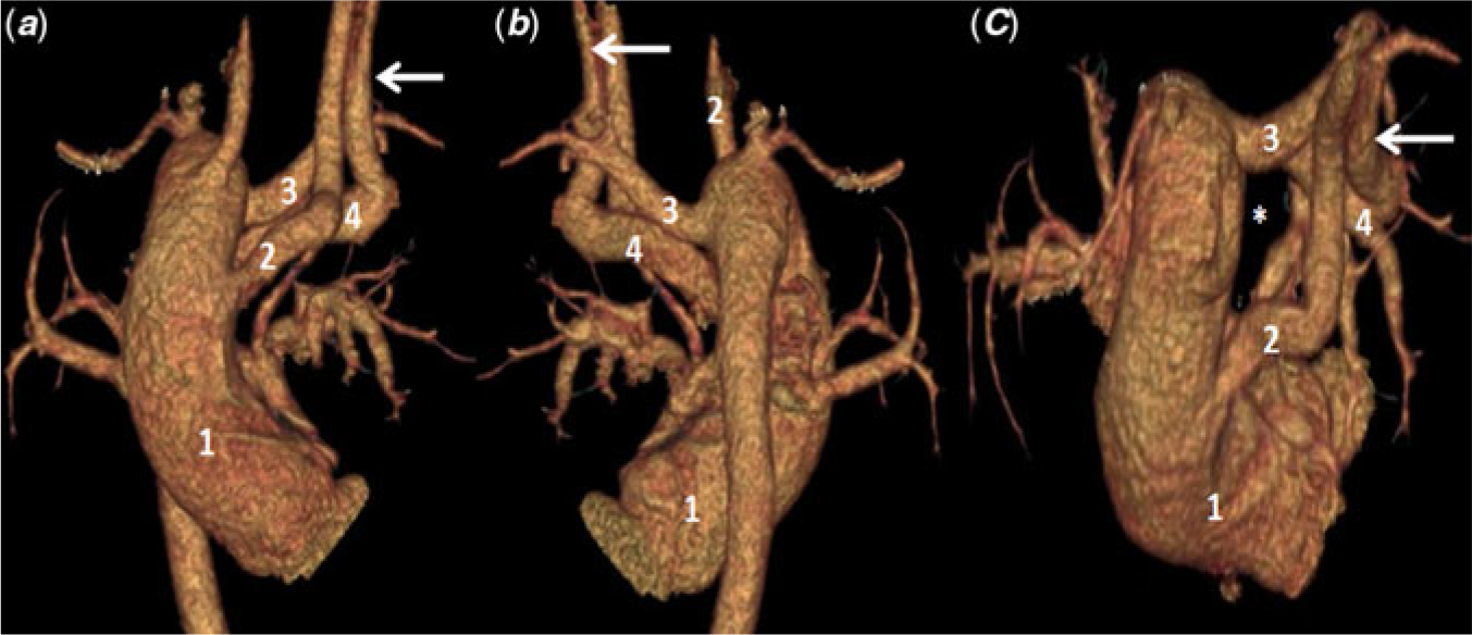
1. Management of vascular ring with trachea and oesophageal compression: Tetralogy of Fallot cases with double aortic arch typically require division of the non-dominant arch (Figure 1). Our series included eight such patients (0.3%). A right aortic arch with aberrant left subclavian artery with left ductus produces a vascular ring, potentially resulting in tracheal and oesophageal compression, and will also typically require division of the ductus (Figure 2).Reference Knight and Edwards17 Though the prevalence of right aortic arch in the general population is 0.1%, in tetralogy of Fallot it approaches 25%. Among these patients, 14% have a vascular ring producing anatomy (10.6% of right aortic arch cases in our series), potentially requiring division. A right aortic arch with retro-oesophageal aortic segment (circumflex aorta) also causes a vascular ring, but was not observed in our series.Reference Knight and Edwards17
2. Planning vascular access for interventional procedures: Common origin of the innominate artery and left common carotid artery or origin of left common carotid artery from the innominate artery is seen in humans (Liechty’s type-2, type-4, type-7, type-8, type-11, type-13, and type-14), which is sometimes referred to as bovine arch or bovine branching pattern, a misnomer.Reference Layton, Kallmes, Cloft, Lindell and Cox16 In cattle, a single great vessel originates from the aortic arch (as the aortic arch is at a fair distance from the thoracic inlet). This brachiocephalic trunk gives rise to bilateral subclavian arteries and a bi-carotid trunk, which then bifurcates into right and left common carotid arteries (true bovine arch). This anatomy has not been reported in humans. Common origin of the common carotid arteries (common carotid trunk) is a relative contraindication for a carotid approach for a ductal stent (for both direct surgical exposure and percutaneous approaches). Guide wire-induced dissection or post-procedure pseudoaneurysm could lead to neurological damage. Totally, 103 (3.8%) of our patients had a common carotid trunk, 100 of which were associated with left aortic arch, and only three with right aortic arch. The prevalence is significantly less than that reported in the normal population with common carotid trunk, which is 29.5%.Reference Liechty, Shields and Anson3
3. Suitability for ductal stenting: Ductal stenting is probably contraindicated in tetralogy of Fallot with right aortic arch with aberrant left subclavian artery and left ductus, as stenting this ductus may exaggerate the tracheo-oesophageal compression. It is better to avoid ductal stenting in tetralogy of Fallot with a double aortic arch for the same reason, though there are isolated case reports of ductal stenting in such lesions.Reference Tola, Ergul, Saygi, Ozyilmaz, Guzeltas and Odemis18
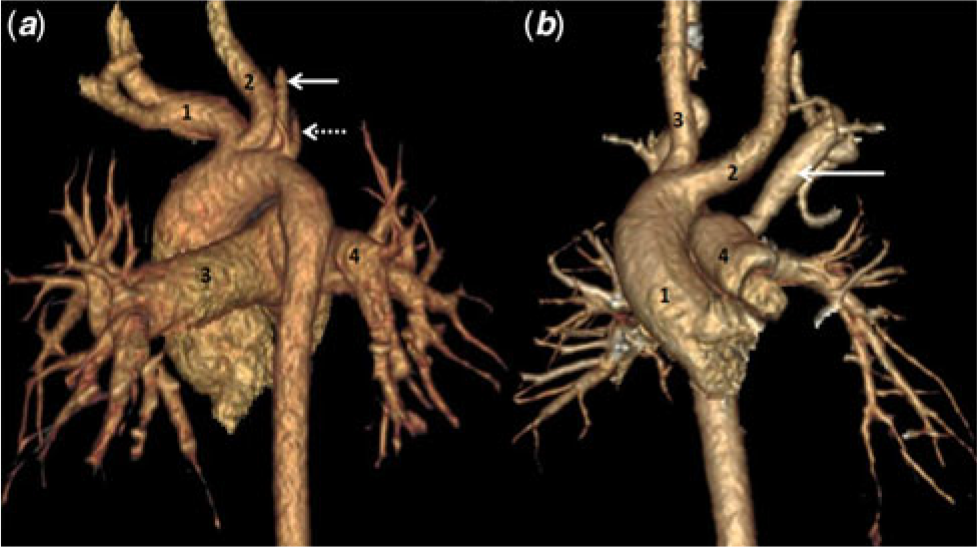
4. Avoiding subclavian steal: Type 3 right aortic arch (isolation of left subclavian artery in right aortic arch) is known to cause subclavian steal syndrome.Reference Shuford, Sybers and Edwards14,Reference Knight and Edwards17,Reference Antia and Ottesen19,Reference Maranhao, Gooch, Ablaza, Nakhjavan and Goldberg20 Type 3 right aortic arch can occur in 0.5% of tetralogy of Fallot patients (Figure 3). A modified Blalock Taussig shunt to this subclavian artery is probably contraindicated as it may not supply adequate flow and also could exaggerate the steal.
5. Siting of monitoring lines for surgical procedures: In type 3 right aortic arch (isolation of left subclavian artery), monitoring line placements in the left radial artery during a surgical procedure will give erroneous readings.Reference Hastreiter, D’Cruz, Cantez, Namin and Licata7 We encountered 14 (0.5%) patients in our series with this anatomy. A subclavian artery arising as the first branch of the aortic arch does not cause a vascular ring, but there are implications when there is associated tetralogy of Fallot. Twelve (0.5%) of our cases had this type of anatomy. Out of these 12, 11 patients had right aortic arch with left subclavian artery as the first branch and one had left aortic arch with right subclavian artery as the first branch. If recognised preoperatively, the arterial line should preferably be placed in the contralateral radial or femoral arteries, because this vessel may be dissected, looped, and/or retracted during the operation.Reference Krishnan, Theodore, Kiran and Neelakandhan21
6. Management of cardioplegia delivery and myocardial protection: 0.45% of our cases had subclavian artery arising as the first branch of aortic arch. When it arises very proximally in the arch, this vessel should be dissected and looped, as mentioned. If the aortic cross-clamp is applied distally to its origin, before delivery of cardioplegic solution, the vessel should be snugged to prevent possible cardioplegia runoff and inadequate myocardial protection.Reference Krishnan, Theodore, Kiran and Neelakandhan21 And as stated above monitoring lines should not be placed on the ipsilateral radial artery.
7. Considerations for trans-cervical cannulation for cardiopulmonary bypass and extra corporeal life support: A common origin of bilateral common carotid arteries (common carotid trunk) is a relative contraindication to trans-cervical veno-arterial extra corporeal life support. In neonates, the risk of neurological injury is similar to those with a normal branching pattern, but the incidence of neurological injury in infants and children is not known.Reference Lamers, Rowland, Seguin, Rosenberg and Reber22 One hundred and three of 2684 (3.8%) patients had a common origin of bilateral common carotid arteries in our series. We did not have a true bovine arch in our series.Reference Layton, Kallmes, Cloft, Lindell and Cox16 Emergency neck vessel cannulation might be required during redo sternotomy in infants and children for re-entry related cardiac laceration. In cases of common origin of bilateral common carotid arteries, soon after sternal re-entry the arterial (inflow) cannula should be relocated to the aorta.
8. Avoidance of confusion during dissection: Knowledge of variants of aortic arch branches is required to avoid confusion during angiography and surgical dissection, especially for redo surgeries in which anatomical definition is difficult due to intra-pericardial adhesions.Reference Boechat, Gilsanz and Fellows23
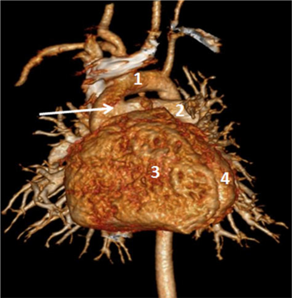
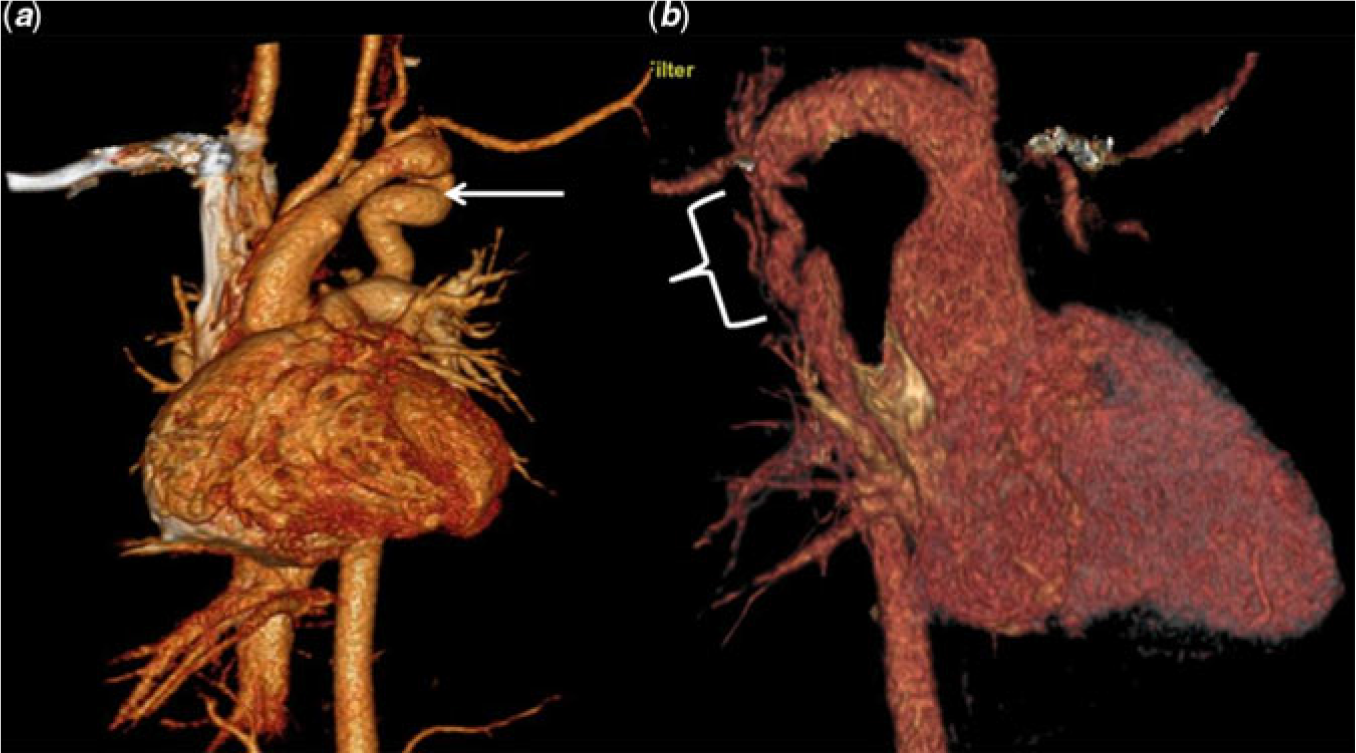
9. Additional arch issues: Aorto pulmonary window and coarctation of aorta are known associations in patients with tetralogy and may require additional procedures for correction.Reference Perdreau, Houyel and Baruteau10,Reference Crawford, Watson and Joransen12,Reference Kothari, Rajani and Shrivastava13 We had one patient each with aorto-pulmonary window (Figure 4) and coarctation (Figure 5) with tetralogy of Fallot in our series. The importance of aorto-pulmonary window in tetralogy of Fallot is that a relatively high pulmonary blood flow may prevent obvious cyanosis, leading to delayed presentation, and severe pulmonary arterial hypertension. The post-operative course can be complicated, especially when there is need of a transannular patch.
10. Systemic to pulmonary artery shunts: Knowledge of the aortic arch anatomy and branching pattern is of paramount importance in constructing a systemic to pulmonary artery shunt. Misinterpretation of branching patterns leading to complications has been reported.Reference Idhrees, Cherian, Menon, Mathew, Dharan and Jayakumar24 When a tetralogy of Fallot patient presents with refractory cyanotic spells and requires emergency surgery, the chances of this might increase.
A. Classical Blalock-Taussig shunt: Ideally, the Blalock-Taussig shunt should be constructed on the side opposite the aortic arch, and the subclavian artery must be divided at its first branch to prevent kinking of subclavian artery origin.Reference de Leval, McKay, Jones, Stark and Macartney25,Reference Litwin and Fellows26 A classical Blalock-Taussig shunt is, however, rarely used in the current era.
B. Modified Blalock-Taussig shunt: This shunt can be performed via either thorax and to either branch pulmonary artery.Reference de Leval, McKay, Jones, Stark and Macartney25 Factors other than arch anatomy may dictate the best approach (e.g., need for ligation of ductus, presence of azygous continuation of inferior vena cava, need for aorto pulmonary collateral ligation, etc.). Failure to recognise the isolated left subclavian artery (type 3 right aortic arch) in tetralogy of Fallot may result in an unsuccessful attempt to create a left Blalock-Taussig shunt.Reference Luetmer and Miller27 Failure to recognise an aberrant subclavian artery might result in construction of the shunt between carotid artery and the ipsilateral branch PA (Figure 2).
C. In cases of tetralogy of Fallot with double aortic arch, in which total correction may not advisable (e.g. very early presentation with tracheobronchial compression due to complete vascular ring), then division of the vascular ring with systemic to pulmonary artery shunt could be performed dividing the non-dominant arch, anastomosing it to the pulmonary artery.Reference Waterston, Pohl, Kallfelz and Kreutzberg11,Reference Chowdhury, Venugopal and Kothari28
D. Modified systemic to pulmonary artery shunts are advised in variations of aortic arch anatomy (complex CHD with decreased pulmonary blood flow with a right- or left-sided aortic arch and right-sided descending thoracic aorta, double aortic arch). The palliation provided by these shunts has been satisfactory, with predictable growth of pulmonary arteries, insignificant distortion in the great majority, and easy take-down.Reference Chowdhury, Venugopal and Kothari28
E. There are case reports in which an aberrant right subclavian artery was used for the construction of a systemic to pulmonary artery shunt with good outcome.Reference Legault, Camilleri, Bailly, Brazzalotto, Lusson and de Riberolles29,Reference Yamaguchi, Obo, Oshima, Ohashi, Hosokawa and Tachibana30 A modified Blalock-Taussig shunt between the aberrant subclavian artery and branch pulmonary artery usually does not pose any problem in palliation of cyanosis. In our series, we had 153 cases of tetralogy of Fallot with aberrant subclavian artery (73 patients of right aortic arch with aberrant left subclavian artery and 80 patients of left aortic arch with aberrant right subclavian artery). Totally, 123 patients underwent intra cardiac repair (121 primary corrections, and two post-shunt repairs, with shunts done elsewhere, both inadvertently constructed between carotid artery and ipsilateral branch pulmonary artery). Thirty patients had systemic to pulmonary artery shunts. Out of these thirty patients, seven had shunts between the aberrant vessel and ipsilateral branch pulmonary artery. Even the shunt from the aberrant vessel has led to good pulmonary artery growth (three had total correction and remaining four patients in follow-up have a McGoon ratio of approximately 2).
11. Significance of persistent fifth arch: Persistent fifth aortic arch is a congenital anomaly that consists of an abnormal vessel originating from the distal ascending aorta proximal to the innominate artery, which is a derivative of the fifth branchial arch vessel. This embryological variation has been classified into four different types based on its connections (systemic to systemic, systemic to pulmonary, pulmonary to systemic, and combined). This variation has been seen in tetralogy of Fallot.Reference Holmes, Holmes, Berman and Yabek31–Reference Gupta, Gulati and Anderson33 We had two (0.07%) patients with a persistent fifth arch (both had left aortic arch type-1 as the primary arch); both of our cases had systemic-to-systemic connections.
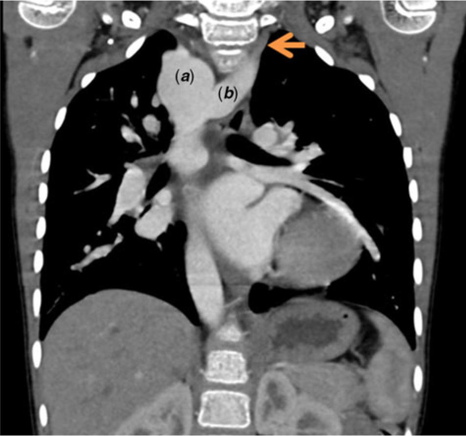
12. Significance of Kommerell diverticulum: Aberrant subclavian artery without an intracardiac lesion is associated with a Kommerell diverticulum. This typically causes indentation of the oesophagus, and when large can cause tracheal compression as well. There are reports of spontaneous dissection involving the diverticulum. In tetralogy of Fallot, during fetal life the flow through the ductus arteriosus is diminished or a ductus arteriosus does not develop; therefore, the dorsal root contralateral to the arch regresses completely or remains small, and there is no diverticulum-like origin of the aberrant left subclavian artery.Reference Velasquez, Nath, Castaneda-Zuniga, Amplatz and Formanek34 Out of 153 tetralogy of Fallot patients with aberrant subclavian artery in our series, only one had a Kommerell diverticulum (Figure 6).
Tetralogy of Fallot with double aortic arch, with dominant left arch (true vascular ring), 1 – ascending aorta, 2 – dominant left arch, 3 – right arch, 4 – left pulmonary artery, 5 – right pulmonary artery, 6 – left atrium, 7 – right atrium (enormously dilated due to severe tricuspid regurgitation), arrow – hypertrophied right ventricle, broken arrow – left ventricle.

Tetralogy of Fallot post modified Blalock-Taussig shunt (arrow) with right aortic arch and aberrant left subclavian artery and left sided ductus causing a vascular ring (*). 1 – ascending aorta, 2 – left common carotid artery, 3 – aberrant left subclavian artery, 4 – left pulmonary artery. The modified Blalock-Taussig shunt is between left common carotid artery and left pulmonary artery.

(a) Tetralogy of Fallot (post right ventricular outflow tract stent) with right aortic arch and mirror image branching, 1 – left innominate artery, 2 – right common carotid artery, 3 – left pulmonary artery, 4 – right pulmonary artery, arrow – right vertebral artery, broken arrow – right subclavian artery. (b) Tetralogy of Fallot with right aortic arch and isolated left subclavian artery (arrow) where ductus is arising from left pulmonary artery and continuing as left subclavian artery, 1 – ascending aorta, 2 – left common carotid artery, 3 – right common carotid artery, 4 – main pulmonary artery.

Tetralogy of Fallot with aorto pulmonary window (arrow), with enormously dilated pulmonary arteries and its branches. 1 – ascending aorta, 2 – left pulmonary artery, 3 – hypertrophied right ventricle, 4 – left ventricle.

Tetralogy of Fallot with cervical aortic arch. (a) Tortuous left-sided descending aorta (arrow) without gradient across the arch (pseudo-coarctation), (b) right-sided descending aorta with coarctation (bracket). The rarity of combined right- and left-sided obstruction is evident, with only one patient of tetralogy of Fallot having a coarctation in this series (0.04%).

Tetralogy of Fallot with right-sided aortic arch (a) and aberrant left subclavian artery (arrow) with Kommerell diverticulum (b). Aberrant subclavian artery associated with Kommerell diverticulum is a rarity in tetralogy of Fallot with only one patient out of 153 in our series (0.04% overall).

Study limitations
1. Control patients (with normal hearts) undergoing cardiac imaging were not available for comparison of CT and MRI.
2. We were not able to assess differences in outcome data for tetralogy of Fallot patients with and without arch anomalies using this retrospective observational study design.
3. Tetralogy of Fallot patients had imaging for a variety of indications, and it cannot be determined if the need for imaging itself may have created bias in the observed incidence of arch anomalies.
4. The surgical implications proposed are theoretical as we were not able to prospectively assess the actual changes in practice for patients with known variations undergoing operations or interventional procedure. This could be the basis for further study.
Conclusion
Knowledge of aortic arch anatomy is important for tetralogy of Fallot patients. Up to 10% patients can have surgically significant anatomical variations which might necessitate changes or additional steps in their surgical procedures (during palliative procedure or total correction of tetralogy of Fallot). Knowledge of aortic arch variations help in deciding appropriate access for interventional procedures, cannulation for extra corporeal life support, placement of monitoring lines, assurance of adequate myocardial protection, and successful construction of systemic to pulmonary artery shunts (among others). We suggest that preoperative imaging beyond the routine trans-thoracic echocardiogram (which is known to have limitations in precise delineation of arch branching patterns) may be necessary to achieve a complete understanding of the aortic arch anatomy.
Acknowledgements
None.
Financial Support
None.
Conflicts of Interest
None.