INTRODUCTION
Okenia is a genus of small-sized goniodoridid nudibranchs comprising 54 species (Bouchet & Gofas, Reference Bouchet and Gofas2013) distributed all over the world, which clearly needs a deep morphological, ecological and molecular revision. In the past ten years, 17 new species have been described, mostly from Indo-Pacific (Gosliner, Reference Gosliner2004) and Australian waters (Burn, Reference Burn1967; Rudman, Reference Rudman2004a, Reference Rudman2007a) but also from the eastern Pacific (Camacho-García & Gosliner, Reference Camacho-García and Gosliner2004; Gosliner & Bertsch, Reference Gosliner and Bertsch2004; Gosliner, Reference Gosliner2010) and West Africa (Edmunds, Reference Edmunds2009). However, only one limited phylogenetic study of this genus based on morphological characters was published (Gosliner, Reference Gosliner2004).
As Rudman (Reference Rudman2007a) and Edmunds (Reference Edmunds2009) stated, there is a considerable variation in external shape and radular morphology within this genus, but many specimens are also almost identical morphologically and need a thorough study to ensure they are conspecific. One of the most problematic species for many years within this group is Okenia zoobotryon (Smallwood, Reference Smallwood1910) (Clark, Reference Clark1984; Edmunds & Just, Reference Edmunds and Just1985; Valdés & Ortea, Reference Valdés and Ortea1995; Rudman, Reference Rudman2004a, Reference Rudmanb; Ortea et al., Reference Ortea, Moro and Espinosa2009; Edmunds, Reference Edmunds2009, among others). Its identity seemed to have been resolved by Clark (Reference Clark1984), who collected further material from the type locality in Bermuda. However, since that paper photographs of many different specimens from Senegal (Poddubetskaia, Reference Poddubetskaia2004, Reference Poddubetskaia2006; Valdés, Reference Valdés2006), Bahamas (Redfern, Reference Redfern2004), Venezuela (Grune, Reference Grune2005), Florida (Ianniello, Reference Ianniello2007) and Australia (Rudman, Reference Rudman2004b; Atkinson & Atkinson, Reference Atkinson and Atkinson2008) identified as Okenia zoobotryon have been published. To date, no molecular studies have been carried out to clarify if these specimens under the name of Okenia zoobotryon belong to a species complex or if it is a true cosmopolitan species.
In 2009, seven specimens identified as Okenia zoobotryon from Bermuda were collected, photographed and preserved for morphological and molecular studies by one of the authors (M.P.). A year later, one specimen identified as Okenia zoobotryon from Nelson Bay (New South Wales (NSW), eastern Australia) also preserved for genetic studies, was sent to the authors. Thus, we started to compile a number of specimens also identified as O. zoobotryon from Senegal and the Canary Islands in order to resolve the ongoing controversy. However, we were not able to amplify the genes of the specimens from Senegal and the Canary Islands, limiting our goal of knowing whether the specimen from Australia belonged to O. zoobotryon or not. In 2012 many more Australian specimens became available and we were able to perform a detailed molecular and morphological study of them, which resulted in the new cryptic species of the genus Okenia here described.
In this study, we provide the first molecular data for the available species of Okenia, showing COI genetic distances among the studied species. The new species is also compared with other species that overlap its distribution area, or with those having different global distribution but that were morphologically similar. We do not attempt to present a molecular phylogeny of the genus Okenia, since material of most species within the genus is missing. But the preliminary molecular data here presented will become useful for future phylogenetic studies when further material properly preserved is available.
MATERIALS AND METHODS
Molecular analysis
TAXON SAMPLING
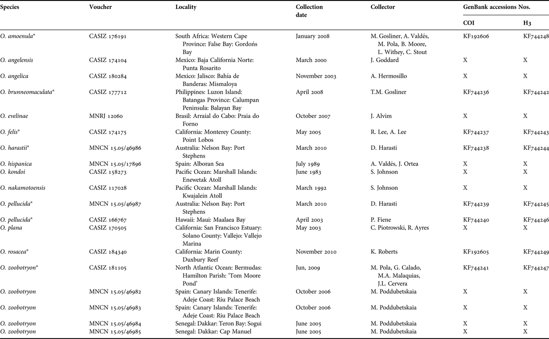
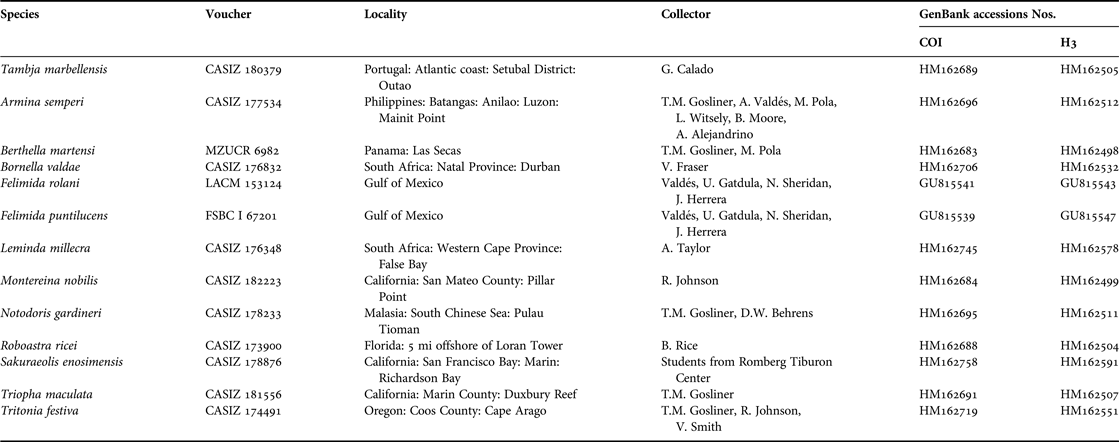
Initial sampling for molecular analysis included 19 specimens representing 13 Okenia species. Only eight of these 19 specimens were successfully amplified, limiting the in-group taxa to seven species (Table 1). Another 12 nudibranch taxa were included from GenBank (Benson et al., Reference Benson, Karsch-Mizrachi, Lipman, Ostell and Wheeler2005) to test the monophyly of the genus (Table 2). The pleurobranchid Berthella martensi (Pilsbry, 1896) was also included as out-group, since members of the Pleurobranchoidea have been shown to be the sister taxon to Nudibranchia (Dinapoli & Klussmann-Kolb, Reference Dinapoli and Klussmann-Kolb2010). Voucher specimens of Okenia species are held either at the Department of Invertebrate Zoology and Geology of the California Academy of Sciences, San Francisco, USA (CASIZ), Museo Nacional de Ciencias Naturales de Madrid, Spain (MNCN), Museu Nacional do Rio de Janeiro, Brazil (MNRJ) or the Australian Museum, Sydney, Australia (AM).
Specimens used for molecular analysis, vouchers, localities, collection dates, collectors and GenBank accession numbers. Those marked with (*) were successfully sequenced.

Species retrieved from GenBank, voucher, collection sites, collector and GenBank accessions numbers.
DNA EXTRACTION, AMPLIFICATION AND SEQUENCING
Genomic DNA was extracted from small pieces of foot tissue using Qiagen DNeasy Blood & Tissue Kits. In cases where the animals were tiny, small pieces from the dorsal process were used. Tissues were lysed in 180 μl of ATL buffer and digested with 20 μl of proteinase K for 4–12 h at 56°C. After incubation, we obtained DNA applying 200 μl of AL buffer, 200 μl of ethanol, 500 μl of AW1 buffer, 500 μl of AW2 buffer, and 300 μl of AE buffer. Partial sequences of mitochondrial cytochrome c oxidase subunit I (COI) and nuclear Histone 3 (H3) were amplified by polymerase chain reaction (PCR) conducted on Labnet Thermocycler (MULTIGENE I) and BioRad Thermocycler (MJ Mini Gradient Thermal Cycler), using the following universal primers: LCO1490 (5′-GGT CAA CAA ATC ATA AAG ATA TTG G-3′) and HCO2198 (5′-TAA ACT TCA GGG TGA CCA AAA AAT CA-3′) (Folmer et al., Reference Folmer, Black, Hoeh, Lutz and Vrijenhoek1994) for COI; H3AD F (5′-ATG GCT CGT ACC AAG CAG ACV GC-3′) and H3BD R (5′-ATA-TCC-TTR-GGC-ATR-ATR-GTG-AC-3′) (Colgan et al., Reference Colgan, McLauchlan, Wilson, Livingston, Edgecombe, Macaranas, Cassis and Gray1998) for H3. The 16S rRNA gene was not successfully amplified for most of the samples and so sequences are not included in this preliminary study. PCR amplifications were carried out in a 50 μl reaction volume with 5 μl of Qiagen buffer (10×), 5 μl of dNTPs (2 mM), 10 μl of ‘Q-solution’ (5×), 7–9 μl of MgCl2 (25 mM) for COI gen and 4–6 μl for H3 gen, 2 μl of each primer (10 μM), 0.5 μl of Qiagen DNApolimerasa and 1 μl of DNA (except for O. pellucida from Australia, in which we used 4 μl of DNA). Standard PCR for COI consisted of an initial denaturing step at 94°C for 2–3 min; 39 cycles based on each in a denaturing at 94°C for 30 s, annealing at 48–54°C for 30 s and a third step at 72°C for 1 min; ending with an extension at 72°C for 5–10 min. Further, we applied another programme consisting of an initial denaturing at 95°C for 3 min; five cycles each based on a denaturing at 94°C for 45 s, annealing at 42°C for 45 s, and a third step at 72°C for 2 min; 35 cycles each based on a denaturing at 94°C for 45 s, annealing at 45°C for 45 s and a third step at 72°C for 2 min; ending with a final extension at 72°C for 10 min. PCR for H3 amplification consisted of an initial denaturing at 94–95°C for 2–3 min; 35 amplification cycles (94–95°C for 30–35 s, 50–52°C for 30–60 s, and 72 °C for 1–1.25 min) and a final step at 72°C for 2–7 min. Amplified products were electrophoresed in a 0.5% TAE agarose gel stained with ethidium bromide. Successful amplified samples were purified with ExoSAP-IT (USB Scientific). The amplified fragments were sequenced in both directions in a DNA sequencer 3730XL (Applied Biosystems).
SEQUENCE ALIGNMENT AND ANALYSIS
H3 and COI sequences were edited and aligned using Genious 4.5.4 (Drummond et al., Reference Drummond, Ashton, Cheung, Heled, Kearse, Moir, Stones-Havas, Thierer and Wilson2009) and checked by eye. Protein-coding sequences were translated into amino acids for confirmation of alignment using MacClade 4.08 (Maddison & Maddison, Reference Maddison and Maddison2005) and MEGA 5.05 (Tamura et al., Reference Tamura, Peterson, Peterson, Stecher, Nei and Kumar2011). A dataset of sequences aligned and ordered in codons was created. Furthermore, another combined molecular dataset was added, and its congruence capacity was determined by an incongruence length difference (ILD) test (Farris et al., Reference Farris, Källersjö, Kluge and Bult1994) conducted using PAUP*4.0 (Swofford, Reference Swofford2002). In order to test the possible saturation types, the number of transitions (Ti) and transversions (Tv) were plotted against the uncorrected pairwise distances between species. Saturation plots were examined, separated from the first, second and third positions of protein-coding genes.
MODEL SELECTION AND PHYLOGENETIC ANALYSES
The evolutionary model that best fitted our data was selected using MrModelTest 2.3 (Nylander, Reference Nylander, Ronquist, Huelsenbeck and Nieves-Aldrey2004) under the Akaike information criterion (Akaike, Reference Akaike1974). To analyse the phylogenetic relationships between specimens, two different methods, maximum likelihood (ML) and Bayesian inference (BI) were used. The analyses were performed using the combined molecular datasets, but each gene was also analysed independently. Support for nodes in the ML analyses was assessed with non-parametric bootstrapping using RAxML v7.04 (Stamatakis et al., Reference Stamatakis, Hoover and Rougemont2008), with 15,000 pseudoreplicates. The combined datasets were also analysed in MrBayes 3.1.2 (Ronquist & Huelsenbeck, Reference Ronquist and Huelsenbeck2003). Analyses were initiated with random starting trees, run for 5 × 10⁷ generations to obtain support, and 10% of trees were discarded. Bayesian posterior probabilities (Pp) of 0.95 or higher are considered strongly supported, whereas ML values of 75 or higher are considered strongly supported. The trees obtained were edited in FigTree v1.3.1. (Rambaut, Reference Rambaut2009) and Adobe Photoshop CS 8.0.1.
Morphological analysis
For the morphological analysis, we studied ten specimens of a species previously identified as Okenia cf. zoobotryon collected in Port Stephens, Nelson Bay, NSW by Dave Harasti, under NSW Fisheries Scientific Research permit P01/0059a. These specimens were erroneously preserved in an inadequate saline solution and thus, they were pretty degraded externally when they arrived at our laboratory. We used photographs of living animals for the description of the colour pattern and some external morphological details. We also studied seven specimens of Okenia zoobotryon collected in Bermuda in 2009 for comparative purposes. The internal features were examined using a dissecting microscope (Nikon SMZ-1500) with an attached camera lucida for drawing anatomical details. The specimens were dissected and the anatomical study was facilitated by making a first dorsal incision. Special attention was paid to the morphology of the reproductive system, including the penis. The buccal mass was removed and dissolved in 10% sodium hydroxide until the labial cuticle and radula were isolated from the surrounding tissue. The labial cuticle and radula were then rinsed in water, dried and mounted for examination under a Hitachi S-3000N scanning electron microscope (SEM). The studied material of O. cf. zoobotryon from NSW is deposited both in AM and MNCN, and that of O. zoobotryon from Bermuda is deposited both in CASIZ and in the University Museum of Bergen, Bergen, Norway (ZMBN).
RESULTS
Amplifications were not successful for most taxa (Table 1). We obtained eight new sequences for H3 and eight for COI, plus 26 sequences from GenBank, 13 for each gene (Table 2). After alignment, 986 bp were used: 328 for H3 and 658 for COI. The saturation plots for both genes showed no saturation for any codon positions (not shown). The best evolutionary model was GTR + G + I. The congruence of the H3 and COI (P > 0.01) allowed these markers to be combined into a single dataset (Cunningham, Reference Cunningham1997). The combined tree provided better resolution than H3 and COI separately.
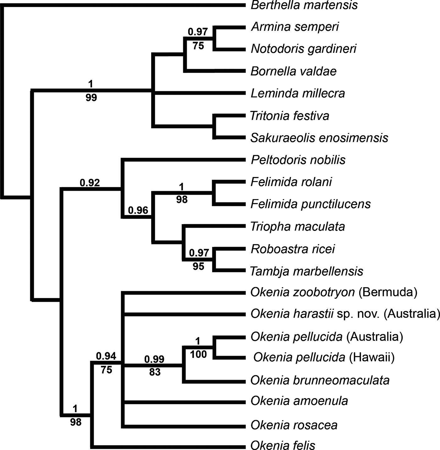
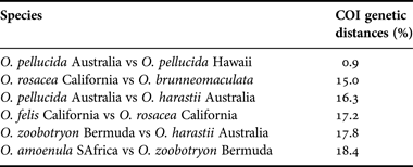
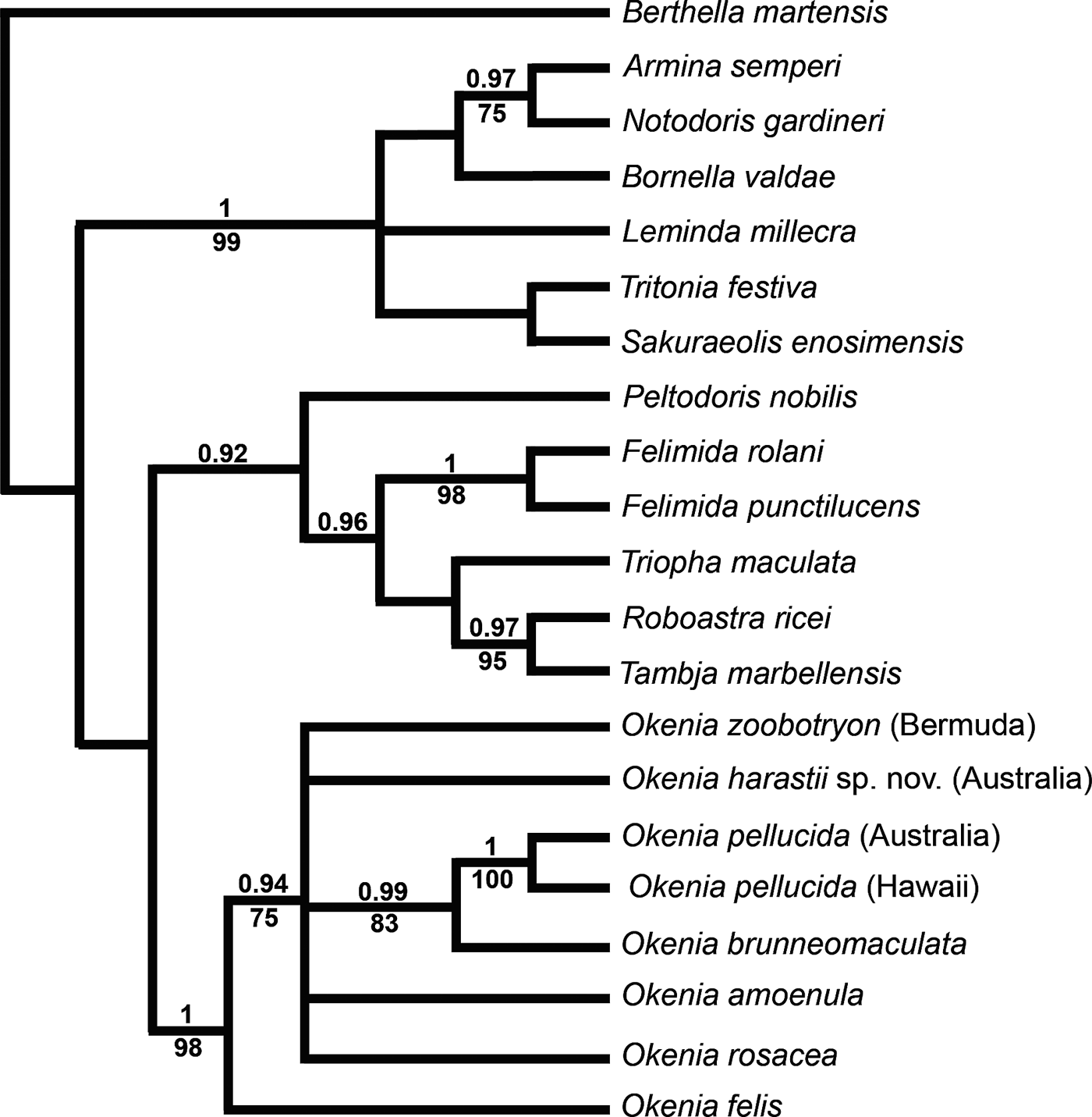
Based on the phylogenetic analysis undertaken, all Okenia species included in this study form a monophyletic clade, highly supported by both BI and ML analysis (Pp = 1; ML = 98) (Figure 1), with Okenia felis Gosliner, Reference Gosliner2010 placed as the basal species of the clade. The remaining studied species of the genus form a supported clade (Pp = 0.94; ML = 75), but without resolution within it. Only a clade including O. brunneomaculata Gosliner, Reference Gosliner2004 and two specimens of O. pellucida Burn, Reference Burn1967 (Pp = 1; ML = 100) is well supported (Pp = 0.99; ML = 83). The COI genetic distances between some of the studied specimens are summarized in Table 3. The pairwise distance analysis done to compare the difference in the protein coding COI gene between the two specimens of O. pellucida, one from Australia (type locality) and one from Hawaii is 0.9% (Table 3). Moreover, the two specimens previously identified as Okenia zoobotryon, one from Bermuda (type locality) and one from Australia, do not cluster together. The COI genetic distance between these two specimens is 17.8%. Such a distance strongly suggests that Australian specimens belong to a different new species, which is described below.
Phylogenetic hypothesis based on the combined dataset (COI + H3) inferred by Bayesian analysis (BI). Numbers above branches represent posterior probabilities from BI. Numbers below branches indicate bootstrap values for ML.
Minimum COI gene pairwise uncorrected p-distances amongst some Okenia species.
SYSTEMATICS
NUDIBRANCHIA Cuvier, 1817
EUCTENIDIACEA Tardy, 1970
Family GONIODORIDIDAE H. Adams & A. Adams, 1854
Genus Okenia Menke, 1830
Okenia harastii sp. nov.
(Figures 2–4)
Okenia harastii sp. nov.: (A–B) photographs of the living animals from Port Stephens, Nelson Bay, New South Wales, Australia. (A) Holotype (AM C.478860): (B) paratype (AM C.478861) (photographs: Dave Harasti): (C–D) photographs of the preserved specimens showing spicules in the mantle: (E) drawing of the living animal showing details of the external morphology. Scale bar: 1 mm.
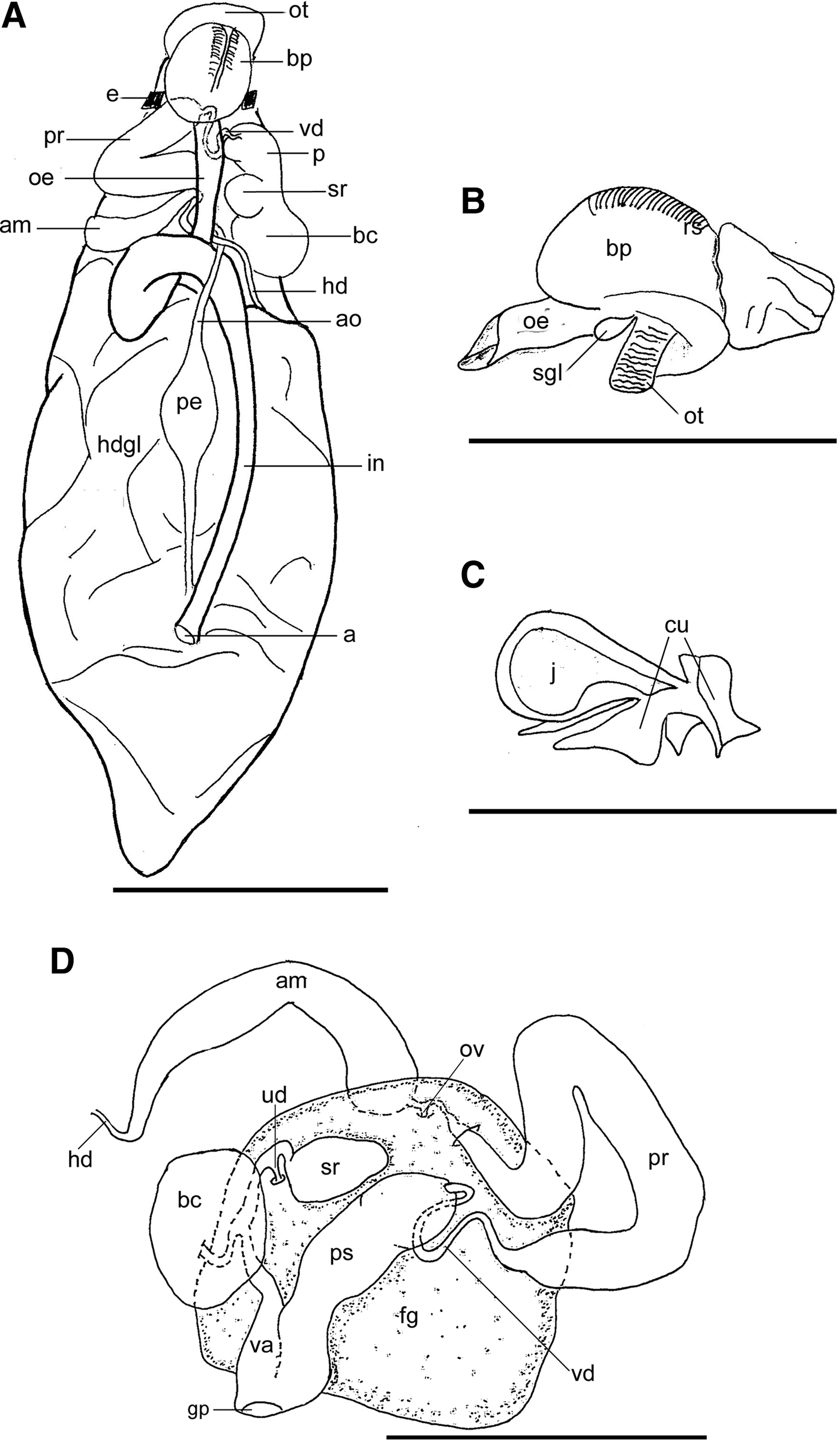
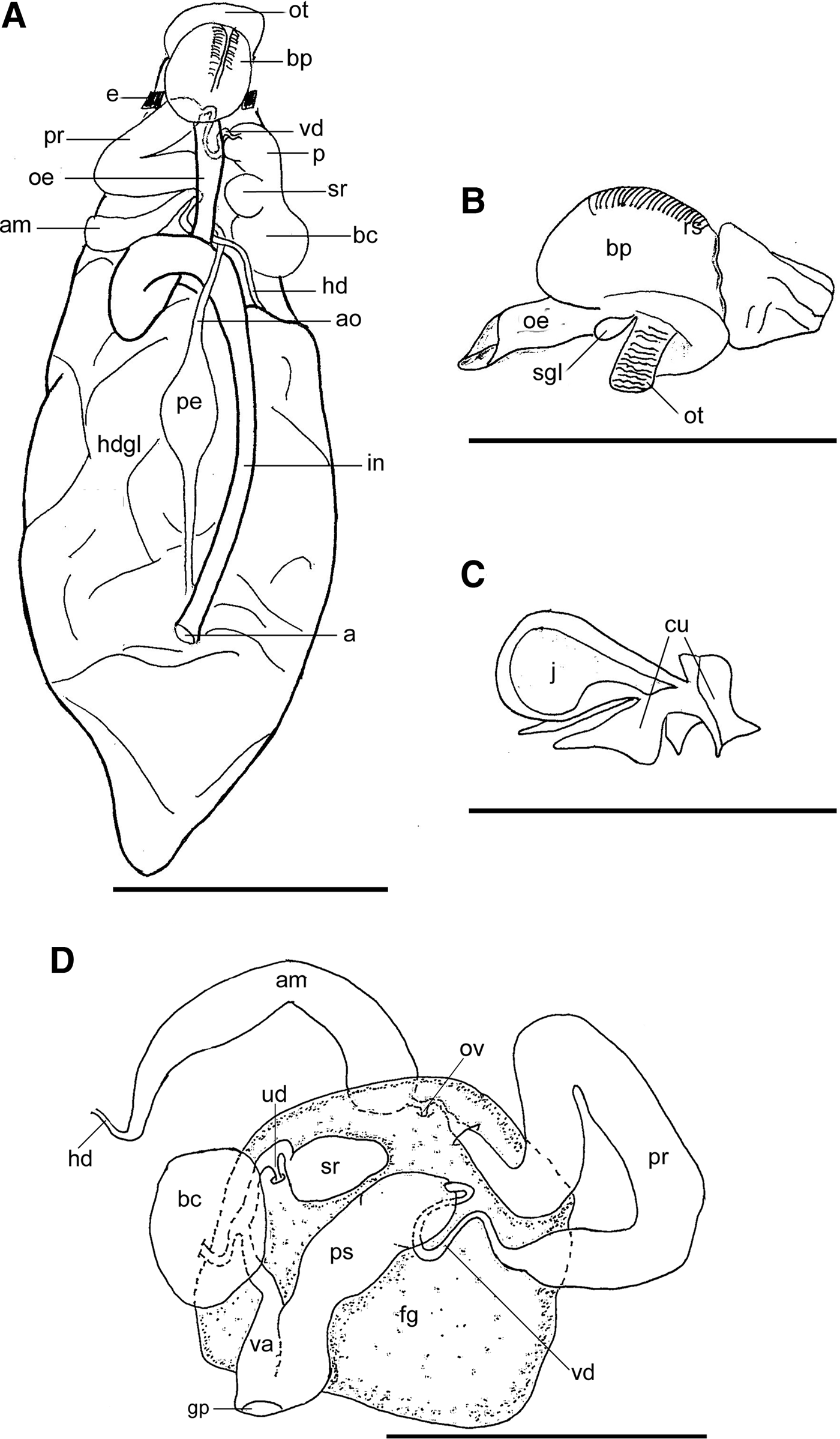
Okenia harastii sp. nov., from Port Stephens, Nelson Bay, New South Wales, Australia: (A) internal anatomy (3 mm specimen, dorsal view): (B) buccal bulb and oral tube (3 mm specimen, lateral view): (C) labial cuticle of a 3 mm specimen: (D) reproductive system (4 mm specimen). Scale bars: A–B, D, 1 mm; C, 2 mm. Abbreviations: a, anus; am, ampulla; ao, aorta; bp, buccal pump; bc, bursa copulatrix; cu, labial cuticle; e, eye; fg, female gland; gp, genital pore; hd, hermaphroditic duct; hdgl, hermaphrodite and digestive gland; in, intestine; j, jaw; oe, oesophagus; ot, oral tube; ov, oviduct; pe, pericardium; pr, prostate; ps, penial sac; rs, radular sac; sgl, salival gland; sr, seminal receptacle; ud, uterine duct; va, vagina; vd, vas deferens.
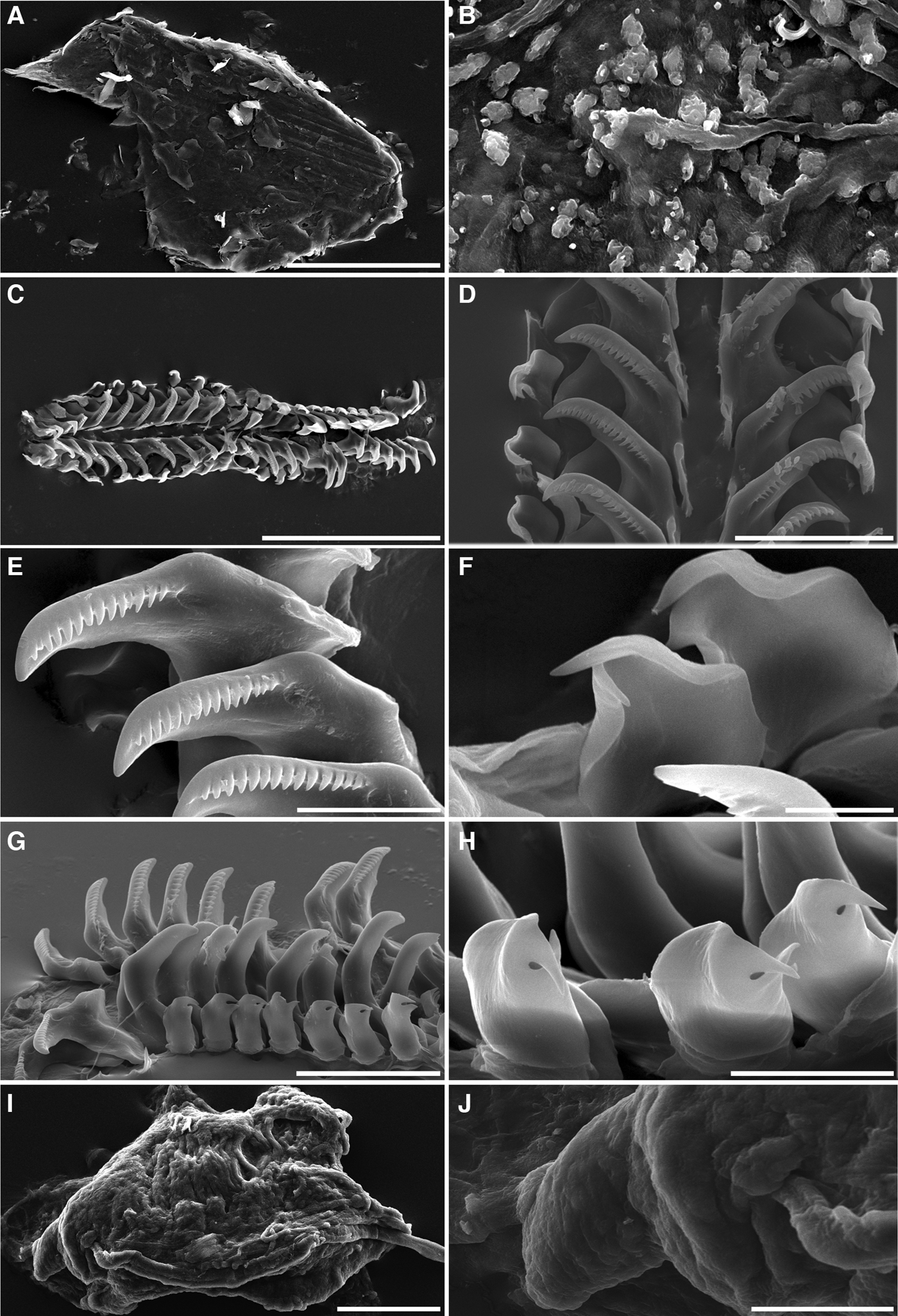
Scanning electron microscopy photographs of Okenia harastii sp. nov. from Port Stephens, Nelson Bay, New South Wales, Australia: (A) jaw; (B) detail of jaw elements; (C) radula; (D) radular teeth; (E) inner lateral teeth; (F) outer lateral teeth; (G) half radula (lateral view); (H) outer lateral teeth (lateral view); (I) penial sac and penis. Scale bars: A, C, 200 µm; B, F, 10 µm; D, G, 50 µm; E, H, 20 µm; I, J, 100 µm.
TYPE MATERIAL
Holotype: 4 mm adult initially fixed in an unknown saline solution and then preserved in 75% EtOH, not dissected. Nelson Bay, Port Stephens, NSW, Australia (32°43′4.63″S 152°008′29.27″E); 5 m depth. Found on bryozoans in a substrate mix of sponges and sand (AM C.478860); coll. D. Harasti, 15 April 2012.
Paratypes: adults of 4 mm completely dissected (AM C.478861), 4 mm not dissected (MNCN 15.05/60084) and 3 mm (AM C.478862) completely dissected, all from the type locality and collected together with the holotype, initially fixed in an unknown saline solution and then preserved in 75% EtOH.
Additional specimens: six specimens of 3 mm (not dissected) (AM C.478865), 3 mm (completely dissected) (MNCN 15.05/66874), 4 mm (completely dissected) (AM C.478864), 4 mm (completely dissected) (MNCN 15.05/66875), 5 mm (not dissected) (AM C.478863) and 5 mm (not dissected) (MNCN 15.05/66876), all from the type locality and collected together with the holotype, initially fixed in an unknown saline solution, and then preserved in 75% EtOH.
COMPARATIVE MATERIAL EXAMINED
Okenia zoobotryon: seven specimens from Tom Moore Pond, Hamilton Parish, Bermuda, North Atlantic Ocean (32°20.767′N 64°42.539′W); 4 m depth; coll. M. Pola, G. Calado, M.A. Malaquias and L. Cervera, 12 June 2009. One adult specimen 6 mm long (preserved), completely dissected and sequenced (CASIZ 181105); one adult specimen 6 mm long (preserved), completely dissected (CASIZ 181107), and five specimens of 4, 5, 5, 5 and 6 mm long (preserved), (ZMBN 83015).
DISTRIBUTION
Known only from the type locality.
ETYMOLOGY
This species is named in honour of Dave Harasti, who kindly collected, photographed and sent the material studied in this work.
EXTERNAL MORPHOLOGY
See Figure 2. The body is oval and elongated, with series of lateral papillae extending along most of first half of the body (Figure 2A–E). The highest and widest point is at the gill level. Behind it, the body narrows into an elongated tail without papillae. The mantle is covered by spicules of different sizes (Figure 2C–D) and bears six long lateral papillae of a length of approximately the width of the body, symmetrically distributed on each side of the mantle edge. The anteriormost pair of papillae is just in front of the rhinophores. The papillae are spindle-shaped when fully extended, being slightly wider in the middle region and tapering distally ending at a rounded tip. The papillae of the three posterior pairs are larger than the others, but with the same shape. There are two dorsal papillae in the midline, one in front of the gill, one behind the rhinophores, and two small papillae on both sides of the latter. The rhinophores are large and perpendicular to the rest of the body, with three or four cup-like lamellae. The gill is formed by four branchial leaves arranged in an arch around the anus, the central leaf divided into two leaves and larger than the other two. The branchial leaves may have small branches. Ventrally, there is a narrow grooved foot. The oral tentacles are two short processes at the anterior part, on both sides of the mouth.
COLOUR PATTERN
The mantle dorsum is light brown with scattered dark brown and white spots of different sizes distributed irregularly (Figure 2). The colour becomes whitish towards the sides and the end of the body, until it becomes translucent at the end of the tail. The papillae, oral tentacles and branchial leaves are translucent, but may have small white spots. The rhinophores are translucent on the lamellar region, whereas in the basal part the colour is like the rest of the body.
FOREGUT ANATOMY
See Figures 3–4. The buccal bulb is large and muscular. The buccal pump is also large and expands dorsally and posteriorly (Figure 3A–B). A pair of short salivary glands is located adjacent to the entrance of the oesophagus (Figure 3B). A labial cuticle surrounds the lips at the opening of the mouth. The cuticle expands as it enters the buccal pump forming the jaw wings (Figures 3C, 4A), which contains irregular polygonal plates (Figure 4B). The radular sac descends ventrally from the buccal mass (Figure 3B). The radular formula is 24 × 1.1.0.1.1 in all specimens examined (Figure 4C). The innermost teeth have a broad base and an elongated and pointed cusp bearing ~15–22 narrow, relatively long, pointed denticles along the cutting edge (Figure 4D, E). These denticles increase in size in the central part. The outer teeth are small, with a broad rectangular base and a large recurved bicuspid tip (Figure 4F–H).
REPRODUCTIVE SYSTEM
The reproductive system (Figure 3D) is located in the anterior third of the body, on both sides of the oesophagus (Figure 3A). The hermaphroditic duct leaving from the ovotestis is thin and elongate and expands into a long and thick kidney-shaped ampulla of pearly white colour. The ampulla divides into a short oviduct that enters the female gland mass and the prostate. The prostate is long, tubular and convoluted near the middle section. It constricts into a thin, long, tubular and muscular vas deferens that forms a slight loop. The distal part of the vas deferens is inside a large thin-walled sac, enclosing the penis, which lacks penial spines (Figure 4I, J). Furthermore, there is a very short, narrow duct coming out from the female gland mass, the uterine duct, which bifurcates into the seminal receptacle and the bursa copulatrix. The seminal receptacle is pyriform and smaller than the bursa copulatrix, which is twice as large and oval. The vagina is relatively long and wide, leading back to the duct which links the bursa copulatrix and the seminal receptacle (Figure 3D).
DISCUSSION
The 54 species of the genus Okenia show a large diversity of forms and colour patterns, but there are also groups of species with a very similar external morphology. Okenia harastii sp. nov. could be included within a set of similar species. Thus, we compare this new species with the similar ones and with those species whose distributions overlap.
To date Okenia harastii sp. nov. is only known from the type locality in NSW, eastern Australia. In that region, and using the colour pattern as a first distinctive character, we can distinguish two groups of species. A first group comprises species with red, pink, orange, violet or blue colours (O. echinata Baba, 1949; O. pellucida Burn, Reference Burn1967; O. hallucigenia Rudman, 2004; O. mellita Rudman, 2004; O. purpurata Rudman, 2004; O. stellata Rudman, 2004; O. vena Rudman, 2004; O. virginiae Gosliner, Reference Gosliner2004 and O. atkinsonorum Rudman, 2007). The other group of species shows background colours that vary among translucent brownish and yellowish with white or dark brown spots (Figure 5). Within this group we found Okenia mija Burn, Reference Burn1967 (Figure 5A) and O. eolida (Quoy & Gaimard, 1832). Okenia harastii could be confused with species of this last group. Okenia eolida has five elongate lateral mantle papillae on each side of the body. Okenia harastii has six papillae on each side and only four in the mantle dorsum, whereas O. mija usually has seven papillae on each side of the body and a median row of dorsal papillae between the gills and the rhinophores and secondary papilla flanking the median row but only one papillae behind the gill. Furthermore, the number of lamellae of the rhinophores in O. mija is six and they have a groove in the posterior midline (Rudman, Reference Rudman2008). Internally, O. mija share with O. harastii the radular formula N × 1.1.0.1.1 and the teeth's shape, although the number of teeth's rows in O. mija (N = 18–22) is smaller than in O. harastii (N = 24). Moreover, the labial cuticle of O. mija is smooth. The reproductive system of O. mija is similar to that of O. harastii, but the vas deferens of O. mija is much longer and coiled, and it is not introduced into a penial sac (see Rudman, Reference Rudman2004a).
Photographs of some species of the genus Okenia that share body shape and colour pattern with Okenia harastii sp. nov.: (A) O. mija, Tasmania (taken from Rudman, Reference Rudman2004a); (B) O. evelinae, Canary Islands (taken from Ortea et al., Reference Ortea, Moro and Espinosa2009); (C) O. impexa, Canary Islands (taken from Ortea et al., 2009); (D) O. zoobotryon, Bermuda (taken by T.M. Gosliner, 2009); (E) O. zoobotryon, Australia (taken from Rudman, Reference Rudman2004a); (F) O. harastii sp. nov., Australia (taken from Atkinson, 2009).
Okenia harastii sp. nov. has a great similarity in body form and colour pattern to species that do not share a priori the same geographical range, like O. zoobotryon, which was originally described from Bermuda (Caribbean), and also O. evelinae Marcus, Reference Marcus1957 (Figure 5B) and O. impexa Marcus, Reference Marcus1957 (Figure 5C), both originally described from Brazil. Okenia evelinae is easily distinguishable because the rhinophores are violet, and the same colour occurs on the sides, the tail, and in small quantities on the back of full-grown animals (Marcus, Reference Marcus1957). These four species have around six lateral papillae on each side of the mantle, with the same position as in O. harastii sp. nov. However, O. impexa can have eight papillae, the last pair arising from a common base, whereas in Okenia harastii all papillae are independent. Furthermore, O. impexa and O. evelinae have claviform papillae and the rhinophores have a higher number of lamellae (seven or more), whereas in O. harastii sp. nov. the papillae are spindle-shaped and the rhinophores have only three to four lamellae. Okenia evelinae has the distal portion of the vagina of globular shape (see Marcus, Reference Marcus1957), whereas O. harastii sp. nov. has a short and narrow vagina.
Okenia zoobotryon is the most problematic species of the genus, as mentioned above. The major problem is that many specimens from many places over the world have been identified as O. zoobotryon and thus, this species seems to have a cosmopolitan distribution. However, there are small differences in the descriptions of specimens from different parts of the world, leaving doubts about this being a single species distributed throughout the world, or whether there are a number of close similar species in different parts of the world (Rudman, Reference Rudman2007b) (Figure 5D–F). We compared O. harastii sp. nov. with the original description of O. zoobotryon from Bermuda (Smallwood, Reference Smallwood1910) and specimens recently collected by ourselves (N = 7) (Figure 5D) (see Comparative material examined). The external morphology of O. harastii sp. nov. is quite similar to that of O. zoobotryon. The body is long and narrow, and both species have four dorsal papillae, one on the midline behind the rhinophores, one in front of the gills and two before and laterals to the latter. Smallwood (Reference Smallwood1910) described the number of lateral papillae in O. zoobotryon as variable; four on each side, or four on one side and five on the other, or five on one side and six on the other. Our specimens from Bermuda have six papillae on each side, but some of them are quite small and could go unnoticed (Figure 5D). All papillae are elongate and clavate (Smallwood, Reference Smallwood1910). In Okenia harastii sp. nov. the number of papillae remains constant, being six on each side, and they are more elongated and spindle-shaped. Moreover, the colour of the papillae in O. zoobotryon is the same as the body ground (Figure 5D), whereas in O. harastii it is translucent. Both species have oral tentacles, groove on the foot and spicules in the tegument. The number of lamellae in the rhinophores in O. zoobotryon varies between three and six, whereas the studied specimens of O. harastii sp. nov. have three or four lamellae. Regarding the branchial leaves, O. zoobotryon may have four or six, whereas O. harastii sp. nov. has four, the central two starting from a common base. Besides, the colour pattern of O. harastii sp. nov. is brownish with scattered white and dark brown spots, whereas O. zoobotryon is whitish, with brown spots irregularly distributed (Figure 5D). Smallwood (Reference Smallwood1912) described in detail the reproductive system of specimens of O. zoobotryon from Bermuda based on dissection and histological sections. Unfortunately, his description does not make any sense, since it seems to confuse the male part with the female part (Rudman, Reference Rudman2004b). Based on our specimens from Bermuda, the reproductive system presents several differences. The ampulla of O. zoobotryon is shorter and more rounded than in O. harastii, not kidney-shaped. In both species the prostate is long and tubular, but in O. harastii it is wider and convoluted near the middle section, whereas in O. zoobotryon it is narrower and forms a semi-circle over the female gland. The seminal receptacle is much larger and elongated in O. zoobotryon, and in this species the vagina is also longer and wider than in O. harastii. The uterine duct of O. zoobotryon is long, narrow and makes two loops before entering the female gland. In O. harastii this duct is quite short.
Finally, the description of specimens from the York Peninsula (near Adelaide, southern Australia) assigned to Okenia zoobotryon by Rudman (Reference Rudman2004a) (Figure 5E), is quite different from the original description of Smallwood (Reference Smallwood1910) and very similar to O. harastii sp. nov., but there are some differences despite the fact that the York Peninsula is about 2000 km west of Nelson Bay. The colour pattern of O. harastii sp. nov. differs from that of the Rudman specimens, which are translucent with white and brown spots. Moreover, the body of the latter is more elongated and the lateral papillae are thicker and also more transparent than in O. harastii. In addition, Rudman specimens do not present the two dorsal smaller papillae that are present on both sides of the prebranchial papillae in O. harastii sp. nov. The radular formula is almost identical (22–25 × 1.1.0.1.1 in Rudman specimens and 24 × 1.1.0.1.1 in O. harastii sp. nov.), as is the teeth morphology. However, the specimens described by Rudman (Reference Rudman2004a) do not have jaw elements, this character being variable among the species of the genus. The reproductive system also shows some small differences. In all dissected specimens of O. harastii sp. nov., the vaginal duct is not directly linked with the bursa copulatrix, as in the Rudman specimens, but to the duct that connects the bursa copulatrix with the seminal receptacle. In both species the prostate gland is elongated and recurved, tapering in a vas deferens, which is longer in O. harastii sp. nov.
One specimen that perfectly matches with O. harastii sp. nov. and from the same location (Port Stephens, NSW) is shown in Figure 5F. However, this is also identified as O. zoobotryon (Atkinson, Reference Atkinson and Atkinson2008; Rudman, Reference Rudman2008).
In summary, based on all the morphological differences discussed above and the high genetic distance (17.8%) in the COI gene regarding O. zoobotryon from Bermuda, we conclude that O. harastii is a new cryptic species of this genus of small Goniodorididae. However, further molecular analysis needs to be done to confirm if the Australian specimens from the York Peninsula belong to O. harastii or to another cryptic and undescribed species of Okenia.
ACKNOWLEDGEMENTS
We are very grateful to Dave Harasti for collecting, photographing and sending us the specimens of Okenia harastii sp. nov. Many thanks are also due to W. Sterrer (Bermuda Aquarium and Museum of Zoology, BAMZ), B. Williams (Bermuda Institute of Oceanographic Sciences) and T.M. Trott (Acting Senior Marine Resources Officer, Bermuda Government), for welcoming, housing and facilitating permits during the expedition in Bermuda. We thank Leila Carmona and J.L. Cervera for hosting and helping Sandra Padilla in the molecular laboratory of the Faculty of Marine and Environmental Sciences at the Faculty of Cádiz. Enrique Rodríguez, of the Electron Microscopy Service of the Universidad Autónoma of Madrid is thanked for SEM photographs. The California Academy of Sciences, the Museo Nacional de Ciencias Naturales de Madrid, the Museu Nacional do Rio de Janeiro and the University Museum of Bergen provided the specimens for the molecular and morphological study. We also thank two anonymous referees for improving the quality of the paper. This is the contribution #213 of the Bermuda Biodiversity Project (BBP), Bermuda Aquarium, Natural History Museum and Zoo, Department of Conservation Services.
FINANCIAL SUPPORT
This work was partially supported by the research project CGL2010-17187 of the Spanish Ministry of Economy and Competitiveness (includes the former Ministry of Sciences and Innovation).