Coagulase-negative staphylococci (CNS) have been considered to be minor pathogens in bovine mastitis. In recent years CNS have predominated in prevalence studies of mastitis in many countries (Pitkälä et al. Reference Pitkala, Haveri, Pyorala, Myllys and Honkanen-Buzalski2004; Haltia et al. Reference Haltia, Honkanen-Buzalski, Spiridonova, Olkonen and Myllys2006; Tenhagen et al. Reference Tenhagen, Koster, Wallmann and Heuwieser2006). CNS have been isolated in cases of subclinical mastitis with moderately increased somatic cell count and in clinical mastitis with mild clinical signs (Jarp, Reference Jarp1991; Waage et al. Reference Waage, Mork, Roros, Aasland, Hunshamar and Odegaard1999; Nevala et al. Reference Nevala, Taponen and Pyörälä2004; Bradley et al. Reference Bradley, Leach, Breen, Green and Green2007). Staphylococcus chromogenes has been frequently isolated before parturition but less frequently after parturition, and Staph. simulans has been typically isolated from cows during later lactation (Taponen et al. Reference Taponen, Koort, Bjorkroth, Saloniemi and Pyorala2007). CNS infections may persist in the mammary gland for several months or over the whole lactation period (Aarestrup & Jensen, Reference Aarestrup and Jensen1997; Taponen et al. Reference Taponen, Simojoki, Haveri, Larsen and Pyorala2006). Virulence factors which could facilitate invasion of the mammary gland and the development of chronic infections by CNS are poorly understood. Surface characteristics such as hydrophobicity, capsule and slime formation, adhesion to fibronectin and collagen, several toxins and enzymes, such as haemolysin, leucocidin, lipase, proteinases, DNase and metalloproteinase with cytoxic activity, have been listed as virulence factors of CNS (Mamo et al. Reference Mamo, Froman and Wadstrom1988; Zhang & Maddox, Reference Zhang and Maddox2000). Bacterial adherence seems to be an essential first stage for the internalization of pathogens in the host cells, which in turn enables them to evade host defences and invade deeper regions of the target organs such as the mammary gland (Finlay & Cossart, Reference Finlay and Cossart1997). Several pathogens of the bovine mammary gland are capable of adhering to and inducing their internalization by mammary epithelial cells (Matthews et al. Reference Matthews, Almeida and Oliver1994; Almeida et al. Reference Almeida, Matthews, Cifrian, Guidry and Oliver1996; Almeida & Oliver, Reference Almeida and Oliver2001; Anaya-Lopez et al. Reference Anaya-Lopez, Contreras-Guzman, Carabez-Trejo, Baizabal-Aguirre, Lopez-Meza, Valdez-Alarcon and Ochoa-Zarzosa2006; Dogan et al. Reference Dogan, Klaessig, Rishniw, Almeida, Oliver, Simpson and Schukken2006). Only one study reported this for CNS isolated from mastitis (Almeida & Oliver, Reference Almeida and Oliver2001).
Bovine lactoferrin (bLf), an iron-binding protein secreted mainly by polymorphonuclear leucocytes, is present in milk and various biological secretions (Lonnerdal & Iyer, Reference Lonnerdal and Iyer1995). Lf belongs to the innate humoral factors in the defence of the mammary gland (Rainard & Riollet, Reference Rainard and Riollet2006). It has many biological functions during infection, including antibacterial, immunomodulatory and anti-inflammatory activities (Brock, Reference Brock2002). The concentration of bLf in the milk increases during intramammary infection (IMI) and depends on the causative bacteria and the severity of the infection. In normal milk the concentration of bLf is 0·02–0·2 mg/ml and in mastitic milk, 0·3–2·3 mg/ml (Kawai et al. Reference Kawai, Hagiwara, Anri and Nagahata1999). In experimentally induced Escherichia coli mastitis, mean concentration of bLf was 2 mg/ml (Hyvönen et al. Reference Hyvonen, Suojala, Orro, Haaranen, Simola, Rontved and Pyorala2006) but in CNS mastitis it was <0·2 mg/ml (Kawai et al. Reference Kawai, Hagiwara, Anri and Nagahata1999). The best known antibacterial property of Lf is iron chelation resulting in iron limitation for bacteria. Bacteria such as coliforms, which have a high requirement for iron, are more susceptible to the bacteriostatic action of Lf; staphylococci have shown variable and strain-dependent susceptibility (Lee et al. Reference Lee, Kawai, Nakamura, Tanaka, Kumura and Shimazaki2004). Some Gram-positive bacteria, such as Staph. aureus, CNS and some streptococci, bind Lf with specific receptors on their surface (Naidu et al. Reference Naidu, Miedzobrodzki, Andersson, Nilsson, Forsgren and Watts1990; Naidu et al. Reference Naidu, Andersson, Miedzobrodzki, Forsgren and Watts1991; Rainard, Reference Rainard1992; de Lillo et al. Reference de Lillo, Quiros and Fierro1997). Lf binding to some specific virulence proteins of staphylococci decreased the ability of staphylococci to adhere to or invade mammalian cells (Naidu et al. Reference Naidu, Miedzobrodzki, Andersson, Nilsson, Forsgren and Watts1990; Naidu et al. Reference Naidu, Andersson, Miedzobrodzki, Forsgren and Watts1991). Biofilm-forming ability facilitates epithelial adherence and colonization of staphylococci in the mammary gland (Oliveira et al. Reference Oliveira, Bexiga, Nunes, Carneiro, Cavaco, Bernardo and Vilela2006). By preventing bacterial biofilm development, Lf decreased the virulence of Pseudomonas spp. (Singh et al. Reference Singh, Parsek, Greenberg and Welsh2002).
The aim of this study was to investigate the capacity of CNS isolated from bovine mastitis to adhere to, invade and replicate in epithelial cells using a bovine mammary epithelial (BME) cell model and study the effect of bLf on these characteristics. CNS isolated from persistent and transient IMI were used to reveal possible differences in the virulence of the strains.
Materials and Methods
Bacterial strains and growth conditions
The 22 strains of CNS used in this study originated from field studies where the characteristics and outcome of CNS mastitis were monitored (Taponen et al. Reference Taponen, Simojoki, Haveri, Larsen and Pyorala2006; Taponen et al. Reference Taponen, Koort, Bjorkroth, Saloniemi and Pyorala2007). Ten strains were isolated from clinical mastitis and twelve strains from subclinical mastitis. Of the subclinical samples, seven were from a persistent and five from a transient IMI. Infection was defined as persistent when CNS with an identical amplified fragment length polymorphism (AFLP) pattern were isolated from the same udder quarter in at least three consecutive milk samples taken at an interval of 1 month. Clinical mastitis was mild in all ten cases: in the majority only changes in the milk appearance, such as clots or flakes, were noted and some cows showed slight swelling of the affected quarter (Taponen et al. Reference Taponen, Simojoki, Haveri, Larsen and Pyorala2006). The strains comprised 10 Staph. chromogenes, 6 Staph. simulans, 3 Staph. haemolyticus and 1 Staph. cohnii urealyticus (Table 1).
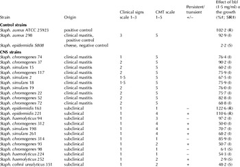
Strains of coagulase-negative staphylococci and Staphylococcus aureus control strains, their origin, clinical signs (scale 1–3) of mastitis, results of California Mastitis Test (CMT) (scale 1–5) of the milk from affected quarters, status of the IMI (persistent/transient) (Taponen et al. Reference Taponen, Simojoki, Haveri, Larsen and Pyorala2006; Taponen et al. Reference Taponen, Koort, Bjorkroth, Saloniemi and Pyorala2007) and the effect of bovine lactoferrin (Lf) (1·5 mg/ml) on growth

† Results of this study on the sensitivity of strains to bLf
‡ SIR-classification: S=sensitive, I=intermediate and R=resistant
Preliminary identification of the species was done using conventional bacteriological methods and later confirmed using AFLP analysis (Taponen et al. Reference Taponen, Koort, Bjorkroth, Saloniemi and Pyorala2007). Staph. aureus ATCC 25923, Staph. aureus 298 and Staph. epidermidis S808 were used as control strains in the assay. The positive control strain, Staph. aureus 298, was isolated from milk of a cow with acute clinical IMI (Saari Unit, Faculty of Veterinary Medicine, University of Helsinki) and the negative control strain Staph. epidermidis S808 from cheese (Finnish Food Safety Authority, Helsinki). The strains were stored at −70°C and thawed at 37°C. The strains were grown in brain heart infusion broth (LabM, Bury, UK) for 18 h at 37°C before exposure. After incubation, the bacterial broth was centrifuged at 3000 g for 10 minutes and washed twice with PBS (Euroclone) and resuspended in 5 ml RPMI 1640 medium (Invitrogen, Paisley, UK). The bacterial count for the assay was adjusted using spectrophotometry (Genesys 10 UV, Thermo Electron USA) at absorbance (A 625) 0·2–0·3.
Bovine lactoferrin
Bovine Lf (Apo-lactoferrin) was purified according to Karpulus et al. (Reference Karpulus, Ulevitch and Wilson1987). Bovine Lf was passed through a Polymyxin B (Sigma, St. Louis MO, USA) and Sepharose 4B (Sigma) preparative affinity column. After eluting, bLf solution was dialysed first against LPS-free PBS and then against LPS-free water and recovered by lyophilization. The concentration of endotoxin was determined by the Finnish Institute of Occupational Health, Kuopio with the quantitative chromogenic Limulus amebosyte Lysate, LAL assay (BioWhittaker, Walkersville MD, USA). The LPS concentration before the purification was 4·0 ng/mg and after the purification, 0·21 ng/mg.
Sensitivity to bovine lactoferrin
The antibacterial effect of bLf was determined using multichannel turbidometry (Bioscreen instrument, Labsystems, Helsinki, Finland). Final concentrations of bLf (Apo-lactoferrin, Biopole SA) in the well of the instrument were 0·1, 0·5, 1·0 and 1·5 mg/ml. Each strain and concentration was tested in triplicate. Control wells contained all components except bLf. The change in turbidity was monitored automatically every hour for 20 h at 37°C and the plates were shaken for 10 min before each measurement. The sensitivity was estimated by comparing the growth curve of the strain without bLf with the growth curve of the strain with bLf. The CNS strain was considered sensitive (S) to bLf if the inhibitory effect was <50%, intermediate (I) 99·9–50% and resistant (R) 100% or >100% (the growth of the strain was the same or increased with bLf).
Bovine mammary epithelial cell culture
A bovine mammary epithelial cell line (BME-UV1) described earlier (Van Oostveldt et al. Reference Van Oostveldt, Tomita, Paape, Capuco and Burvenich2002) was grown in 25 cm3 cell culture bottles (Sarstedt, Nümbrecht, Germany) using Ham's F12 medium (40%) (Invitrogen) made up of 23% RPMI 1640 (Invitrogen), 20% NCTC 135 (Invitrogen), 10% fetal bovine serum (fetal bovine serum, Sigma), 1% lactose (α-lactose monohydrate, Sigma), 1% lactalbumin hydrolysate (Lactalbumin Enzymatic Hydrolysate, Sigma), 2% antibiotic-antimycotic solution (antibiotic antimycotic mix, Sigma), 2% hydrocortisone (hydrocortisone, Sigma), 0·5% l-ascorbic acid (ascorbic acid, Sigma), 0·03% mm-GSH (l-glutathione reduced, Sigma) and 0·01% insulin (insulin, Sigma). Cells were incubated at 37°C in a 5% CO2 atmosphere. The culture medium was replaced every 2–3 d.
Internalization assay
The internalization assay model was validated with control strains: Staph. aureus ATCC 25923, Staph. aureus 298 isolated from acute mastitis and Staph. epidermidis S808 from cheese. In 24-well cell culture plates (Sarstedt), confluent monolayers of BME cells of the fifth to seventh passage were subcultured once in antibiotic-free medium before bacterial exposure. Adhesion, invasion and replication were determined as described by Almeida & Oliver (Reference Almeida and Oliver2001) with modifications. After adjusting the bacterial count with spectrophotometry, 500 μl of staphylococcal suspension (1×10⁸ cfu/well) was added per well. Analyses were carried out in triplicate for each strain tested including positive and negative controls. Co-cultures, one 24-well cell culture plate for adhesion, one for invasion and one for replication, were incubated at 37°C in 5% CO2:95% air for 30 min (adhesion), 2 h (invasion) and 21 h (replication).
To define the adherence of CNS strains to BME cells, co-cultures were washed five times with PBS and monolayers were treated with 500 μl cold 0·1% Triton-X-100 on ice for 10 min. Cell lysates were serially diluted 10-fold, plated in duplicate on tryptone soy agar (TSA, LabM) and counted after overnight incubation at 37°C; cfu/ml was then determined. The number of adherent bacteria was determined by subtracting the number of internalized bacteria from the number of cell-associated bacteria.
To define internalization of CNS strains into BME cells and determine their intracellular replication rate, co-cultures were washed three times with PBS, and 500 μl gentamicin (100 μg/ml, Sigma) was added to monolayers and incubated for 1·5 h at 37°C to kill extracellular bacteria. After incubation, co-cultures were washed once with 1 ml PBS, treated with 500 μl cold 0·1% Triton-X-100 on ice for 10 min and cfu/ml in lysates calculated as described.
To ascertain the replication rate of CNS strain into BME cells, monlayers were treated with gentamicin (100 μg/ml) for 1·5 h at 37°C. After incubation, co-cultures were washed and further incubated with a lower concentration of gentamicin (500 μl of solution containing 5 μg of gentamicin/ml) for 21 h at 37°C. After incubation monolayers were washed with PBS, lysed, and cfu/ml in lysate calculated as described. Adhesion and invasion, values as percentages, were calculated using the number of adherent/invaded bacteria per original cfu/ml added on monolayers multiplied by one hundred. Replication rate is the staphylococcal cfu number after 21 h incubation divided by the cfu number of the invaded bacteria.
To test the effect of bLf on the adhesion, invasion and intracellular replication, a series of assays described above, including bLf (2 mg/ml) in the wells, was also performed.
Statistical analysis
The correlation of the exposure was analysed using the Spearman test. For the analysis of the inhibitory effect of bLf on adhesion, invasion or intracellular replication, a t test was used: the data were log-transformed and the variations in environmental circumstances were eliminated using the percentage proportion of the strain with or without bLf. The tests were carried out with SPSS-software 14.0.1. for Windows.
Results
Sensitivity to bovine lactoferrin
The effect of bLf (1·5 mg/ml) on the growth of CNS strains is shown in Table 1 as a percentage of the growth without bLf. Three CNS strains were sensitive (S, <50%) to bLf and the inhibitory effect was intermediate (I, 99·9–50%) for 17 CNS strains. Two strains were resistant (R, >100%).
Adherence and internalization
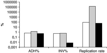
The validation of the BME cell model was carried out with the control strains. In co-cultures of BME cell monolayers, the control strains, Staph. aureus ATCC 25923, Staph. aureus 298 and non-pathogenic Staph. epidermidis S808, adhered similarly, but both Staph. aureus strains invaded better than Staph. epidermidis (Fig. 1). The intracellular replication rate of Staph. aureus isolated from clinical mastitis was higher than that recorded for Staph. aureus ATCC 25923 and for the non-pathogenic Staph. epidermidis. CV for the internalization assay were determined for the Staph. aureus ATCC 25923 strain. The inter-assay and intra-assay CV for adhesion were 20% and 59%, for invasion 28% and 56%, and for replication 24% and 53%. Because of the high variation the two control strains were always included in each bacterial exposure.
The adhesion, ADH (%), invasion, INV (%) and replication rate of control strains Staph. aureus ATCC 25923 (white), Staph. aureus 298 (grey) and Staph. epidermidis S808 (black) in validation of the bovine mammary epithelial cell model.

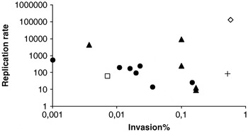
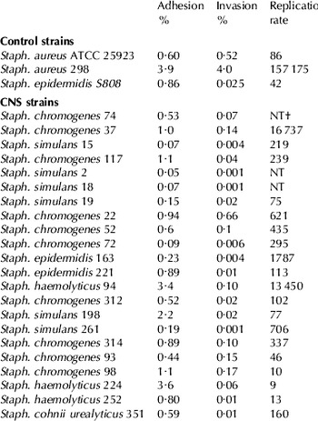
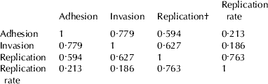
The intensity of adhesion and invasion of CNS strains was generally weaker than that of Staph. aureus strains (Table 2). The adhesion percentage of CNS strains varied between 0·05% and 3·6%, for Staph. aureus 298 it was 3·9% and for Staph. aureus ATCC 25923, 0·60%. Some CNS strains, e.g. Staph. haemolyticus strains 94 and 224, were almost as adhesive as Staph. aureus 298 but their ability to invade was markedly weaker. The invasion percentages of CNS strains were 0·001–0·7% and that of Staph. aureus 298, 4·0%. The intracellular replication of CNS strains was lower than for the Staph. aureus 298 strain, but 13 of 19 CNS strains had higher intracellular replication rates than that of the reference strain Staph. aureus ATCC 25923. The highest replication rates for the CNS strains were approximately 10% of that for Staph. aureus 298. Adhesion and invasion, as well as replication and replication rate, were strongly and positively correlated during the exposure (Table 3). No differences in adhesion, invasion or replication rates were established between the persistent and transient CNS strains (Fig. 2).
Replication rate and invasion (%) of the persistent (•) and transient (▴) CNS strains and the control strains, Staphylococcus aureus ATCC 25923 (+) and Staph. aureus 298 (◊) and Staph. epidermidis S808 (□).

Adhesion (%), invasion (%) and replication rate of CNS and control strains in bovine mammary cell model

† NT, not detected
Spearman correlation coefficients between adhesion, invasion and replication during the bacterial exposure
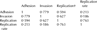
† Replication is the number of intracellular bacteria after 21-h incubation (cfu/ml)
Effect of bLf on internalization assay
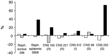
The effect of bLf on adhesion, invasion and intracellular replication was tested with all nineteen CNS strains in bLf at 2 mg/ml. bLf did not have a significant effect on the number of adhered bacteria during the exposure. Degree of the adherence established in the assay did not correlate with the susceptibility of the strain to bLf (Tables 1 and 2). bLf significantly decreased the intracellular replication (P=0·013) and replication rates (P=0·012) of the CNS strains. The effect of bLf on adhesion, invasion and replication rates of the control strains Staph. aureus 298, Staph. epidermidis S808 and five CNS strains, resistant (R), partly susceptible (I) and susceptible (S) to bLf, are shown in Fig. 3.
The effect of bovine lactoferrin (bLf) (2 mg/ml) on the growth of control strains, Staphylococcus aureus 298 and Staph. epidermidis S808, and five CNS strains in a bovine mammary epithelial cell model. The effect on adhesion % (white), invasion % (grey) and replication rate (black) is positive or negative compared with growth without bLf. CNS 163 and CNS 221 strains were resistant (R), CNS 312 moderately susceptible (I) and CNS 98 and 252 susceptible (S) to bLf (1·5 mg/ml). The control strains Staph. aureus 298 was moderately susceptible (I) and Staph. epidermidis S808 susceptible (S) to bLf. CNS strains 221, 312 and 252 were persistent and 163 and 98 transient.

Discussion
This study is the first that has used a cell model to demonstrate intracellular persistence of CNS. The results indicate that CNS that cause bovine IMI can be characterized on the basis of their invasion of the mammary gland cells, although the intensity of invasion of CNS was clearly weaker than that of Staph. aureus, a major mastitis pathogen. While the capacity for internalization varied among the CNS strains, all of the CNS species examined, Staph. chromogenes, Staph. simulans, Staph. epidermidis, Staph. haemolyticus and Staph. cohnii urealyticus, had an adhesive ability equal to that of Staph. aureus. The antagonistic effect of bLf on the adhesion and internalization of CNS strains was weak and strain-dependent, but bLf significantly decreased intracellular replication and replication rates of CNS.
In their internalization study, Almeida & Oliver (Reference Almeida and Oliver2001) reported that three CNS species included adhered equally to BME cells. The degree of adhesion was of the same magnitude as found in our study, ranging from 0·27% to 3·5% compared with 0·05% to 3·6%. In their experiment, adherence varied substantially according to the length of exposure and CNS species. A strong correlation between adhesion and invasion, as well as between replication and replication rate, was found in our study (Table 2) confirming previous in-vitro findings indicating that invasion is a frequent consequence of adherence (Hensen et al. Reference Hensen, Pavicic, Lohuis and Poutrel2000). Here, replication rates of CNS strains varied substantially; the highest rates were a thousand times higher than the lowest rates. Staph. aureus 298 had a ten-fold higher replication rate than the CNS strains with the highest values. CNS strain 37, with the highest replication rate and adhesion and invasion percentages among the CNS strains, was able to induce IMI with mild clinical signs in an experimental challenge study (H. Simojoki, unpublished observations).
Bacterial infection in vivo is a complex process, the first step being an intimate colonization of epithelia through non-specific physical interactions and specific ligands to host receptors (Van Belkum et al. Reference Van Belkum, Kools-Sijmons and Verbrugh2002). Most studies on CNS adhesion have focused on bacterial aggregation and binding to biomaterials and on the role of specific adhesins and slime production in the pathogenesis of CNS infections (Kloos & Bannerman, Reference Kloos and Bannerman1994). Production of capsular polysaccharide adhesin and slime has been suggested to essentially determine the persistence of bacteria in biofilms (Cucarella et al. Reference Cucarella, Tormo, Ubeda, Trotonda, Monzon, Peris, Amorena, Lasa and Penades2004). Accordingly, Aguilar et al. (Reference Aguilar, Amorena and Iturralde2001) reported that bacterial slime is an important effector of staphylococcal binding to ovine mammary epithelial cells. In clinical staphylococcal infections in humans, slime-producing staphylococci have been found to form cell clusters and biofilms mediated by an anionic intercellular polysaccharide adhesin. In Staph. aureus, biofilm formation is associated with ica locus (Melchior et al. Reference Melchior, Vaarkamp and Fink-Grimmels2006). Vasudevan et al. (Reference Vasudevan, Nair, Thirunavukkarasu and Venkitanarayanan2003) reported a high prevalence of ica genes among Staph. aureus isolated from bovine mastitis. However, for CNS isolated from mastitis no data are available on the relationship between the ica genes, biofilm formation and recurrent mastitis. Expression of adhesins is strongly affected by the environment, including the chemical composition of the growth medium. Staphylococci may show stronger adhesion to damaged udder tissue cells than to the cultured host cells used in the in-vitro models, which may, for example, express different receptors to cells in the mammary gland (Cifrian et al. Reference Cifrian, Guidry, O'Brien, Nickerson and Marquardt1994). Extrapolation of the in-vitro findings directly to in-vivo circumstances should be done with caution (Van Belkum et al. Reference Van Belkum, Kools-Sijmons and Verbrugh2002) but, for example, using the in-vitro assay described here, differences in the capacity for internalization between bacterial species or strains can be investigated.
Intracellular staphylococci can cause subclinical, chronic IMI (Hensen et al. Reference Hensen, Pavicic, Lohuis and Poutrel2000; Kerro Dego et al. Reference Kerro Dego, van Dijk and Nederbragt2002). It has also been suggested that resistance of chronic infections to host immune defence and antibiotic therapy could be due to the intracellular presence of these bacteria (Aarestrup et al. Reference Aarestrup, Larsen and Jensen1999; Chaffer et al. Reference Chaffer, Leitner, Winkler, Glickman, Krifucks, Ezra and Saran1999; Hebert et al. Reference Hebert, Sayasith, Senechal, Dubreuil and Lagace2000). CNS strains in this study were selected from field material, about half of them representing CNS IMI with a history of persistence for extended periods during lactation (Taponen et al. Reference Taponen, Koort, Bjorkroth, Saloniemi and Pyorala2007). The replication time allowed here may have been too short, only 24 h. In further studies it would be worthwhile to extend the time to study the true intracellular survival of CNS in the BME cells. Presence or absence of bLf did not affect the adhesion, invasion and intracellular replication, and there were no differences between strains originating from persistent or transient infections. This finding may support the key role of the host-microbe interaction in the genesis of infection. In the study of Dogan et al. (Reference Dogan, Klaessig, Rishniw, Almeida, Oliver, Simpson and Schukken2006) both transient and persistent Escherichia coli strains adhered, but persistent strains invaded, survived and replicated in mammary cell culture to a much greater extent than the transient strains. The pathogenesis of Esch. coli and staphylococcal IMI probably differs. Host response may be significant or even decisive in determining the persistence of the IMI. This is, however, an aspect that is very difficult to study with in-vitro models.
Mastitis-causing staphylococci appear to be generally more resistant to bLf than Esch. coli (Rainard, Reference Rainard1986). In the study of Lee et al. (Reference Lee, Kawai, Nakamura, Tanaka, Kumura and Shimazaki2004) most CNS strains were not inhibited by bLf (2004). In our in-vitro sensitivity and internalization analyses, the role of bLf was mainly bacteriostatic probably by chelating iron and binding to the specific receptors on the bacterial cell surface. While bLf inhibited growth of the CNS strains in vitro, the inhibition was not complete and was dependent on the concentration of bLf. According to Naidu et al. (Reference Naidu, Miedzobrodzki, Andersson, Nilsson, Forsgren and Watts1990) specific receptors for bLf are present on the cell surface of CNS associated with IMI. Specific binding of Lf, indicating the presence of Lf receptors, has been demonstrated also for various types of mammalian cells including mammary epithelial cells (Rochard et al. Reference Rochard, Legrand, Lecocq, Hamelin, Crepin, Montreuil and Spik1992; Rejman et al. Reference Rejman, Turner and Oliver1994). Lf inhibited adhesion of some oral bacteria to epithelial cells both via specific (adhesin-ligand) and non-specific charge-dependent interactions (Alugupalli & Kalfas, Reference Alugupalli and Kalfas1997). Singh et al. (Reference Singh, Parsek, Greenberg and Welsh2002) reported that in the presence of subinhibitory concentrations of Lf, Pseudomonas bacteria attached and multiplied but failed to form microcolonies or differentiated biofilm structures. The possible effect of Lf on staphylococcal biofilm formation has not been investigated. However, iron-binding capacity and antibacterial activity of bLf against staphylococci is stronger in mastitic secretions than under normal physiological circumstances or in-vitro cell culture assay (Komine et al. Reference Komine, Komine, Kuroishi, Kobayashi, Obara and Kumagai2005). This may explain why in this in-vitro BME cell model bLf had no apparent effect on adhesion, although the concentration of bLf (2 mg/ml) used was comparable with the concentrations recorded in mastitic milk. In a previous study bLf had no significant effect on the colonization of mammary epithelial cells by Staph. aureus, but it decreased the invasion (Diarra et al. Reference Diarra, Petitclerc, Deschenes, Lessard, Grondin, Talbot and Lacasse2003). In our study, bLf decreased the intracellular replication and replication rates of CNS strains significantly irrespective of their extracellular sensitivity or resistance to bLf. Recognition of invading pathogens via receptors is the first decisive step in initiating immune defence, generating signals which induce response in cytokine and Lf coding mRNA expression during the first day after exposure (Griesbeck-Zilch et al. Reference Griesbeck-Zilch, Meyer, Kühn, Schwerin and Wellnitz2008). Berlutti et al. (Reference Berlutti, Schippa, Morea, Sarli, Perfetto, Donnarumma and Valenti2006) reported that Lf had an anti-inflammatory effect by down-regulating pro-inflammatory cytokines, which could be one mechanism to impair the intracellular replication of CNS. Accordingly, bLf may play a role in the defence of the mammary gland also in IMI caused by CNS. Further investigations are needed to elucidate the possible mechanisms of Lf in IMI.
Financial support from Walter Ehrström Foundation and the Mercedes Zachariassen Foundation is gratefully acknowledged. We thank Professor C Burvenich, Faculty of Veterinary Medicine, Ghent University, Belgium, for providing us with the BME cells. The skilful technical assistance of Eeva-Liisa Palkispää, Terhi Nissinen and Joel Jukka in the laboratory of Nutrition and Food Biotechnology, University of Kuopio is gratefully acknowledged.