Introduction
Tetralogy of Fallot, the most common cyanotic CHD, involves four components: large ventricular septal defect, overriding of the aorta, narrowing of the right ventricle outflow tract, and right ventricle hypertrophy.
Tetralogy of Fallot repair was originally performed through a large right ventricular incision with a transannular patch to relieve right ventricle outflow tract obstruction. However, this surgical procedure often results in significant pulmonary regurgitation and may lead to right ventricular dilation, dysfunction, arrhythmias, and eventual pulmonary valve replacement.Reference Gott1 Many operative technique modifications have been developed to avoid ventriculotomy and preserve the pulmonary valve annulus.
The current surgical strategies for the relief of right ventricle outflow tract obstruction that aim to prevent pulmonary regurgitation in patients with corrected tetralogy of Fallot may result in a certain degree of residual pulmonary stenosis. Van der Hulst et al. reported that mild residual pulmonary stenosis (peak pressure gradient 15–30 mmHg through the right ventricle outflow tract and pulmonary valve) in corrected tetralogy of Fallot reduced the risk of pulmonary valve replacement.Reference van der Hulst, Hylkema and Vliegen2 However, the authors did not describe changes in the pulmonary stenosis gradient over the study period. Even though residual pulmonary stenosis immediately after surgery has been acceptable, the long-term changes of pulmonary stenosis are not well known. The aims of this study were to observe the natural course of postoperative acceptable residual pulmonary stenosis in corrected tetralogy of Fallot patients and to investigate factors related with pulmonary stenosis progression.
Methods
From January 2006 to December 2012, patients who underwent tetralogy of Fallot total correction and received follow-up for a minimum of 5 years after surgery at Samsung Medical Center in Seoul, South Korea were identified. Patients with pulmonary atresia, absence of pulmonary valve syndrome, and those who underwent the Rastelli (right ventricle to pulmonary artery conduit) operation were excluded.
A retrospective chart review was conducted to collect the following patient characteristics: age and body weight at surgery, sex, follow-up duration, and previous palliation history. If re-intervention was performed during the follow-up period, the last follow-up time was defined as just before re-intervention. Operation data, including surgical method (transannular or non-transannular) and the ratio of right ventricle pressure to aorta pressure after weaning off cardiopulmonary bypass at the end of surgery, were reviewed.
In addition, the preoperative, immediate postoperative, and serial postoperative echocardiography images were reviewed. Immediate postoperative echocardiography was usually performed within 10 days of the operation prior to discharge. On echocardiography, the peak pressure gradient over the right ventricle outflow tract, including the pulmonary valve, was measured by continuous wave Doppler. Acceptable pulmonary stenosis immediately after surgery was defined as peak gradient between 15 and 45 mmHg, and patients with no (<15 mmHg) or significant (>45 mmHg) pulmonary stenosis were excluded.Reference van der Hulst, Hylkema and Vliegen2 The main level of residual pulmonary stenosis was assessed. If right ventricle outflow tract muscle hypertrophy was visible and colour aliasing occurred from beneath the pulmonary valve, the pulmonary stenosis was classified as mainly sub-valvar pulmonary stenosis. If the pulmonary valve motion was restrictive and colour aliasing occurred from the pulmonary valve opening, the pulmonary stenosis was classified as valvar pulmonary stenosis. The preoperative and postoperative pulmonary valve annulus z-score and severity of pulmonary regurgitation were assessed as well. Echocardiography was performed by a singular experienced paediatric cardiologist, and the average of the data from three measurements of each Doppler study was calculated. All patients underwent immediate postoperative echocardiography and subsequent echocardiography exams more than 3 times during the follow-up period.
We used the latent class linear mixed model to differentiate the trajectories of residual pulmonary stenosis after tetralogy of Fallot surgery. Instead of a priori grouping of the study participants using their baseline characteristics, the latent class linear mixed model allowed us to group study participants a posteriori using the longitudinal trajectory patterns of main interest. A detailed explanation and application of latent class linear mixed model models are published elsewhere.Reference Verlinden, van der Geest, de Bruijn, Hofman, Koudstaal and Ikram3 We used link function with quadratic spline (using five equidistant nodes) to divide the entire patient sample into two groups. The specific model was selected based on the Akaike Information Criterion and Bayesian Information Criterion. Random intercept and slope over time (1 year after tetralogy of Fallot surgery) were included in the model. A total of 385 annual follow-up sonography studies from 52 patients were used to group blood flow trajectory patterns. Latent class linear mixed model function in the “latent class linear mixed model” R package was used in our analysis.
Differences between pulmonary stenosis progression group and no progression group were assessed with the t-test for continuous variables and with the chi-square test for categorical variables. A receiver operating characteristic curve was used to determine the cut-off value of immediate postoperative pulmonary stenosis gradient between the progression and no progression groups. A p value <0.05 was regarded as significant. Variables were expressed as the mean (±standard deviation) or median (range). Data analysis was performed using the SPSS software version 25 (IBM Corporation, Armonk, NY).
The study protocol was approved by the Institutional Review Board at Samsung Medical Center (IRB No. 2018-12-020). The need for informed consent was waived by the board.
Results
A total of 134 patients underwent tetralogy of Fallot total correction, and 82 patients were excluded for the following reasons: loss to follow-up (14 patients), death during follow-up (6 patients), no residual pulmonary stenosis (53 patients), and significant pulmonary stenosis immediately after surgery (9 patients). As a result, 52 patients who had acceptable pulmonary stenosis were included in this study.
There were 32 (61.5%) males in the study, and the median age at the time of surgery was 158 days (range, 64–3108). All but two patients underwent total correction before 12 months of age; one patient underwent surgery at 8 years and the other at 2 years. The median follow-up duration was 110 months (range, 6–141). A previous palliative procedure had been performed in 10 patients (21.2%) and 11 procedures; there were 10 cases of shunt operation and 1 case of balloon valvuloplasty. Thirty-six (69.2%) patients had a bicuspid pulmonary valve, and 47 (90.4%) patients underwent non-transannular repair. Five (9.6%) patients underwent transannular repair with monocusp homograft patch insertion. All patients experienced a pressure ratio (right ventricle/aorta) less than 0.7 in the operation room. The mean postoperative residual pressure gradient was 27.4 mmHg (range, 15.3–43.3).
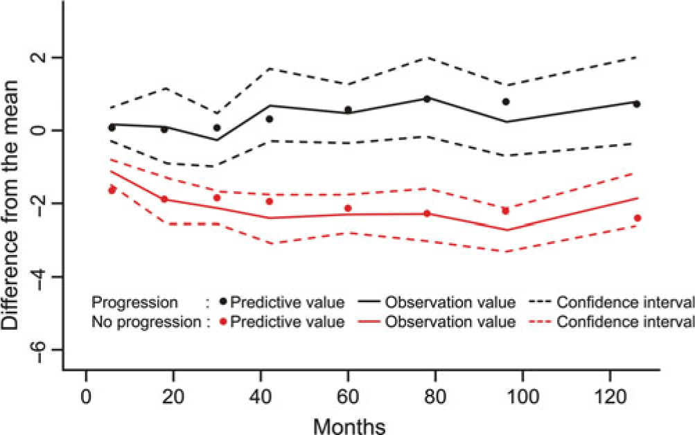
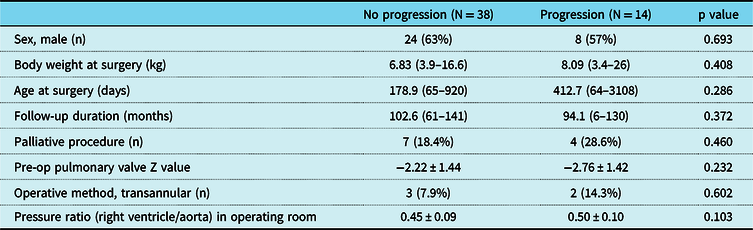
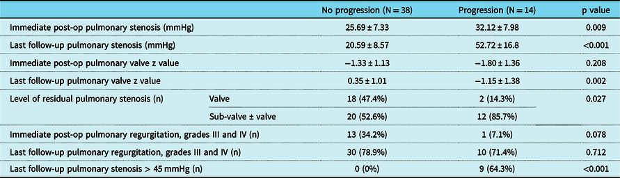
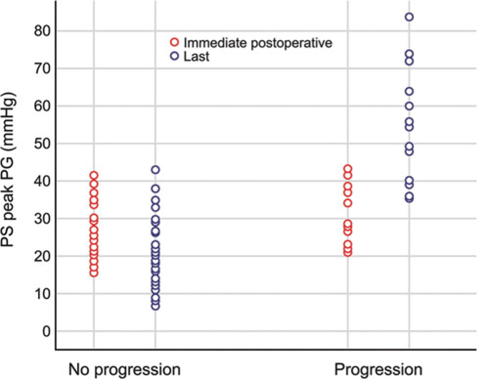
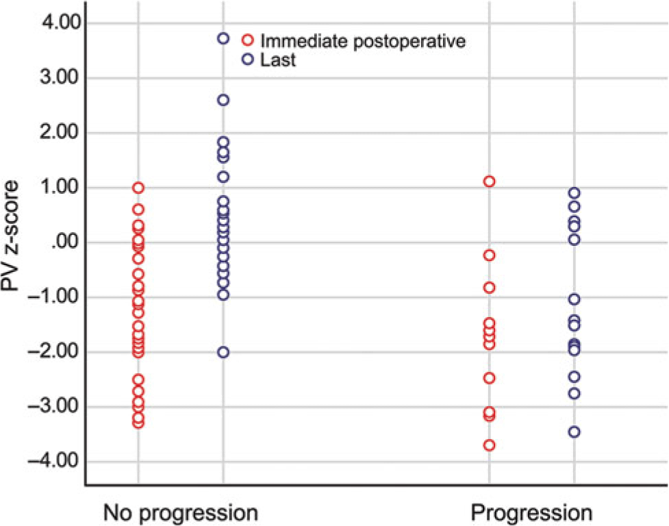
The residual pressure gradient progressed in 14 (27%) patients according to the Latent class linear mixed model statistical method (Fig 1). There were no statistically significant differences in sex, body weight, and age at surgery, follow-up duration, palliative surgery, preoperative pulmonary valve annulus z-score, operative method, and pressure ratio (right ventricle/aorta) in the operating room between the two groups (Table 1). Table 2 shows the postoperative characteristics of the two groups. The immediate and last follow-up postoperative pressure gradient of the progression group were significantly higher than those of the no progression group (32.12 ± 7.98 versus 25.69 ± 7.33, p = 0.009 and 52.72 ± 16.8 versus 20.59 ± 8.57, p < 0.001, respectively) (Fig 2). There was no difference in the immediate postoperative pulmonary valve annulus z-score between the two groups. However, the last pulmonary valve annulus z-score was significantly smaller in the progression group than in the no progression group (−1.15 ± 1.38 versus 0.35 ± 1.01, p = 0.002) (Fig 3). The pulmonary valve annulus z-score significantly increased in the no progression group from −1.33 to 0.35, whereas there was no significant change in the progression group.
The result of latent class mixed model division into two groups (progression and no progression).

Preoperative and perioperative characteristics of the two groups.

Postoperative characteristics of the two groups.

Distributions of immediate postoperative pulmonary stenosis gradient and last pulmonary stenosis gradient of the two groups.

Distributions of immediate postoperative pulmonary valve annulus z-score and last pulmonary valve annulus z-score of the two groups.

The number of patients with residual pulmonary stenosis remaining mainly at the sub-valvar area was significantly higher in the progression group than in the no progression group (85.7% versus 52.6%, respectively; p = 0.027). All patients who underwent a transannular repair showed residual pulmonary stenosis predominantly in the sub-valve area.
Although the difference was not statistically significant, there was a greater percentage of patients with immediate postoperative pulmonary regurgitation greater than grade III in the no progression group compared to the progression group (34.2% versus 7.2%, respectively; p = 0.078). At the last follow-up echocardiography, 40 (76.9%) patients had pulmonary regurgitation greater than grade III, and there was no significant difference between the two groups.
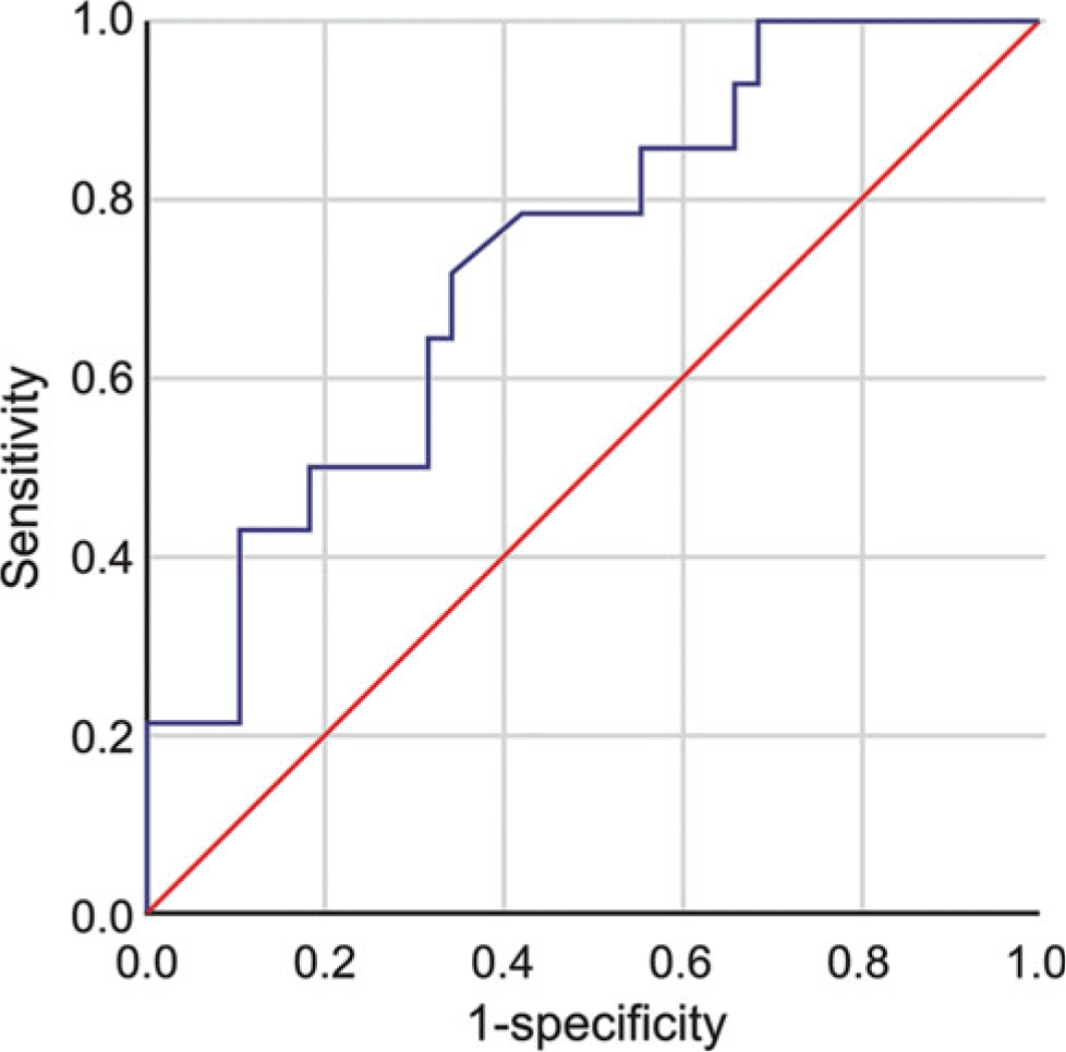
To forecast which patients would progress to an unacceptable level of pulmonary stenosis, a receiver operating characteristic curve was calculated, and it showed that the cut-off value of immediate pulmonary stenosis that could progress was 26.8 mmHg (sensitivity = 65.3%, specificity = 65.8%) (Fig 4).
Receiver operating characteristic curve of immediate pulmonary stenosis in between the progression and no progression groups.

A pressure gradient over 45 mmHg on the last echocardiography was found in nine patients, all of whom were in the progression group. Their median value of immediate postoperative pressure gradient was 36.8 mmHg (range, 23–43.3), and median time interval until the pressure gradient reached over 45 mmHg was 72 month (range, 6–120). Among the nine patients, eight patients had sub-valvar pulmonary stenosis.
Two patients received re-intervention during the follow-up period. The first patient underwent total correction (non-transannular method) at the age of 2 months, and the immediate postoperative residual pulmonary stenosis was 26.8 mmHg predominantly in the sub-valve area. The patient’s pulmonary stenosis progressed up to 73.9 mmHg over a period of 80 months after the operation. Balloon dilatation of right ventricle outflow tract was performed, but it was not effective. Right ventricle outflow tract muscle resection and pulmonary valve replacement were simultaneously performed due to combined severe pulmonary regurgitation. The second patient underwent a palliative shunt operation at 4 months of age and total correction (non-transannular method) at the age of 10 months. The immediate postoperative residual pulmonary stenosis was 26.4 mmHg, and the pulmonary stenosis rapidly progressed to 49.4 mmHg over a period of 6 months after the operation. Eventually, the patient underwent right ventricle outflow tract muscle resection.
Discussion
In this study, we observed that acceptable pulmonary stenosis immediately after tetralogy of Fallot total correction progressed during a follow-up period in 27% of patients, and the main lesion of pulmonary stenosis in the progression group was located in the sub-valvar area. These patients had a significantly higher value of immediate residual pulmonary stenosis pressure gradient compared to those with no progression. We suggest an optimal cut-off value of immediate pulmonary stenosis to be 26.8 mmHg.
As expected, mild residual pulmonary stenosis on the right ventricle outflow tract contributed to preventing excessive right ventricle dilatation by limiting the amount of pulmonary regurgitation.Reference van der Hulst, Hylkema and Vliegen2 However, it is difficult to establish the optimal range of mild residual pulmonary stenosis, and we are not convinced that residual pulmonary stenosis does not progress.
Our study investigated the change of acceptable residual pulmonary stenosis immediately after tetralogy of Fallot total correction over time. Unlike other studies that focused on postoperative pulmonary regurgitation and re-intervention after tetralogy of Fallot total correction, our study was limited to progression of residual pulmonary stenosis regardless of intervention.
The primary reasons for reoperation in North America and Europe were residual ventricular septal defect and/or pulmonary regurgitation.Reference Nollert, Fischlein, Bouterwek, Bohmer, Klinner and Reichart4,Reference d’Udekem, Ovaert and Grandjean5 However, progression of pulmonary stenosis is another indication for reoperation. A group in Japan performed the first nationwide survey on the indications for reoperation in tetralogy of Fallot.Reference Mizuno, Niwa and Matsuo6 Pulmonary stenosis (31.7%) was the most common reason, followed by pulmonary regurgitation (29.0%), combined pulmonary stenosis and regurgitation (11.2%), and residual ventricular septal defect (9.4%). The authors explained that the reason for this difference was operation technique. However, there was no detailed information about the characteristics of pulmonary stenosis or the interval to reoperation in each patient.
Stewart et al. reported that seven (6.9%) patients required reoperation due to residual pulmonary stenosis, and their median duration to reoperation was 8 months (range, 12 days – 38 months). Among them, the residual stenosis was predominantly sub-valvar in four patients.Reference Stewart, Backer, Young and Mavroudis7 Similarly, two patients in our study with acceptable immediate pulmonary stenosis after total correction required re-intervention for residual stenosis mainly at the sub-valvar area.
Nowadays intraoperative trans-oesophageal echocardiogram became popular in tetralogy of Fallot surgery. In addition, many studies have considered the adequate pressure ratio (right ventricle/aorta) in tetralogy of Fallot total correction. A Chicago studyReference Stewart, Backer, Young and Mavroudis7 demonstrated that pressure ratio (right ventricle/aorta) for successful pulmonary valve preservation is less than 0.7, and a Michigan study found that a pressure ratio (right ventricle/aorta) greater than 0.7 was a risk factor for reoperation.Reference Hirsch, Mosca and Bove8 Hoashi et al. analysed long-term outcomes after repair of tetralogy of Fallot with preservation of the pulmonary valve annulus. The mean follow-up duration was 15.8 years, and only 1 of the total 84 patients required reoperation for recurrent right ventricle outflow tract obstruction. They reported that aggressive right ventricle outflow tract muscle resection contributed to the prevention of recurrent right ventricle outflow tract obstruction, even though patients had a pressure ratio (right ventricle/aorta) >0.7 and a small pulmonary valve annulus.Reference Hoashi, Kagisaki and Meng9 Similarly, we admitted the concern about sub-valvar stenosis on echocardiogram in operating room, but we could not suggest any cut-off pressure ratio in operating room from our study. There are limitations in evaluating pressure gradient or right ventricular pressure in operating room. The pressure gradient on echocardiogram and pressure data immediate after bypass might be biased. And from our Table 1, there was no differences in pressure ratio (right ventricle/aorta) between groups.
If residual pulmonary stenosis remains at the sub-valvar area after surgery, we can suppose that pulmonary stenosis would progress and re-intervention would be likely. More aggressive right ventricle outflow tract muscle resection may protect against progression of pulmonary stenosis. Boni et al. reported that when infundibular and supra-valve stenoses are fully removed, residual obstruction, only due to the hypoplastic annulus, does not affect the growth of the pulmonary valve or right-sided heart development.Reference Boni, Garcia and Galletti10 In our study, there was no difference in the immediate postoperative pulmonary valve annulus between the pulmonary stenosis progression and no progression groups. However, the pulmonary valve annulus was significantly smaller on the last echocardiography exam in the progression group. Data from these patients imply that the pulmonary valve can grow if sufficient blood flows through it after relief of right ventricle outflow tract obstruction, even though the pulmonary valve annulus is small.
There are several studies on the natural course of isolated mild pulmonary valvar stenosis, and results show that the natural history of mild isolated pulmonary valvar stenosis tends to be even more favourable. Vogel and LuekerReference Vogel and Lueker11 showed that patients aged >6 months whose gradient was below 40 mmHg at the time of diagnosis remained stable and required no treatment during a 7-year follow-up period. AtikReference Atik12 showed that among 13 patients who had mild pulmonary valvar stenosis lower than 41 mmHg, progression was observed in just 1 patient (7.6%). Although the authors included no detailed information about pulmonary valve morphology and pulmonary annulus size, the findings suggest that proper forward flow through the pulmonary valve protects against progression of mild isolated pulmonary valvar stenosis.
By the way, it was interesting in our study that there was no significant difference in PR degree at the latest follow-up between transannular group and non-transannular groups. This finding showed that ‘annulus preservation’ didn’t guarantee a competent pulmonary valve. This was also demonstrated in the study by HoashiReference Hoashi, Kagisaki and Meng9 by showing that the freedom from moderate or greater pulmonary regurgitation at 20 years after ‘annulus-preserving’ repair of TOF was only 35.7%.
There are several limitations to our study, including its retrospective design, the small number of patients, and the short follow-up period. In addition, although we divided patients into either a progression group or no progression group according to change of pulmonary stenosis pressure gradient over time after surgery, the follow-up duration was different between the groups, creating the possibility that a participant’s grouping could change with longer follow-up. Also, we cannot explain the direct causal relationship between relating factors and progression because this study was cross-sectional. Although the echocardiography study was carried out by a single experienced professional, measurement error was inevitable. We could not analyse the mean pressure gradient because we did not have the data for all patients. Well-designed, prospective, long-term studies across multiple centres are necessary to obtain reliable and precise outcomes.
We conclude that acceptable immediate residual pulmonary stenosis (less than 45 mmHg) in corrected tetralogy of Fallot patients can progress, and higher pressure gradient than 26.8 mmHg and stenosis that originated from the sub-valvar area were significant factors in progression.
Financial support
This research received no specific grant from any funding agency, commercial or not-for-profit sectors.
Conflicts of interest
None.
Author contributions
Study design: J Song, Y-S Kim
Data collection: Y-S Kim, J Song, J Huh, I-S Kang, J-H Yang, T-G Jun
Data analysis: Y-S Kim, J Song, J Huh,
Manuscript drafting: Y-S Kim, J Song, I-S Kang, J-H Yang, T-G Jun
Critical revision: J Song, J Huh
Final review/approval: J Song, J Huh, I-S Kang, J-H Yang, T-G Jun