High hydrostatic pressure (HHP) is an emerging technology developed as an alternative to heat treatment. HHP has been applied experimentally to milk, though some side effects have been reported, such as disruption of casein structure, whey protein denaturation and changes in viscosity (Johnston et al. Reference Johnston, Murphy and Birks1994; Huppertz et al. Reference Huppertz, Alan and Patrick2002). However, HHP has been reported as a treatment that improves the quality of some milk products, such as cheese and yogurt (Trujillo et al. Reference Trujillo, Capellas, Saldo, Gervilla and Guamis2002; Harte et al. Reference Harte, Luedecke, Swanson and Barbosa-Canóvas2003; Penna et al. Reference Penna, Subbarao-Gurram and Barbosa-Canovas2007).
In addition, a considerable amount of information is available on the effect of HHP on some whey proteins. In the review by Considine et al. (Reference Considine, Patel, Anema, Singh and Creamer2007) it is stated that β-lactoglobulin is one of the most pressure sensitive proteins and α-lactalbumin one of the most pressure resistant (Huppertz et al. Reference Huppertz, Fox and Kelly2004; Mazri et al. Reference Mazri, Sánchez, Ramos, Calvo and Pérez2012b).
However, there is not much information available about the effect of high pressure on minor milk proteins, such as lactoferrin, a protein with valuable biological properties. Lactoferrin (LF) is an 80 KDa iron binding glycoprotein present in many external secretions of mammals, with antimicrobial, anti-inflammatory, antitumoral and immuno-modulatory activities (Brock, Reference Brock2002). These functions make bovine LF (LFb) a valuable protein to industry. Actually, LFb is already used in some countries as a natural bioactive ingredient in food and health products and it has been recently authorised in Europe to be placed on the market as a novel food ingredient (Commission Implementing Decision 2012/725/EU).
We have investigated in previous studies the effect of heat treatment on the structure and activity of LF (Sánchez et al. Reference Sánchez, Peiró, Castillo, Pérez, Ena and Calvo1992; Mata et al. Reference Mata, Sánchez, Headon and Calvo1998; Conesa Reference Conesa, Sánchez, Rota, Pérez, Calvo, Farnaud and Evans2008a, Reference Conesa, Rota, Castillo, Pérez, Calvo and Sánchez2009a, Reference Conesa, Rota, Castillo, Pérez, Calvo and Sánchezb). The results showed that LFb started to lose its structure, determined by its immunoreactivity, after heat treatments above 77 °C, the treatment at 72 °C for 15 s being rather protective (Sánchez et al. Reference Sánchez, Peiró, Castillo, Pérez, Ena and Calvo1992). In the same study, we observed that LFb devoid of iron was much more sensitive to heat treatment than the iron-saturated form, and that its thermoresistance was lower in milk than in phosphate buffer. Therefore, when considering the use of LF as a bioactive ingredient is very important to determine the influence of the technological conditions on its activity, especially when using new preservation treatments.
The effect of high pressure on some milk proteins has been previously investigated in our group. We have studied the kinetics of denaturation by high pressure of LFb, lactoperoxidase, α-la, β-lg and immunoglobulins (Mazri et al. Reference Mazri, Sánchez, Ramos, Calvo and Pérez2012a, Reference Mazri, Sánchez, Ramos, Calvo and Pérezb, Reference Mazri, Ramos, Sánchez, Calvo and Pérezc). We have also investigated the effect of high pressure on human LF from transgenic rice (Franco et al. Reference Franco, Castillo, Pérez, Calvo and Sánchez2011). In the case of bovine native (LFb-nat), our previous study showed the influence of the composition of medium in the decrease of LF immunoreactivity after high pressure, by determining the kinetic parameters of the denaturation process (Mazri et al. Reference Mazri, Sánchez, Ramos, Calvo and Pérez2012a).
The objective of the present study was to investigate the effect of HHP on LFb considering three levels of iron-saturation and three different media of treatment, by measuring several characteristics related to its structure and also by evaluating its iron-binding capacity and antibacterial activity against Escherichia coli O157:H7.
Materials and Methods
Sample preparation
Bovine lactoferrin (LFb-nat) with an iron saturation of around 26% was kindly donated by Fina Research (Seneffe, Belgium). Iron-depleted lactoferrin (LFb-apo) was obtained according to the method of Mazurier & Spik (Reference Mazurier and Spik1980). Iron-saturated lactoferrin (LFb-holo) was prepared by saturating a solution of 50 mg LFb-nat/ml physiological saline solution with ferric nitriloacetic acid as described by Conesa et al. (Reference Conesa, Sánchez, Rota, Pérez, Calvo, Farnaud and Evans2008a). Iron saturation of LFb-holo was estimated at about 98% by measuring the absorbance ratio between 280 and 465 nm. LFb samples for HHP treatments were prepared at a concentration of 2 mg/ml, using three different diluents: 15 mm potassium phosphate buffer containing 150 mm NaCl pH 7·4 (phosphate-buffered saline, PBS), bovine whey and bovine skimmed milk.
High-pressure treatments
Samples of LFb at 2 mg/ml were introduced into 2·5-ml polyethylene vials, eliminating headspace, and the vials were introduced in polyethylene bags that were heat-sealed before pressurisation. Samples were pressurised using a HHP equipment model FPG 11500.B, Stansted Fluid Power (Eurotherm Automation, Lyon, France), with water as transmitting medium. Pressure was raised to 400, 500 or 650 MPa and maintained at those values for 15 min. The temperature of the samples was controlled by circulating water at 20 °C through a jacket surrounding the pressure vessel. High-pressure treatments were performed at least twice on different days and samples were kept at 4 °C until analysed.
Fluorescence spectroscopy measurement
Samples of LFb after HHP treatment were diluted to a concentration of 0·1 mg/ml in PBS. This concentration was estimated by spectrophotometric determination at A280 using the extinction coefficients (1 mg/ml) of 1·27 for LFb-apo and LFb-nat, and 1·51 for LFb-holo (Brown & Parry, Reference Brown and Parry1974). Fluorescence spectra were acquired at 20 °C on a spectrofluorimeter Aminco SLM 4800C (Rochester, NY, USA) with a thermoregulated cell holder. Samples of LFb were placed in 1·0 cm cuvettes vs. a blank of buffer, and fluorescence quenching spectra were obtained at excitation and emission wavelengths of 290 nm and 300–400 nm, respectively. The absorbance was read and a spectral scanning curve was made.
Differential scanning calorimetry (DSC) analysis
Samples of LFb after being subjected to HHP treatment were concentrated by using a centrifugal ultrafiltration device with a nominal molecular weight limit of 3000 Da (Amicon Ultra-15 3 K, Billerica, MA, USA). LFb samples were centrifuged at 4000 g at 4 °C to a concentration of 100 mg/ml, which was estimated by measuring the absorbance at 280 nm, using a molar extinction coefficient (E1%) of 1·51 ml/cm/g and 1·27 ml/cm/g for the saturated and native forms of the protein, respectively. Samples of 10 μl were introduced into aluminium pans (TA Instruments, New Castle, DE, USA) and sealed for analysis. The reference pans consisted of the same volume of PBS. DSC of LFb samples was performed in a DuPont thermal analyzer (Model DSC 10, Neu-Isenburg, Germany), using a Thermal analyst 2000 system. DSC scans were programmed in a temperature range of 35–110 °C at a heating rate of 10 °C/min. Samples were analysed at least in triplicate.
Analysis by inductively coupled plasma-optical emission spectrometry (ICP-AES)
Ultrafiltration was used to separate any ferric ions that could have been released from LFb during HHP treatment. Solutions of LFb-nat and LFb-holo in PBS (4 ml) after treatment were centrifuged using an ultrafiltration device as described above. Volumes of 2 ml of PBS were added consecutively to the concentrated protein and centrifuged; this procedure was repeated four times. To the filtrate (10 ml) was added nitric acid to reach a 2% concentration, and the ferric ion content was determined using inductively coupled plasma-optical emission spectrometry, by using a Thermo Elemental IRIS Intrepid ICP-AES, Radial Spectrometer (Thermo Electron Corporation, Waltham, MA, USA). Similar samples of LFb not subjected to ultrafiltration were also analysed by ICP-AES to determine the initial iron bound to LF in the three forms. ICP-AES analyses were conducted in duplicate.
Iron-saturation assay by spectrophotometry
The iron saturation ability of LFb-apo after being subjected to HHP treatment was determined by adding sequentially 5 μl-volumes of 2 mm Fe3+ as ferric nitriloacetate (FeNTA) solution. The absorbance of the solution at 465 nm increased with addition of iron until complete saturation. The ability of LFb to bind iron after being subjected to thermal treatments of 72 °C for 15 s and 85 °C for 2 min was determined to compare the effects of high pressure and heat treatment.
Non-competitive ELISA assay
Antisera against LFb-nat were obtained in rabbits according to Conesa et al. (Reference Conesa, Rota, Pérez, Calvo and Sánchez2008b). The specific antibodies were obtained by immunoadsorption using LFb insolubilised in Sepharose-4B (Sigma, MO, USA). The anti-lactoferrin antibodies were conjugated with peroxidase as described by Catty & Raykundalia (Reference Catty, Raykundalia and Catty1989). The non-competitive ELISA to determine LFb concentration was performed according to the procedure described by Franco et al. (Reference Franco, Castillo, Pérez, Calvo and Sánchez2010). The wells were coated with anti-LFb antibodies (20 μg/ml) and the samples and LFb standards (10, 50, 100, 250, 500, 750 and 1000 ng/ml) were added to wells and afterwards, the anti-LFb antibodies conjugated with peroxidase were added. Finally, 3,3′−5,5′tetramethylbenzidine was used as substrate and the absorbance measured at 450 nm using a plate reader (Multiskan MS, Labsystem, Helsinki, Finland).
Antibacterial assays
Esch. coli O157:H7 CECT 4267 (Colección Española de Cultivos Tipo, Valencia University, Spain) was used to study the antibacterial activity of LFb subjected to HHP treatment. Bacterial cultures were obtained by transferring a loop of stock culture, stored in skim milk at −20 °C, into 10 ml tryptic soy broth (TSB). This culture was incubated overnight and afterwards, a loop was transferred to tryptic soy agar (TSA). After an overnight incubation, an isolated colony was transferred to 10 ml TSB and incubated at 37 °C for 16–20 h. These cultures were used for the antibacterial assays that were carried out in microtitre plates. Esch. coli O157:H7 suspension in TSB was diluted in 1% sterile peptone water to achieve 104–105 CFU/ml. A volume of 100 μl of the final bacterial suspension was added to the wells of a 96-well plate with 100 μl LFb solutions prepared in the three media assayed, PBS, whey or skim milk, to a final concentration of 1 mg/ml. The wells used as controls contained 100 μl of the bacterial suspension added to 100 μl of the media used in the assay, PBS, whey or skim milk. The plates were incubated at 37 °C for 24 h, and appropriate dilutions of the samples in the wells were made using 1% peptone water, spread on TSA and incubated at 37 °C. Colonies were counted after incubation of agar plates for 24 h. The antibacterial activity was expressed as the difference between the log number of colonies grown on the plates inoculated with the samples from the control wells and the log number of colonies from the samples of wells with different lactoferrins.
Results and discussion
High pressure treatment affects the secondary, tertiary and quaternary structures of proteins to different extents. Some investigations have indicated reversible changes under pressure below 300 MPa, though the use of higher pressures can induce irreversible denaturation (Rastogi et al. Reference Rastogi, Raghavarao, Balasubramaniam, Niranjan and Knorr2007). The application of elevated pressures (>700 MPa) causes irreversible denaturation of proteins due to the breakdown of the hydrogen bonds that stabilise the secondary structure (Balny & Masson, Reference Balny and Masson1993). The changes induced by the application of intermediate pressures (400–650 MPa) to bovine LF were studied in the present investigation by DSC analysis. The parameters of the maximum temperature of denaturation and the change in denaturation enthalpy obtained from DSC analysis indicate the thermal stability of a protein that is related to the integrity of its structure.
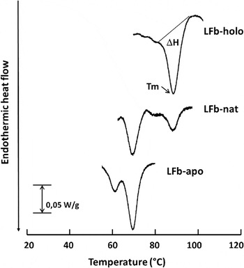
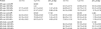
The results obtained in the present study by DSC analysis of HHP-treated LFb with different degrees of iron saturation are shown in Table 1. The endotherms corresponding to LFb in the apo, native and holo forms treated at 500 MPa are presented in Fig. 1 to illustrate the results of calorimetry. The DSC parameters of HHP-treated LFb were compared with those of non-treated LFb with respect to the main peaks of the endotherms. The endotherms obtained for the apo form of LFb presented two peaks when the protein was subjected to treatments of 500 and 650 MPa. The endotherm corresponding to LFb-apo presented the minor peak with a temperature of maximum absorption of about 61 °C, that is a value close to that reported previously presented in Table 1 as the control (Sánchez et al. Reference Sánchez, Peiró, Castillo, Pérez, Ena and Calvo1992). The value of enthalpy obtained for this first peak is very low, probably due to the presence of a low number of molecules with the native structure and thermoresistance. The second peak appearing in the endotherm, that is not present in the control, showed a temperature of maximum absorption of about 70 °C and a denaturation enthalpy value of around 10 J/g, close to the value corresponding to the reference LF that was 8·60 J/g. The values of the maximum temperature and denaturation enthalpy of the second peak were very similar for LFb in solution treated at the three levels of pressure. The appearance of the second peak in the endotherm corresponding to pressure-treated LFb probably indicates that the molecules are aggregated and therefore, the temperature of maximum heat absorption is higher than that of non-treated LFb. LFb in the apo form treated at 400 MPa could be aggregated to a greater extent since the first peak was absent. Similar results were observed by Paulsson et al. (Reference Paulsson, Svensson, Kishore and Naidu1993), though studying the effect of heat treatment on LF structure by calorimetry. They subjected LFb after being heated at 70 °C during 15 s, to DSC and found a shift in the maximum temperature of the endotherm. However, the changes in DSC parameters found in that study were higher than those obtained in the present study, probably due to the greater effect of heat treatment on protein structure compared with high pressure.
DSC thermograms obtained for LFb-apo (a), LFb-nat (b) and LFb-holo (c) treated by high pressure at 500 MPa. The protein concentration was 100 mg/ml. The parameters obtained from the thermograms were the peak maximum temperature (Tm) and the enthalpy change of denaturation (ΔHcal).

DSC thermal parameters obtained in the denaturation of HHP treated LFb in three iron-saturated forms. Heating rate applied was 10 °C/min. Each value represents the mean±sd of three or four replicates

† Onset temperature. ‡Peak maximum temperature. §Enthalpy change of denaturation. n.d.: non-detected. n.det.: non-determined
¶Sánchez et al. (Reference Sánchez, Peiró, Castillo, Pérez, Ena and Calvo1992)
The endotherm obtained for LFb-nat (Fig. 1) showed a maximum temperature at 70 °C for all the high-pressure treatments coincident with that of the respective control. The first peak corresponds to those molecules of lactoferrin that are non-saturated with iron, while the second peak corresponds to iron-saturated LFb molecules. The values of the denaturation enthalpy obtained for LFb-nat at all pressures were about 70–80% lower than that of the control. The second peak of the endotherm presented some differences between the control and the treated samples, the maximum temperature being 3 °C higher when the protein was treated at 400 and 500 MPa, and resulting in a shift from 89 °C to 76·5 °C when the protein was treated at 650 MPa. The values of the denaturation enthalpy for the second peak were very low and similar for the samples treated at the three levels of pressure.
The temperatures of maximum heat absorption for the holo form of LFb treated at different pressures were 4–5 °C higher than that reported for the non-treated protein (Table 1), though the endotherm presented only one single peak as for the non-treated holo form of LFb (Fig. 1). Furthermore, the values of denaturation enthalpy corresponding to the endotherms of LFb-holo were 45–55% lower with respect to the control. The results found in the present study show that the stability of LFb at high pressure followed the order LFb-apo<LFb-nat<LFb-holo. These differences in the stability of LF depending on its iron-saturation are in agreement with the results previously reported for LFb subjected to heat treatment or pulsed electric fields (Sánchez et al. Reference Sánchez, Peiró, Castillo, Pérez, Ena and Calvo1992; Paulsson et al. Reference Paulsson, Svensson, Kishore and Naidu1993; Brisson et al. Reference Brisson, Britten and Pouliot2007; Sui et al. Reference Sui, Roginski, Williams, Versteeg and Wan2010). Taking into account that the conformation of LF is more exposed when is devoid of iron this could explain why that form is more affected by high pressure.
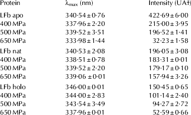
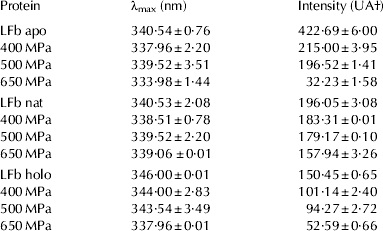
The effect of HHP treatment on the conformation of LFb was also studied by measuring the intrinsic fluorescence emission of tryptophan (Table 2). The three forms of LFb (apo, native and holo) exhibited an emission maximum wavelength (λmax) between 340 and 346 nm. The highest maximum of fluorescence intensity was obtained for the apo form of LFb that can be explained by its more open conformation due to the absence of iron. A similar effect has been also observed for bovine α-la since the maximum of fluorescence intensity increases when calcium is removed from its native form (Wehbi et al. Reference Wehbi, Pérez, Dalgalarrondo, Sánchez, Calvo, Chobert and Haertlé2006). The fluorescence of LFb did not show differences in λmax after HHP treatment with respect to the non-treated protein. However, there was a decrease in the maximum of fluorescence intensity of the treated samples with the increase in the pressure applied, the decrease being greater for LFb-apo than for LFb-nat and LFb-holo. The results obtained in the DSC experiments agree with those obtained in the analysis of fluorescence of LFb-apo after high pressure treatment, since the aggregation of molecules revealed by the appearance of the second peak in the endotherm could explain the decrease in fluorescence intensity. With respect to LFb-nat, the effect of high pressure on structure could be mainly detected after treatment at 650 MPa since there was a shift in the temperature of maximum absorption of the second peak, and also a decrease in fluorescence intensity. LFb-holo was the most resistant to high pressure compared with the other forms, though a certain shift of the maximum temperature of heat absorption and a decrease in fluorescence intensity were observed at all pressures.
Fluorescence spectroscopy data for LFb in three iron-saturation forms after HHP treatment. Each value represents the mean±sd of three replicates
† UA: arbitrary units
The results obtained for the effect of HHP on LFb can be compared with those we obtained in a similar study on the effect of HHP on recombinant human LF obtained from rice (Franco et al. Reference Franco, Castillo, Pérez, Calvo and Sánchez2011). On the whole, the calorimetric study of pressure treated recombinant human LF showed less changes in the thermal parameters than those found for LFb, though the apo form of the recombinant protein did not give any peak after treatment at 600 MPa. For both bovine and human recombinant LF, there was a decrease in the maximum of fluorescence intensity with the increase of pressure applied and that decrease was higher for the apo form.
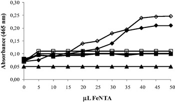
The iron retention capacity of LFb-nat and LFb-holo after HHP treatments was determined by ICP-AES. Iron was not detected in the aqueous phase of any of the high-pressure treated samples after ultrafiltration. Consequently, HHP treatment at 400, 500 and 650 MPa, does not seem to produce a release of the iron bound to LFb. This finding would explain that the change in the structure of LF molecule caused by high pressure can have an effect on tryptophan exposure and consequently on fluorescence, though it would not affect the forces involved in the iron binding site. With respect to the iron saturation capacity (Fig. 2), there was a similar behaviour of LFb-apo subjected to 400 MPa compared with the non-treated protein. However, when HHP treatment increased to 500 and 650 MPa, LF lost completely the iron binding capacity, similarly to the effect found for LF treated at 85 °C. These results suggest that the change in the structure of LF under treatments at pressures of 500 MPa and higher can impede the accessibility of iron to the metal-binding site.
Uptake of Fe (III) from Fe-NTA in LFb-apo after different HHP and heat treatments. The protein concentration is 2 mg/ml in PSS, pH 7·4. Symbols: (⧫) LFb apo, (■) 72 °C 15 s, (▲) 85 °C 2 min, (◊) 400 MPa, (□) 500 MPa, (△) 600 MPa.

The susceptibility of LFb to high pressure is similar to that found for human recombinant LF from rice, as both proteins maintained the iron-binding capacity only after treatment at 400 MPa (Franco et al. Reference Franco, Castillo, Pérez, Calvo and Sánchez2011). Some authors have also studied the effect of non-thermal treatments such as pulsed electric field processing (Sui et al. Reference Sui, Roginski, Williams, Versteeg and Wan2010) and they showed that iron was not released from the protein in a relatively low ionic strength environment. However, when those authors applied several thermal treatments to native bovine LF, they found that under treatment at 90 °C during 10 min there was a 20% release of iron from the protein. Similar results were found by Kawakami et al. (Reference Kawakami, Tanaka, Tatsumi and Dosako1992) who reported a 15–30% iron release from native LF, after treatment at 90 °C for 4 min.
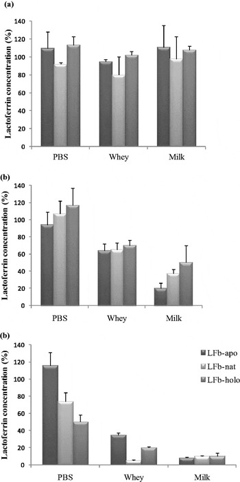
The effect of high pressure on LFb structure was also studied by determining the concentration of immunoreactive protein in its three iron-saturated forms, since a decrease of reactivity with its specific antibodies is related to a loss of the conformational structure. The immunoreactivity of LFb was determined by ELISA after HHP treatment in PBS, whey and milk (Fig. 3a–c). The percentage of immunoreactive LFb after applying 400 MPa (Fig. 3a) was relatively high for all forms of LFb (apo, native and holo) in the three treatment media. When the pressure increased to 500 MPa (Fig. 3b), the immunoreactivity of LFb decreased around 30–40% when treated in whey, the concentration of LFb-apo treated in milk showed a decrease of 80% and no decrease was observed for the concentration of any LFb form when treated in PBS. After treatment at 600 MPa, the immunoreactivity of LFb was maintained high only in PBS, compared with that treated in whey and milk (Fig. 3c). Surprisingly, LFb-apo maintained its whole immunoreactivity in PBS, while for LFb-nat and LFb-holo around 30 and 50% of immunoreactivity was lost, respectively. When LFb was treated in whey and milk at 600 MPa, the immunoreactivity practically disappeared, except for the apo form in whey that maintained around 30% of immunoreactivity.
Immunoreactivity of bovine lactoferrin analysed by ELISA after treatments of 400 MPa (2a), 500 MPa (2b) and 600 MPa (2c) in three different media, expressed as percentage of the initial immunoreactivity. Each value represents the mean±sd of 3 replicates from 3 independent experiments.

The same influence of the treatment media was observed in bovine LF resistance to heat (Sánchez et al. Reference Sánchez, Peiró, Castillo, Pérez, Ena and Calvo1992) and pressure treatment (Mazri et al. Reference Mazri, Sánchez, Ramos, Calvo and Pérez2012a), as in those studies it was found to be more sensitive when treated in milk than in phosphate buffer. It is possible that LFb after being denatured by heat or high pressure treatments, could form aggregates with other milk proteins. Therefore, the interaction of LFb with its antibodies would be hampered because part of its epitopes could be hidden by milk proteins, not being accessible. There are precedents for this where HP has been shown to cause binding between casein and β-lg (Huppertz et al. Reference Huppertz, Fox and Kelly2004).
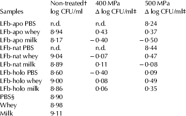
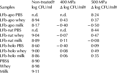
The two mechanisms used by LF to exert antibacterial activity are the capacity to sequester iron and the adsorption to bacterial surface, depending on the microorganism. It has been shown that LF may have specific interaction with many bacteria, including Esch. coli and other members of the family Enterobacteria (Naidu et al. Reference Naidu, Erdei, Czirók, Kalfas, Gado, Thoren, Forsgren and Naidu1991; Kishore et al. Reference Kishore, Erdei, Kalfas, Forsgren and Naidu1992). In the present work, the antibacterial activity of LFb in the three iron saturation forms after being subjected to 400 and 500 MPa treatments was studied on Esch. coli O157:H7. LFb showed antibacterial activity only in its apo and native forms in PBS, the reduction being of about 9 log cycles CFU/ml (Table 3). The antibacterial effect was not exerted by LFb-apo and LFb-nat in milk and whey or by LFb-holo in all media. After treatment at 400 MPa, LFb-apo and LFb-nat maintained the same antibacterial activity as the non-treated protein, being effective only when tested in PBS. However, when pressure increased to 500 MPa, the antibacterial activity of LFb-apo and LFb-nat was completely lost. These results agree with those obtained from the previous experiments of the effect of high pressure on LFb iron-binding ability and indicate that the change in the structure of LFb caused by pressures above 500 MPa can impede the entrance of iron to the metal-binding site, thus neutralising the antibacterial activity of LFb.
Antibacterial activity of bovine lactoferrin (2 mg/ml) in three different media after 400 and 500 MPa treatments against Esch. coli O157:H7. All data derived from means of 3 replicates from 3 independent experiments
† Antibacterial activity of samples before HHP treatment
‡ The antibacterial activity is expressed as the decrease in log units (log CFU/ml before HHP-log CFU/ml after HHP) after 24 h of incubation at 37 °C
§ The control wells contained the bacterial suspension and the treatment media in the same proportion as the test samples to evaluate if there was an effect due to dilution of the culture medium
The initial count of bacterial suspension in the test wells was about 0·5×104–105 CFU/ml
n.d.: <1 colony detected per plate
Our previous studies on recombinant human LF from rice also showed that treatment of the apo form at 400 MPa for 15 min preserved its antibacterial activity against Esch. coli O157:H7 (Franco et al. Reference Franco, Castillo, Pérez, Calvo and Sánchez2011). Likewise, Masschalck et al. (Reference Masschalck, Van Houdt and Michiels2001) reported that LF showed antibacterial activity at low concentration (0·5 mg/ml) against some species of Salmonella and Escherichia when subjected together, bacteria and protein, to high pressure treatment at 400 MPa for 15 min.
The application of high pressure treated LF as natural preservative in foods still presents some controversy. Thus, Del Olmo et al. (Reference Del Olmo, Calzada and Nuñez2011) have recently studied the effect of LFb and its derivatives against Gram-positive bacteria using HHP (400 MPa for 10 min) in meat, and they found a limited potential of LFb for pathogen inactivation. However, other studies have shown a synergistic microbial inactivation in milk by combination of high pressure and natural antimicrobial proteins, such as LF and lysozyme, or peptides such as nisin, against Gram-positive and Gram-negative species (Black et al. Reference Black, Kelly and Fitzgerald2005; Iucci et al. Reference Iucci, Patrignani, Vallicelli, Guerzoni and Lanciotti2007). Consequently, more studies are needed to evaluate the right combinations of LF, iron-saturation, treatment media and high pressure conditions to have antimicrobial activity on different pathogens before application in food and functional products. Anyway, we can say that high pressure can be considered as a good alternative to heat treatment for LFb.
Conclusions
The use of high pressure partially affects the structure of LFb, the effect being more pronounced at 500 and 650 MPa. After treatment at 400 MPa, LFb retains many of its structural characteristics intact. Therefore, a treatment of 400 MPa for 15 min can be recommended for treatment of products supplemented with LFb, especially because its apo and native forms subjected to those conditions maintain their antimicrobial activity.
This study was supported by a CICYT grant (AGL2005-05494) from the Ministerio de Ciencia y Tecnología (Madrid, Spain), by Social European Fund and by Universidad de Zaragoza. Indira Franco (from Gorgas Memorial Institute for Health Studies, Panamá) was awarded with a research-fellow grant from Secretaría Nacional de Ciencia y Tecnología (SENACYT) of Gobierno de Panamá.