Introduction
Livestock embryo production in in vitro systems has been highlighted due the emergence of embryo stem cells (ESC). ESC potency and the wide prospects for application have been recognized in medicine, fundamental research fields and markets due to ESC totipotency or pluripotency and self-renewal. Research in ESC isolation and line foundation has progressed, such as for human ESC (Thomson & Marshall, Reference Thomson and Marshall1998; Shamblott et al., Reference Shamblott, Axelman, Wang, Bugg, Littlefield, Donovan, Blumenthal, Huggins and Gearhart1998; Thomson et al., Reference Thomson, Itskovitzeldor, Shapiro, Waknitz, Swiergiel, Marshall and Jones1998; Park et al., Reference Park, Kim, Oh, Moon, Roh, Kim and Yoon2003), non-human primates (Pau & Wolf, Reference Pau and Wolf2004) and livestock, such as bovine (Cherny et al., Reference Cherny, Stokes, Merei, Lom, Brandon and Williams1994; Mitalipova et al., Reference Mitalipova, Beyhan and First2001), porcine (Chen et al., Reference Chen, Shiue, Bertolini, Medrano, BonDurant and Anderson1999), equine (Saito et al., Reference Saito, Ugai, Sawai, Yamamoto, Minamihashi, Kurosaka, Kobayashi, Murata, Obata and Yokoyama2002) and in experimental animals (for example, mouse, rat, zebrafish, rabbit and so on) ESC lines (Martin, Reference Martin1981; Evans & Kaufman, Reference Evans and Kaufman1981; Doetschman et al., Reference Doetschman, Williams and Maeda1988; Graves and Moreadith Reference Graves and Moreadith1993; Sun et al., Reference Sun, Bradford, Ghosh, Collodi and Barnes1995; Hong et al., Reference Hong, Winkler and Schartl1996). In addition, some progress has been achieved in ESC directional induction and differentiation in human and mice (Lavon & Benvenisty, Reference Lavon and Benvenisty2003; Watabe et al., Reference Watabe, Nishihara, Mishima, Yamashita, Shimizu, Miyazawa, Nishikawa and Miyazono2003; Schmitt et al., Reference Schmitt, de Pooter, Gronski, Cho, Ohashi and Zuniga-Pflucker2004). However, only a few papers on livestock ESCs have been published because of species specificities, culture conditions in vitro and the difficulty of blastula sources, particularly for sheep ESCs. However, sheep are an important livestock around the world. Besides its conventional usages, sheep, in recent years, have become an important research tool for new and advanced technologies, such as transgenic bioreactors, cloning livestock and as a source of bio-products, for all of which sheep ESCs are important platforms. Therefore, the foundation of ovine ESC lines is currently a hot topic for focus in these fields. Since natural blastula material is difficult to isolate, ‘test-tube embryos’ – embryos produced in in vitro systems – are of increasing interested by researchers.
Our embryo in vitro production system (IVP) includes three steps for oocyte in vitro maturation (IVM), in vitro fertilization (IVF) and embryos in vitro cultivation (IVC), which has been applied in assisted reproduction techniques and bio-techniques. ‘Test-tube baby’ emergence by IVF provides a solution for procreation obstacles in humans. For animals and livestock that are valuable and have a long regeneration time, many offspring can be born by the IVP system and embryo transplantation, aiming at shortening regeneration intervals and increasing the number of progeny (Armstrong, Reference Armstrong, Kotaras and Earl1997).
Blastulas, as an ESC source, have been paid great attention in emerging ESC studies. Blastula preparation by in vitro production systems displays great advantages, particularly for solving litter size and season limitation in large livestock. Armstrong (Reference Armstrong, Kotaras and Earl1997) reported that there are no significant differences between natural mature eggs and ones derived from in vitro maturation and between natural blastulas and those derived from IVF/IVC in cattle and sheep, a fact that is strong supported by sheep ESC isolation from blastulas and in vitro production. In order to investigate a route for ovine ESC isolation and for cell line foundation from blastulas by in vitro production, we have established a production system for ovine blastulas in vitro. Sufficient blastulas can be obtained for the isolation of ES-like cells and ESC-like colonies were passaged several times and identified by AKP staining and morphology.
Materials and methods
Collection of ovine ovaries and testicles
Fresh ovine ovaries and testicles were collected at a local slaughterhouse. Ovaries were kept in 37–39 °C in saline solution (0.9% NaCl, 250 µg/ml penicillin, 250 µg/ml streptomycin); testicles were stored in ice water. Both samples were carried to laboratory within 3 h.
Culture in vitro of cumulus oocyte complexes (COC)
The procedure for COC maturation in vitro has been documented in the literature (Chen et al., Reference Chen2000; Qin et al., Reference Qin, Li, Tan, Luo, Tian and Li2001; Tremoleda et al., Reference Tremoleda, Stout, Lagutina, Lazzari, Bevers, Colenbrander and Galli2003) and modified. The main procedure was as follows: on arrival at the laboratory, for sterilization ovaries were washed with 20–30 ml 75% ethanol by shaking for 3–5 s (ovaries in ethanol for a longer time would lead to COC death) and washed with normal saline plus 100 IU/ml penicillin and 100 µg/ml streptomycin three times and were transferred to M2 medium (Nagy et al., Reference Nagy, Gertsenstein, Vintersten and Behringer2003) supplemented with 10 µg/ml of heparin (Sigma, H-3393). Cumulus–oocyte complexes were released by scrapping the inside of the follicles with a sharp knife and transferred using an anatomical lens, Cells were washed five times with maturation medium: M199 (Hyclone) plus 10% FBS (Hyclone); penicillin 100 IU/ml (Hyclone); streptomycin 100 µg/ml (Hyclone); glutamine 2 mM (Hyclone); NEAA (non-essential amino acids) 0.1 mM (Hyclone); sodium pyruvate 1 mM (Hyclone); follicle-stimulating hormone (FSH, Ningbo Hormone Products Co. Ltd) 10 µg/ml; luteinizing hormone (LH, Ningbo Hormone Products Co. Ltd) 10 µg/ml; estradiol (E2, Sigma) 1 µg/ml; and cysteine (cys, Sigma) 20 mM; then transferred into pre-equilibrated droplets of maturation medium (at 38.5 °C) under paraffin oil and were incubated for 23 h at 38.5 °C in an atmosphere of 5% CO2 for oocyte in vitro maturation (IVM).
Sperm capacitation and fertilization in vitro (IVF)
The procedure was as reported in the literature (Chen et al., Reference Chen2000; Qin et al., Reference Qin, Li, Tan, Luo, Tian and Li2001) and the main procedure was as follows: SOFM (see Walker et al., Reference Walker, Heard and Seamark1992) and HSOF – SOFM plus HEPES 20 mM (Merk); FBS 2% (Hyclone); and hypotaurine 20 µg/ml (Sigma) – and fertilization solution – SOFM with FBS 15%; hypotaurine 20 µg/ml (Sigma); penicillin 100 IU/ml; streptomycin 100 µg/ml; glutamine 2 mM, EAA 1%; NEAA 0.1 mM – were freshly prepared and pre-equilibrated at 38.5 °C in an atmosphere of 5% CO2. Before 3 h of fertilization, sperm capacitation in vitro was started. A testis was washed quickly in 75% ethanol and transferred into normal saline with 100 IU/ml penicillin, 100 µg/ml streptomycin and washed three times with this solution, then moved into a polystyrene culture dish (60 mm) with 3 ml HSOF and cut with sterile scissors until the solution became a white viscous liquid that contained effluent sperm. The viscous liquid was cultured at 38.5 °C in an atmosphere of 5% CO2 for 30 min, moved into a 10 ml test tube and supplemented with a half volume of HSOF, mixed well and centrifuged at 1000 rpm for 6 min. After removing the supernatant, the sediment was repeatedly washed with HSOF, two or three times. Then, the middle sperm fluid obtained from the sediment was taken to make a large droplet (1 cm diameter) and covered with paraffin oil in a petri dish and incubated at 38.5 °C in an atmosphere of 5% CO2 for 30 min for sperm capacitation in vitro.
After 23 h culture, the first polar body could be observed in mature oocytes, which usually are surrounded by expanded fluffy zona granulosa of twice the oocyte diameter. The oocytes were separated from their cumulus cells by washing in fertilization solution and transferred into droplets of pre-equilibrated fertilization solution at 10 to 20 eggs per droplet. Fertilization in vitro (IVF) was carried out by adding 3–10 µl of the capacitated sperm liquid into each oocyte droplet and culturing in an incubator in 5% CO2 at 38.5 °C for 16 to 22 h.
Embryo in vitro culture (IVC)
After 16–22 h co-incubation with sperm, the fertilized eggs were washed with IVC medium (M199 plus 10% FBS, penicillin 100 U/ml, streptomycin 100 µg/ml, glutamine 2 mM, NEAA 0.1 mM, sodium pyruvate 1 mM; pre-equilibrated at 38.5 °C) to remove sperm. Then the eggs were cultured in IVC medium droplets at 38.5 °C in an atmosphere of 5% CO2 for 6–8 days. The medium was replaced at 2-day intervals. State of fertilization, cleavage and embryonic development were observed under inverted phase-contrast microscopy (Olympus) from the second day of fertilization.
Isolation of inner cell mass and culture of ES-like cells
Feeder cell layers were prepared by a regular procedure, as in the literature (Spector et al., Reference Spector, Goldman and Leiwand1998) and the main procedure was as follows: ovine embryonic fibroblasts (OEF) (passage 5) were revived and grown in DMEM (Hyclone) plus 10% FBS (Hyclone), penicillin 100 IU/ml, streptomycin 100 µg/ml, and glutamine 2 mM. One day before ICM isolation, the fibroblast cells were treated with mitomycin C (Sigma) at 95% cell confluence and collected with trypsin–EDTA (Hyclone) and transferred to droplets, covered with mineral oil and incubated in 5% CO2 at 38.5 °C. On the second day, feeder droplets were washed twice with DMEM and ES-DMEM added: DMEM plus 20% FBS, penicillin 100 IU/ml, streptomycin 100 µg/ml, glutamine 2 mM, sodium pyruvate 1 mM, β-mercaptoethanol 0.1 mM (Sigma), LIF 1000 U/ml (r-mLIF, Chemicon International), SCF 1000 U/ml (rhSCF, US Biological). Blastulas were formed after 6–8 days of culture, removed from IVC medium and washed three times with PBS, then zona pellucida were removed by pronase (Sigma) and cells were transferred quickly into DMEM and again washed six times with DMEM. The blastulas were moved to the feeder droplets and cultured with ES-DMEM for 6–8 days, or longer, until ES-like cell colonies appeared (G0 generation), while the medium was replaced at 2-day intervals.
Passage and AKP staining of ES-like cell colonies
ICM grew on OEF feeder cells for about 1 week and formed colonies. The colonies were dispersed into small cell mass by trypsin digestion after washing three times with PBS and the reaction was stopped by ES-DMEM, then cells were delivered into fresh OEF feeder cell droplets and cultured continually until new colony generation. ES-like cell colonies were identified by colony morphology and AKP staining following the AP-Red kit protocol (AP-Red kit, Beijing Zhong-Shan Biotechnique Co.). Generally, the positive red signal would appear after staining for 10–30 min at room temperature.
Results
Oocytes from IVM and fertilization in vitro
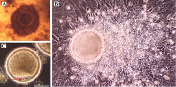
There are varied oocyte morphologies among different mammal species. However typical standards of oocyte maturation usually are similar, such as expanded fluffy cumulus oophorus cells, germinal vesicle breakdown (GVBD) and the first polar body released. Because livestock oocyte plasma is generally dark, GVBD is hard to be seen without orcein staining (Chen et al., Reference Chen2000). We did observe GVBD of the mature oocytes derived from IVM by orcein staining (data not show). However the oocytes died soon after the staining and the experiment was stopped. So we used the other two standards, including first polar body release and cumulus oophorus expansion, at least twice (Fig. 1b, c), to determine oocyte maturation, by which we could get not only oocyte maturation data, but also live eggs that could continue to the next fertilization step in vitro. The first polar body could be observed in mature oocytes without zona granulosa. The mature oocytes were usually surrounded by expanded fluffy zona granulosa with twice the oocyte diameter. The ratio of oocyte maturation currently reached 65%. In addition, we found that for very young oocytes if we prolonged IVM for an additional 1 day we could also obtain mature eggs. However ova nuda were discarded due to their differential status. We tried two kinds of medium for IVM, M199 and DMEM/F12 and also compared these with or without progesterone. The results suggested that there was no significant difference between the media.
Ovine oocyte maturation by IVM. (a) An immature COC just isolated from an ovary. (b) A mature oocyte with twice the zona granulosa extension after IVM. (c) A mature oocyte with an extruded first polar body (indicated by an arrow) after IVM. The bar represents 35 µm.

A comparison of BO medium, mDM, TALP, RSCM and SOF was done for sperm capacitation (data not shown). The results suggested that the SOF system was best for sperm capacitation and was used in the later experiments. In addition, sperm quality is closely related to testis quality and its transfer temperature. Those testes can still be used when kept in an ice bath for 2–3 days; in contrast, when testes were kept at the normal temperature, sperms could not be capacitated.
Fertilized eggs developed in vitro into blastulas
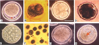
Most ovine fertilized eggs, after culture in vitro for about 24 h postfertilization, were able to extrude second polar bodies (Fig. 2a), or even three polar bodies (data not shown), which was seen clearly under a phase-contrast microscope. Among the three polar bodies, two probably derived from division of the first polar body and another from the second division of a fertilized egg (Chen et al., Reference Chen2000). Cleavage phenomenon could be observed after the second day following fertilization (Fig. 2b–f). Two-cell embryos occurred at 20–24 h postfertilization and most of these were in a uniform rhythm. Blastocysts appeared at the sixth to eighth day (Fig. 2f, g), with a thin pellucid zone (ZP) for some. We did not observe blastulas hatching spontaneously, which is distinct from the natural process in vivo. Ovine eggs and blastulas look very like bovine ones in morphology and pigmentation (Chen et al., Reference Chen2000), but are clearly different to the structures of mouse eggs and embryos. The blastula ratio generally can reach more than 50% (the fertilized eggs as the denominator, Table 1).
Ovine embryos developed in in vitro culture. (a) 1-cell zygote, a red arrow indicates a second polar body. (b) 2-cell embryo. (c) 3-cell embryo. (d) 4-cell embryo; (e, f) Morulas (g) A blastula. (h) A mature blastula. The red arrow indicates ICM. Both the white bar (a–e, g, h) and the blue bar (f) represent 40 µm.

Development of in vitro ovine embryos

In addition, there are still some abnormal and developmentally arrested embryos (Table 1). Most of the arrested embryos stopped at the 4-cell and 8-cell stages, which is consistent with ‘blockage of early embryonic development’ and usually occurred at the 8–16-cell stage for ovine MZT (Qin et al., Reference Qin, Li, Tan, Luo, Tian and Li2001). There are some, hitherto unknown, reasons for hypoevolutism, such as a few embryos blocked at morula stage after 6–8 days’ culture in vitro.
Primary ESC-like colony isolation and culture
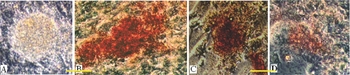
Identification of ES-like cells was by cell colony morphology and AKP staining. A total of 90% of blastulas adhered and grew on OEF feeder cells after removal of the ZP by pronase. ES-like colonies appeared compact and raised cell mass after culture for 2–4 days (Fig. 3a), which means the foundation of primary ES-like colonies (G0). The colonies were passaged to new feeders and new colonies again grew up, until the third generation of ES-like cells (Fig. 3b, c). Meanwhile, a few colonies stained by AKP kit and showed positive alkaline phosphatase activity (Fig. 3b–d). However, we found a few colonies could be sustained for 13 days (Fig. 3d) or longer on the original feeder layer and still show AKP-positive staining, but their morphology was non-adherent and cell numbers reduced and these cells did not reproduce new ESC-like colonies when passaged to new feeder cells, which means that the colonies had differentiated. In fact, most of the ESC-like colonies differentiated and disappeared when cultured longer than 10 days on the original feeder cells, even though LIF and SCF were replaced every 2 days. The results suggest that feeder cells are more important than LIF and SCF in sustaining ES-like cell growth. So for ES-like cell passage, the colonies were generally dissected and delivered to new OEF feeder cells after 4–7 days’ growth. The results indicate that OEFs as feeder cells with LIF and SCF can initiate and support primary ES-like cell growth and the first two passages for ESC-like colonies. However, differentiation phenomena and slow rates of growth are still two problems that cannot be ignored. Other feeders are now being tried to improve ESC growth and to increase their totipotency.
Ovine ESC-like colonies. (a) ICM implanted and grown on OEF feeder cells for 2 days and became a primary ESC-like colony (G0). (b) A primary ESC-like colony grown on OEF feeder cells for 5 days. (c) The third passage of an ESC-like colony at 5 days. (d) A primary ESC-like colony grown on OEF feeder cells for 13 days and beginning to differentiate. (b–d) Stained with AKP reagent. Bar = 50 µm in (a, b). Bar = 30 µm in (c, d).

Discussion
Since the 1980s, reports on livestock IVP embryos has been accumulating and have founded a basis for embryo IVP and ovine blastula sources (Cheng & Polge, Reference Cheng and Polge1986; Yoshida, Reference Yoshida, Ishizaki and Kawagishi1990; Walker et al., Reference Walker, Heard and Seamark1992). Based on the data, using fresh ovaries and testes from a local slaughterhouse, we have modified the ovine embryo IVP system and have proved it suitable for supplying blastocysts for ICM isolation, therefore ovine blastocyst source will not be a limit or a barrier to ESC isolation. However, whether each embryonic IVP experiment succeeds, or not, is greatly dependent on the quality of ovaries and testes used each time, so the collection of ovaries and testes is a key step that requires technicians who have extensive collecting experience and accurate isolation of the sheep material.
Another problem is how to promote rapid ES-like cell growth but meanwhile control the random cell differentiation. We found in this study, ES-like cells grew slowly after the third passage and easily disappeared. There are two probable reasons for this. Firstly, OEF may not be the most suitable feeder cells for ovine ESC growth after several passages, though they are better than the mouse SNL cell line for ovine blastocyst adherence on feeder layers and initiating primary ESC-like colony growth (data not show). Secondly, perhaps only a few pluripotent cells were present in IVP blastocysts (Boquest et al., Reference Boquest, Abeydeera, Wang and Day1999), as it is hard to get vigorous population growth on OEF feeder cells, so that ES-cells cannot proliferate quickly but differentiate or die due to unknown factors.
In summary, we demonstrated that the modified IVP system can supply blastocysts for ICM isolation, from which ovine primary ESC-like colonies can be obtained and passaged several times on OEF feeder cell layers. By this system, the rates of oocyte maturation and blastulas reached 65 and 50% respectively and provided sufficient blastulas for ICM isolation. However, we could not generate immortalized ES-cell lines by this method, so it will be necessary to improve culture conditions and obtain better feeder cells for ovine ESC growth.
Acknowledgements
We thank Professor Miao Du, Dayuan Chen and Ruizhen Zheng for useful discussion and suggestions. We thank Dr Jiaxin Zhang and Beijing Glorious Land Agricultural Company Ltd for supporting the application and initiation of the project. We thank students Jiazhong Duan, Tao Ning, Yong Li and Peiwu Qin for transferring ovine fresh samples weekly. This work was supported by grants from the Beijing Natural Science Foundation (no. 5021002) and the CAS Leading Field Programme of Knowledge Innovation.