Introduction
Positron emission tomography-computed tomography (PET-CT) with fluorine-18 fluorodeoxy-D-glucose (‘18F-FDG’) has a major role in the investigation of patients with head and neck cancer.Reference Purohit, Ailianou, Dulguerov, Becker, Ratib and Becker 1 Cancer cells and normal cells metabolise fluorodeoxy-D-glucose, a glucose analogue, in direct proportion to their glycolytic rates. As tumour cells have higher glycolytic rates than normal cells, this increased uptake can assist in the detection of cancers, particularly in the setting of an unknown primary presenting as metastatic cervical lymph nodes.Reference Purandare, Rangarajan and Shah 2 , Reference Subramaniam, Truong, Peller, Sakai and Mercier 3
Fluorine-18 fluorodeoxy-D-glucose is not a tumour-specific tracer and can also accumulate in benign tumours, inflammatory conditions and during normal physiology, for example when speaking and within brown fat (prevalent in the neck). Benign, non-physiological fluorine-18 fluorodeoxy-D-glucose uptake has been seen in 25 per cent of whole-body PET-CT examinations, and, therefore, fluorine-18 fluorodeoxy-D-glucose uptake can mimic malignant tumours.Reference Metser, Miller, Lerman and Even-Sapir 4 There can be normal physiological glucose uptake in the tongue, pharyngeal musculature and larynx if the patient talks, coughs or chews after injection of the isotope.Reference Heller, Meltzer, Fukui, Rosen, Chander and Martinelli 5 This uptake can vary in intensity, but it is generally symmetrical.Reference Purandare, Rangarajan and Shah 2
Asymmetrical increased fluorodeoxy-D-glucose uptake in the larynx should raise suspicion of primary neoplastic or inflammatory vocal fold pathology. Impaired movement or paralysis of a vocal fold can also cause asymmetrical increased uptake on the contralateral side.Reference Heller, Meltzer, Fukui, Rosen, Chander and Martinelli 5 Referral to an ENT team for laryngoscopy is recommended in cases of asymmetrical uptake on PET-CT, to assess for pathology and function in the context of asymmetrical glucose uptake of fluorine-18 fluorodeoxy-D-glucose.
In essence, PET-CT scan interpretation difficulties are common in the head and neck, which can produce false-positive results. This audit aimed to investigate patients detected as having abnormal vocal fold uptake on fluorine-18 fluorodeoxy-D-glucose PET-CT.
Materials and methods
Positron emission tomography-computed tomography scans were identified between January 2015 and March 2016 where reports contained the words ‘vocal cord’ ‘laryngeal’ or ‘larynx’. The PET-CT scans were found on the Trust’s picture archiving and communication system. These scans were filtered, and scan reports that mentioned any evidence of unilateral vocal fold uptake or bilateral abnormal uptake, paralysis, or vocal fold pathology were analysed.
Patients’ hospital notes were reviewed, and from these the following criteria were investigated: indication for the PET-CT scan; any known vocal fold paralysis or pathology prior to the PET-CT scan; follow up in the ENT department after the PET-CT scan and results of flexible nasendoscopy; and documented radiology advice for ENT follow up based on the scan report and whether this was implemented by the clinical team.
Patient preparation and scan protocol
All patients were nil by mouth 6 hours prior to the injection of fluorine-18 fluorodeoxy-D-glucose. The PET-CT imaging was performed 1 hour after injection, with patients having been instructed to relax and not speak. Scans were acquired using a standard protocol on a Siemens 64-slice PET-CT scanner from the skull base to the upper thighs, with or without dedicated head and neck acquisition, without intravenous contrast.
Scan interpretation
Each scan was originally interpreted by two consultants experienced in PET-CT. Scan reports were re-reviewed by a consultant radiologist with 11 years of PET-CT experience. The results were compared to the clinical notes, including biopsy and histology, when available.
Results
A total of 46 patients were identified between January 2015 and March 2016. These patients were flagged as having abnormal vocal fold uptake on PET-CT. Patient age ranged from 22 to 81 years.
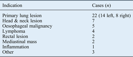
The indications for PET-CT on the clinical information or radiology request forms are outlined in Table I.
Indications for PET-CT on clinical information or radiology request forms

PET-CT = positron emission tomography-computed tomography
In primary lung cancers, metastatic nodal involvement of the left recurrent laryngeal nerve is more common, because of its longer course. Therefore, vocal fold paralysis detected on a PET scan is likely due to left recurrent laryngeal nerve involvement by left-sided lung lesions. Impaired movement or paralysis of the vocal fold can cause asymmetrical increased uptake on the contralateral side.
Known vocal fold pathology
Fourteen of 46 patients were known to an ENT department prior to the PET-CT scan. Eight patients were referred for the PET-CT scan by ENT following laryngoscopy, and six patients had been treated previously for head and neck pathology. Eight of the 14 patients had vocal fold paralysis and 6 patients had vocal fold tumours.
Patients followed up by ENT
Twenty-three patients were not followed up by ENT following scanning. Of these patients, the reporting radiologist recommended ENT follow up for six patients, which was not carried out. The remaining 23 patients (50 per cent) were followed up by ENT after PET-CT scanning and these patients make up the study group.
Radiologist's follow-up advice
Of the 23 patients followed up by ENT, the reporting radiologist recommended ENT follow up for 7 patients. Of these, three patients were already known to the ENT department and four patients were subsequently referred to ENT.
True- or false-positive scans
A PET-CT scan was considered true-positive in patients with verified vocal fold abnormality (either tumour or paralysis) before or after the scan, which was visualised by laryngoscopy. The PET-CT scan was considered false-positive if paralysis or pathology was not seen on laryngoscopy.
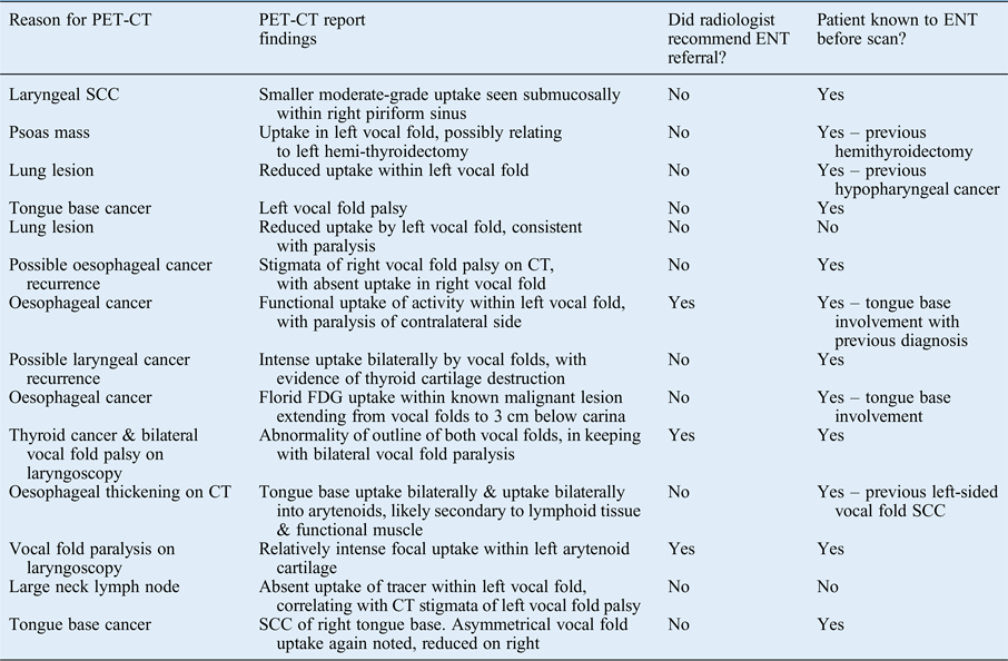
Of the 23 patients who underwent PET-CT and flexible laryngoscopy, 14 had true-positive PET-CT scan results. Twelve of these patients had a known head and neck pathology. The other two patients were referred to ENT based on their PET-CT findings, and were found to have unilateral vocal fold paralysis, as outlined in Table II.
Patients with true-positive FDG uptake with vocal fold pathology or paralysis on laryngoscopy

FDG = fluorodeoxy-D-glucose; PET-CT = positron emission tomography-computed tomography; SCC = squamous cell carcinoma; CT = computed tomography
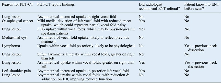
Nine patients had false-positive scan results. They were referred to ENT following unilateral uptake on the PET-CT scan. All nine patients underwent laryngoscopy in the out-patient department (Table III).
Patients with false-positive FDG uptake referred to ENT with normal vocal folds on laryngoscopy

FDG = fluorodeoxy-D-glucose; PET-CT = positron emission tomography-computed tomography
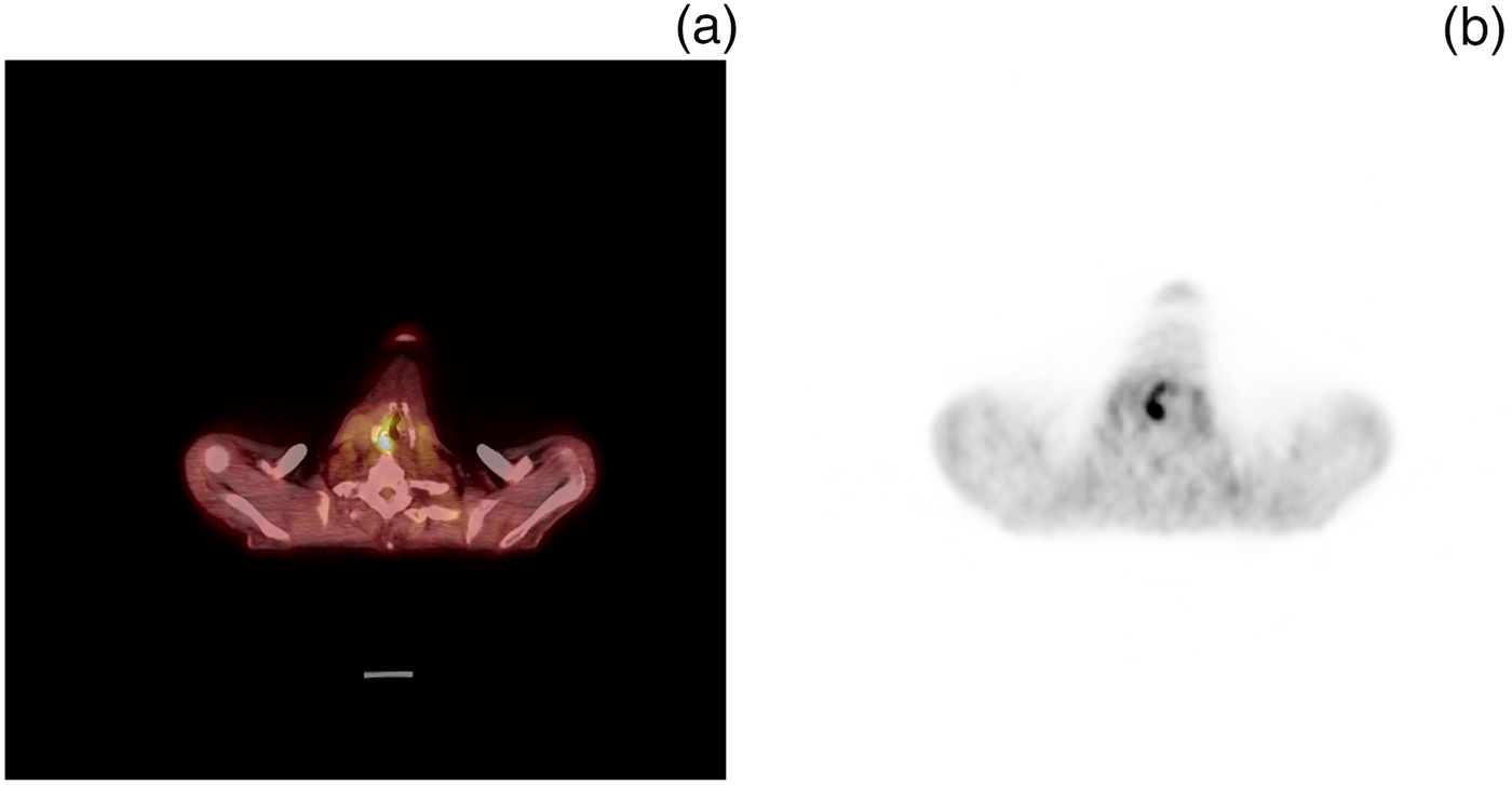
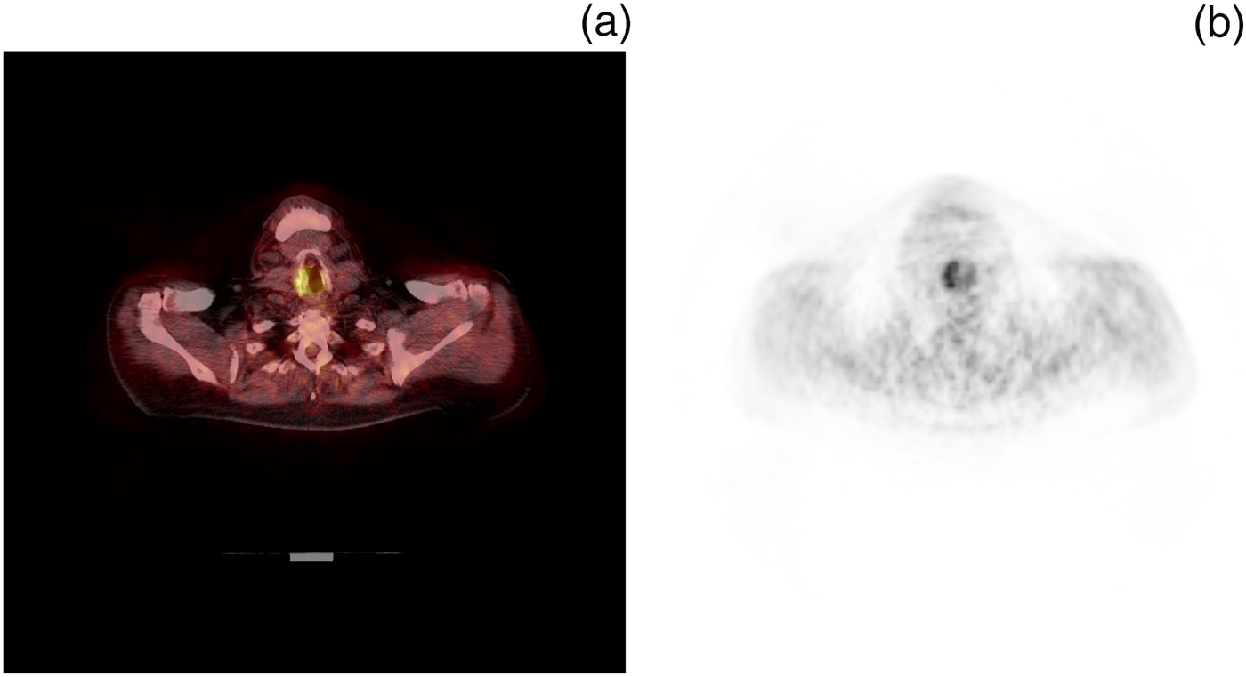
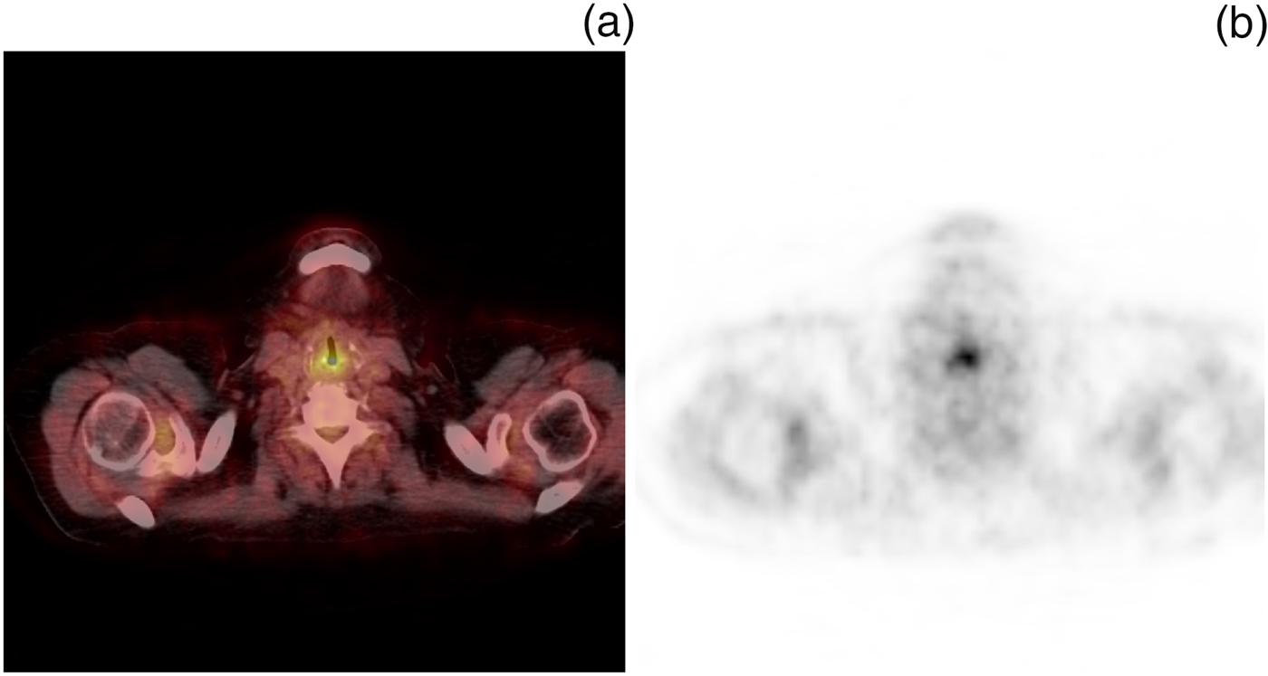
Figures 1–3 show PET-CT vocal fold images of true-positive, false-positive and true-negative findings, respectively.
True-positive result on positron emission tomography (PET)-computed tomography: patient with tongue base cancer, with marked differential vocal fold uptake greater on right than left on (a) fused and (b) PET-only image, with left vocal fold paralysis found on flexible laryngoscopy.

False-positive result on positron emission tomography (PET)-computed tomography: patient with increased uptake in right vocal fold on (a) fused and (b) PET-only image, with normal laryngoscopy. Patient was confirmed to have normal nasendoscopy findings in clinic.

True-negative result on positron emission tomography (PET)-computed tomography: patient reported as speaking during scanning, with symmetry of increased uptake at the cricoarytenoid junction on (a) fused and (b) PET-only image. The reporting radiologist noted that the patient was speaking, and laryngoscopy findings were normal.

Discussion
This audit aimed to investigate patients reported to have abnormal vocal fold uptake of fluorodeoxy-D-glucose during PET-CT scanning. The true- and false-positive rates of these scans were analysed. This work will add to the literature by outlining how ENT consultants can manage patients referred to them for suspected increased fluorine-18 fluorodeoxy-D-glucose vocal fold uptake. There were no other similar works found on a literature search for true- or false-positive laryngeal fluorine-18 fluorodeoxy-D-glucose uptake on PET-CT imaging.
Vocal fold paralysis is mainly the result of traumatic, neurological or malignant causes. Before the 1980s, the main cause of bilateral true vocal fold palsy was iatrogenic, occurring during surgery; however, because of improvements in surgical techniques, the incidence of recurrent laryngeal nerve injury has now decreased.Reference Lee, Ramaswamy, Lilien and Nathan 6 One of the more common causes of unilateral vocal fold paralysis is extralaryngeal malignancy, most commonly a primary lung tumour.Reference Lee, Ramaswamy, Lilien and Nathan 6 This was supported in our series; most of the patients with abnormal vocal fold uptake on PET-CT scan had a primary lung lesion (22 out of 46). Nevertheless, the majority of patients were not referred for ENT follow up, perhaps because of poor prognosis.
True-positive scan results
The second largest subgroup of patients referred for PET-CT scan were seven patients with known head and neck cancers (Table II).
Fourteen patients in total had true-positive PET-CT scans. Of this group, 12 (86 per cent) were already known to have head and neck pathology by the ENT department. Only two patients in the cohort were referred to ENT based on PET-CT findings and were found to have unilateral vocal fold paralysis that was previously unknown. Hence, most patients referred to ENT did in fact have head and neck pathology, but the vast majority of these patients were already known to the ENT department.
False-positive scan results
Nine of the 23 patients (39 per cent) referred to ENT had false-positive scan results. They were referred to ENT following abnormal uptake on the PET-CT scan. Four of these patients were recommended to have laryngoscopy by the PET-CT reporters. These patients underwent laryngoscopy in the out-patient department, which showed no abnormality.
Other series have indicated the main cause of false-positive fluorodeoxy-D-glucose uptake in PET-CT scans to be increased physiological uptake of fluorodeoxy-D-glucose with normal vocal fold use. The site of laryngeal uptake differs depending on the activity of intrinsic laryngeal muscles, and on variation in adduction or abduction of the vocal folds.Reference Kostakoglu, Wong, Barrington, Cronin, Dynes and Maisey 7 The amount of glucose taken up in muscles is directly proportional to the muscle work. This is especially a problem in the head and neck, where there are many small muscle groups at work during swallowing and speech.Reference Fujimoto, Itoh, Tashiro, Yamaguchi, Kubota and Ohmori 8
False-positive PET-CT scans can initiate possible redundant investigations and cause patient anxiety. However, in this series, 23 patients were reported to have abnormal uptake of fluorodeoxy-D-glucose on vocal folds, but were not referred for subsequent ENT referral. From a retrospective analysis of patients’ notes, the reasons for this included: likely physiological uptake associated with patients’ swallowing or coughing, advanced disease status, and likely uptake of fluorine-18 fluorodeoxy-D-glucose due to recurrent laryngeal nerve involvement based on known extralaryngeal disease distribution.
Questions raised
The questions raised by this study, and the suggested answers, include the following. First, should the ENT department see all patients with unilateral PET-CT uptake in the larynx? As true-positive rates (61 per cent) were higher than false-positive rates (39 per cent), patients with unilateral laryngeal uptake should be seen for laryngoscopy in the ENT clinic. Second, how would this happen logistically? This would involve direct referral or discussion from the reporting radiologist. Third, 50 per cent of patients with unilateral laryngeal uptake were not referred to ENT; is this an issue? Potentially this is an issue, as patients with laryngeal pathology may not be investigated. However, analysis of the patients’ notes shows that the reasons for non-referral to the ENT team included advanced disease status, or reports of the patient speaking or chewing during the scan.
-
• Positron emission tomography-computed tomography with fluorine-18 fluorodeoxy-D-glucose is important in head and neck cancer investigation
-
• Cancer cells and normal cells metabolise fluorine-18 fluorodeoxy-D-glucose, which is a glucose analogue
-
• Cancer cells have a higher glycolytic rate than normal cells, which increases uptake
-
• Normal cells can mimic cancer cells, especially in the neck, where there are complex small muscle groups and brown fat cells
-
• Uptake of fluorine-18 fluorodeoxy-D-glucose into normal cells can therefore produce false-positive results
-
• True- and false-positive rates of laryngeal uptake were analysed, and management advice was given
Limitations
Out of 46 patients, only 23 (50 per cent) underwent laryngoscopy; therefore, our sample size was limited. Our case data were retrospectively collected, and so for those patients who underwent both tests there was a time delay between the laryngoscopy and PET-CT, which could account for some differences in the results.
Conclusion
Positron emission tomography-computed tomography scanning plays a vital role in the diagnosis of laryngeal cancers, alongside laryngoscopy. Most patients (61 per cent) referred to ENT because of abnormal findings on PET-CT scans had true-positive findings, but the vast majority of these patients were already known to ENT. Asymmetrical fluorine-18 fluorodeoxy-D-glucose uptake should raise suspicion of vocal fold pathology or paralysis, accepting a false-positive rate of approximately 40 per cent. Patients with increased or unilateral laryngeal uptake should be referred or discussed with an ENT team.
Acknowledgements
Thank you to Mr Meredydd Harries for the original paper concept, and for support and advice. Thank you also to Dr Burkill for the expert input of head and neck PET-CT imaging, and advice with the imaging.