INTRODUCTION
Three Urobatis species have been reported for the Gulf of California: U. halleri (Cooper, 1863), U. concentricus Osburn & Nichols, 1916 and U. maculatus Garman 1913 (De La Cruz-Agüero et al., Reference De La Cruz-Agüero, Arellano-Martínez, Cota-Gómez and De La Cruz-Agüero1997). These stingrays present eyes almost as large as their spiracular diameter. The length and width of their disc are very similar, hence, they are commonly known as round stingrays (Castro-Aguirre & Espinosa-Pérez, Reference Castro-Aguirre and Espinosa-Pérez1996).
The round stingray Urobatis halleri is distributed from Monterey Bay, California, to Panama, including the Gulf of California (Babel, Reference Babel1967; De La Cruz-Agüero et al., 1997). This benthic stingray inhabits sandy and muddy bottoms near the coast, mainly associated with bays, lagoons, and estuaries, down to depths of 15 m (Allen & Robertson, Reference Allen and Robertson1994). The diet of U. halleri is primarily composed of benthic invertebrates, such as stomatopods, amphipods, decapods, brachyurids, portunids, and in fewer quantities, polychaetes and fish (Valadez-Gonzalez et al., Reference Valadez-González, Aguilar-Palomino and Hernandez-Vazquez2000).
Several authors have reported malformations in batoids (Castro-Aguirre & Torres-Villegas, Reference Castro-Aguirre and Torres-Villegas1979; Bornatowski & Abilhoa, Reference Bornatowski and Abilhoa2008; Escobar-Sánchez et al., Reference Escobar-Sánchez, Galván-Magaña, Downton-Hoffmann, Carrera-Fernández and Alatorre-Ramírez2008). However, there are no teratologic records describing ocular malformations or black mucus for the round stingray U. halleri. In this context, the aim of this work is to report the first description of ocular malformations, as well as black mucus occurrence in U. halleri in the Gulf of California.
MATERIALS AND METHODS
Using different fishing gear, thirteen adult round stingrays (Urobatis halleri) were captured at La Paz Bay, Baja California Sur, Mexico, in May and June of 2009. The specimens were assessed and deposited in the Ichthyologic Collection at Centro Interdisciplinario de Ciencias Marinas-IPN (Catalogue numbers: CICIMAR-CI 6674, CICIMAR-CI 6675, CICIMAR-CI 6676, CICIMAR-CI 6677 and CICIMAR-CI 6678).
The stingrays were weighed, photographed, and identified to the species level according to Allen & Robertson (Reference Allen and Robertson1994) and Castro-Aguirre & Espinosa-Perez (1996). In addition, the coloration patterns and morphometric measures were recorded, including the distance between the tip of the tail and the centre of the cloaca, and the distance between the tip of the snout and a tangent to the anterior margin of both ocular orbits.
RESULTS
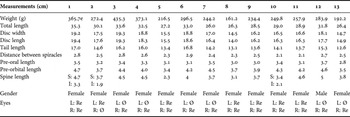
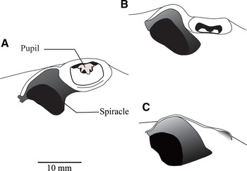
All of the thirteen specimens captured were adults with malformed or absent eyes (anophthalmia), with total lengths between 263 and 353 mm (Table 1). In completely anophthalmic individuals, the eyes were substituted by smooth without reminiscence or folding. The ocular malformations consisted in the lack of the pupils and reduction of the ocular globe (Figure 1).
Morphometric characteristics of thirteen specimens with ocular malformations of Urobatis halleri.

e, eviscerated, L, left, R, right, S, superior, I, inferior, Re, reminiscence, Ø, absent.
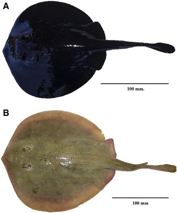
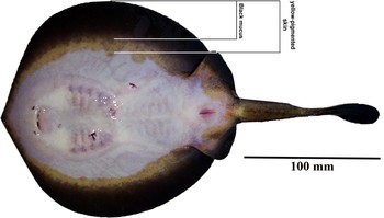
All individuals presented a thick black layer of mucus that completely covered the dorsal region of the disc, tail and caudal fin lobe (Figure 2). This mucus spreads to the ventral region, covering approximately 2/3 of a strip of yellow-pigmented skin that surrounds the disc and portions of the pelvic fins of U. halleri (Figure 3). Underneath the mucus there was a soft skin, which, in the dorsal region, had a greyish or dark brown background, with small yellow dots overlaid, similar to those described by Babel (Reference Babel1967) for U. halleri. In some cases, these dots were closely spaced, forming rosettes. The yellow dots became smaller toward the periphery of the disc.
Dorsal view of Urobatis halleri. (A) Presenting a black, thick mucus; (B) normal.

Side-view of the ocular zone of Urobatis halleri. (A) Normal eye; (B) reminiscent eye; (C) absent eye.

Ventral view of a specimen of Urobatis hallerii presenting black mucus (A) on skin with yellowish pigment (B).

DISCUSSION
Several types of malformations in elasmobranchs have been reported, such as lordosis, scoliosis, bicephalia, cranial malformations, and albinism, among others. Usually, these anomalies are associated with embryos or juveniles which, because of unfavourable environmental pressures, do not reach adulthood (Castro-Aguirre & Torres-Villegas, Reference Castro-Aguirre and Torres-Villegas1979; Heupel et al., Reference Heupel, Simpfendorfer and Bennet1999; Sandoval-Castillo et al., Reference Sandoval-Castillo, Mariano-Meléndez and Villavicencio-Garayzar2006; Bornatowski & Abilhoa, Reference Bornatowski and Abilhoa2008; Escobar-Sánchez et al., Reference Escobar-Sánchez, Galván-Magaña, Downton-Hoffmann, Carrera-Fernández and Alatorre-Ramírez2008).
The periscopic position of the stingray eyes allows them to detect predators, thus any ocular malformation may render them vulnerable to predation (Kalmijn, Reference Kalmijn1971). However, the feeding strategy of U. halleri could explain how the individuals reported here reached adult stages. As an elasmobranch, the round stingray uses its electroreceptor system to detect prey on, or within sediment. In this way, absent or malformed eyes may not directly affect the feeding capability or, therefore, the growth of the individual into adulthood (Kalmijn, Reference Kalmijn1971).
The malformations can be attributed to several causes, one of them being water pollution due to wastewater discharges from urban zones and generating stations (Haaker, Reference Haaker1977; Hoisington & Lowe, Reference Hoisington and Lowe2005), which both occur in La Paz Bay, mainly at the Ensenada and surrounding areas, the same locations as where the specimens were extracted (Mendoza-Salgado et al., Reference Mendoza-Salgado, Lechuga-Deve'ze and Ortega-Rubio2005). Some authors (Heupel et al., Reference Heupel, Simpfendorfer and Bennet1999) also think that lesions, tumours, and parasitic infections can cause these anomalies. However, we did not observe any of these in our specimens, although it is possible that one or more of such causes could have been present in earlier stages of the individual's development. Alternatively a genetic alteration should not be dismissed as a possible cause for the abnormalities observed (Lodi, Reference Lodi1978).
On the other hand, the black coloration of the mucus can be attributed to hyperpigmentation due to overproduction of melanin (Chavin, Reference Chavin1969). It is interesting to note that the pelagic stingray Dasyatis violacea has probably similar dark mucus (Wilson & Beckett, Reference Wilson and Beckett1970). But the prevalence of individuals presenting the malformations described here is notable. Nevertheless, in order to determine the cause, additional studies addressing both the malformed eyes and black mucus are needed.
ACKNOWLEDGEMENTS
We wish to thank the Ichthyologic Collection of CICIMAR-IPN for all the materials provided for this study. We also thank Dr José de La Cruz Agüero for his useful comments and Juan Pablo Partida Marquez for editing the English language version of this document. Last but not least, we thank MSc Luis Burnes Romo for the technical support provided.