INTRODUCTION
In parasitic Platyhelminthes there is a family of calcium-binding proteins which, thus far, have not been found in any other group of organisms. The proteins consist of an N-terminal domain which comprises two EF-hand structures and a C-terminal dynein light chain-like (DLC-like) domain (Russell and Timson, Reference Russell, Timson and Yamaguchi2014). Their physiological function is currently unknown, but it is assumed that they play a role in calcium signalling in the organism. The only known binding partner is a DLC (Hoffmann and Strand, Reference Hoffmann and Strand1997), suggesting that at least some members of this family could be involved in calcium-mediated signalling to the cytoskeleton. Given the central importance of the cytoskeleton to living cells and the uniqueness of this group of proteins, it has been suggested that antagonising them may be a viable strategy for chemotherapeutic control of trematode infections. It has been shown that, in a number of species, members of this protein family are secreted and induce IgE-mediated immune responses in the host (Santiago et al. Reference Santiago, Hafalla, Kurtis, Aligui, Wiest, Olveda, Olds, Dunne and Ramirez1998; Fitzsimmons et al. Reference Fitzsimmons, Stewart, Hoffmann, Grogan, Yazdanbakhsh and Dunne2004, Reference Fitzsimmons, McBeath, Joseph, Jones, Walter, Hoffmann, Kariuki, Mwatha, Kimani, Kabatereine, Vennervald, Ouma and Dunne2007, Reference Fitzsimmons, Jones, Stearn, Chalmers, Hoffmann, Wawrzyniak, Wilson, Kabatereine and Dunne2012 Zhang et al. Reference Zhang, Xu, Gan, Zeng and Hu2012). Consequently considerable efforts have been made to develop these proteins as potential vaccines. While immunization with some family members does provide a degree of protection against infection, none are yet in clinical or veterinary use as a vaccine (Pacifico et al. Reference Pacifico, Fonseca, Chiari and Oliveira2006).
The best characterized members of the family are the Schistosoma mansoni tegumental allergen-like (SmTAL) proteins, especially SmTAL1 (Sm22·6), SmTAL2 (Sm21·7) and SmTAL3 (Sm20·8) (Jeffs et al. Reference Jeffs, Hagan, Allen, Correa-Oliveira, Smithers and Simpson1991; Francis and Bickle, Reference Francis and Bickle1992; Mohamed et al. Reference Mohamed, Shalaby, LoVerde and Karim1998; Lopes et al. Reference Lopes, Paiva, Martins, Cardoso, Rajao, Pinho, Caliari, Correa-Oliveira, Mello, Leite and Oliveira2009; Fitzsimmons et al. Reference Fitzsimmons, Jones, Stearn, Chalmers, Hoffmann, Wawrzyniak, Wilson, Kabatereine and Dunne2012; Thomas et al. Reference Thomas, Fitzsimmons, Dunne and Timson2015). Similar proteins have been found in Schistosoma japonicum and Schistosoma haematobium, including Sj20·8, SjTP22·4, Sj22·6 and Sh22·6 (Waine et al. Reference Waine, Becker, Scott, Kalinna, Yang and McManus1994; Fitzsimmons et al. Reference Fitzsimmons, Stewart, Hoffmann, Grogan, Yazdanbakhsh and Dunne2004; Zhang et al. Reference Zhang, Xu, Gan, Zeng and Hu2012; Xu et al. Reference Xu, Ren, Xu, Chen, Li, Gan, Zhang, Zhan and Hu2014). Biochemical characterization of family members has also been carried out on proteins from the Chinese liver fluke (Clonorchis sinensis), the carcinogenic liver fluke (Opisthorchis viverrini) and the liver flukes Fasciola hepatica and Fasciola gigantica (Ruiz de Eguino et al. Reference Ruiz de Eguino, Machin, Casais, Castro, Boga, Martin-Alonso and Parra1999; Vichasri-Grams et al. Reference Vichasri-Grams, Subpipattana, Sobhon, Viyanant and Grams2006; Kim et al. Reference Kim, Yoo, Lee, Kim, Lee, Kang, Na and Ju2012; Orr et al. Reference Orr, Kinkead, Newman, Anderson, Hoey, Trudgett and Timson2012; Senawong et al. Reference Senawong, Laha, Loukas, Brindley and Sripa2012; Subpipattana et al. Reference Subpipattana, Grams and Vichasri-Grams2012; Banford et al. Reference Banford, Drysdale, Hoey, Trudgett and Timson2013). Interestingly, despite considerable sequence similarity and a common domain arrangement to all the family members, differences have been found in the ion-binding, dimerization and drug-binding properties of these proteins. For example, the F. hepatica family member FhCaBP3 interacts with calcium and manganese ions and the binding of calcium ions favours the monomeric state of the protein (Banford et al. Reference Banford, Drysdale, Hoey, Trudgett and Timson2013). In contrast, another family member from the same species, FhCaBP4, interacts with a wider range of divalent cations and calcium ion-binding favours the dimeric state of the protein (Orr et al. Reference Orr, Kinkead, Newman, Anderson, Hoey, Trudgett and Timson2012). These biochemical differences between apparently similar proteins have led to the hypothesis that these proteins have different (although possibly overlapping) roles in the liver fluke (Banford et al. Reference Banford, Drysdale, Hoey, Trudgett and Timson2013; Russell and Timson, Reference Russell, Timson and Yamaguchi2014).
In recent years, there has been increased interest in the biochemical characterization of proteins from F. hepatica and F. gigantica. These organisms are the main causative agents of fascioliasis. This zoonotic, infectious disease afflicts an estimated 7 million humans, mostly in the developing world (Robinson and Dalton, Reference Robinson and Dalton2009). Fascioliasis has been designated as a neglected tropical disease by the World Health Organization. In addition to the human burden of disease, infection of farm animals by these parasites causes billions of pounds of losses to the global agricultural industry annually (Boray, Reference Boray1994; Schweizer et al. Reference Schweizer, Braun, Deplazes and Torgerson2005). There is a safe and effective drug to treat fascioliasis, triclabendazole; however, resistance to this drug is now common in liver fluke populations in all inhabited continents (Brennan et al. Reference Brennan, Fairweather, Trudgett, Hoey, McCoy, McConville, Meaney, Robinson, McFerran, Ryan, Lanusse, Mottier, Alvarez, Solana, Virkel and Brophy2007). Resistance to an alternative chemotherapeutic agent, albendazole, has also been reported (Sanabria et al. Reference Sanabria, Ceballos, Moreno, Romero, Lanusse and Alvarez2013). Although the vast majority of drug-resistant flukes have been isolated from farm animals, it is only a matter of time before triclabendazole treatment begins to fail in the human population too. Indeed, one case of a human infected with triclabendazole-resistant F. hepatica has been reported recently in the Netherlands (Winkelhagen et al. Reference Winkelhagen, Mank, de Vries and Soetekouw2012). Therefore, one of the driving forces behind the investigation of liver fluke proteins has been the search for potential novel drug targets. Proteins, such as this family of calcium-binding proteins, which are unique to fluke and not present in the host's cells are especially attractive since antagonists are more likely to be selective for the parasite.
In this paper, we describe a thorough biochemical characterization of another family member from F. hepatica. The gene sequence was previously deposited in the GenBank database (AJ003821) and the corresponding protein named FH22-2. Here we refer to it as FhCaBP2 in order to be consistent with the naming of the equivalent proteins in F. gigantica. The corresponding protein in this organism (FgCaBP2) has been described although there is no experimental demonstration that this is a calcium-binding protein (Subpipattana et al. Reference Subpipattana, Grams and Vichasri-Grams2012). Here, we evaluated the FhCaBP2's ability to interact with various divalent cations and drugs. We dissected the role of the two domains in ion-binding and dimerization and located the calcium-binding site to a specific EF-hand.
MATERIALS AND METHODS
Bioinformatics and molecular modelling
An initial molecular model was generated using Phyre2 (Kelley and Sternberg, Reference Kelley and Sternberg2009). This model was energy minimized using YASARA (Krieger et al. Reference Krieger, Joo, Lee, Lee, Raman, Thompson, Tyka, Baker and Karplus2009). A calcium ion was added to the second EF-hand by aligning to structure to calcium bound Reps1 EH domain (PDB: 1FI6) (Kim et al. Reference Kim, Cullis, Feig and Baleja2001) and inserting the calcium ion from the structure. A final, calcium-bound structure of FhCaBP2 was generated by energy minimization in YASARA. Structures were visualized using PyMol (http://www.pymol.org) and the calcium free and calcium bound model structures are provided in supplementary data to this paper. Sequence alignments were carried out using MEGA 6·06 (Kumar et al, Reference Kumar, Nei, Dudley and Tamura2008; Tamura et al, Reference Tamura, Stecher, Peterson, Filipski and Kumar2013).
Expression and purification of full length FhCaBP2 and domains
The FhCaBP2 coding sequence was polymerase chain reaction (PCR) amplified from cDNA derived from adult liver flukes, using primers based on the GenBank sequence (AJ003821). The amplicon was inserted into pET 46 Ek/LIC (Merck, Nottingham, UK) according to the manufacturer's protocol. Correct insertion was confirmed by PCR and DNA sequencing (GATC Biotech, London, UK). The recombinant plasmid was transformed into Escherichia coli HMS174 (DE3) cells. Protein expression and purification was performed essentially as previously described for FhCaBP3 and FhCaBP4 (Orr et al. Reference Orr, Kinkead, Newman, Anderson, Hoey, Trudgett and Timson2012; Banford et al. Reference Banford, Drysdale, Hoey, Trudgett and Timson2013). Briefly, bacterial cells (1 L) were grown until mid-log phase, induced with IPTG (1·3 mm), grown for a further 2–3 h, harvested by centrifugation (4200 g , 15 min, 4 °C), resuspended in cell resuspension buffer (50 mm Hepes-OH, pH 7·5, 150 mm NaCl, 10% (v/v) glycerol) and stored frozen at −80 °C. For purification, cell suspensions were thawed, disrupted by sonication on ice (three pulses at 100 W for 30 s with 30 s gaps for cooling) and clarified by centrifugation (20 000 g , 20 min, 4 °C). The supernatant was applied to a cobalt agarose column (1 mL, His-Select, Sigma, Poole, UK) which had been pre-equilibrated in buffer A (cell resuspension buffer except 500 mm NaCl) and allowed to pass through by gravity. The column was washed with 40 mL of buffer A and the protein eluted with three 2 mL aliquots of buffer B (buffer A plus 250 mm imidazole). Protein containing fractions were identified by SDS-PAGE and dialysed overnight at 4 °C against cell resuspension buffer supplemented with 1 mm DTT. FhCaBP2 was stored frozen at −80 °C in 20 μL aliquots.
The EF-hand domain (residues 1–98) and DLC-like domain (residues 99–189) were generated by PCR amplification of the appropriate parts FhCaBP2 coding sequence, insertion of these amplicons into pET46 Ek/LIC and the same expression/purification protocol as used for the full length protein. Site-directed mutagenesis using the QuikChange method (Wang and Malcolm, Reference Wang and Malcolm1999) was used to generate plasmids encoding full length and EF-hand domain with the D58A variant (EF-D58A). These proteins were also purified using the same method as the full length wild-type.
Native gel electrophoresis
FhCaBP2, FhCaBP2 with Asp-58 altered to alanine (FhCaBP2-D58A), EF-hand domain, DLC-like domain and EF-D58A were resolved by discontinuous native gel electrophoresis (Ornstein and Davis, Reference Ornstein and Davis1964). Proteins (50 μ m) were incubated at 20 °C for 30 min in the presence of EGTA (1 mm) or EGTA (1 mm) plus a range of cations (2 mm) diluted in cell resuspension buffer. An equal volume (10 μL) of non-denaturing native loading buffer (120 mm Tris HCl pH 6·8, 20% (v/v) glycerol, 5% (w/v) bromophenol blue, 1% (w/v) DTT) was added to each reaction mixture before electrophoresis (20 mA constant current for 60 min) on 10% polyacrylamide gels at pH 8·8 with native running buffer (25 mm Tris-HCl pH 8·3, 250 mm glycine). Gels were stained with Coomassie blue and destained with 0·75% (v/v) acetic acid and 0·5% (v/v) ethanol.
Limited proteolysis
Proteins (50 μ m) were incubated at 37 °C for 30 min in cell resuspension buffer supplemented with calcium chloride (1 mm) and various drugs (250 μ m). Vehicle-only controls were incubated in an equivalent concentration of DMSO (1% (v/v)). The proteases chymotrypsin (150 nm) or protease X (50 nm) were added to reaction mixtures and incubation was continued at 37 °C for a further 45 min prior to the addition of an equal volume (10 μL) of SDS-loading buffer (120 mm Tris HCl pH 6·8, 4% (w/v) SDS, 20% (v/v) glycerol, 5% (w/v) bromophenol blue, 1% (w/v) DTT). Proteolysis products were resolved by 15% SDS-PAGE.
Protein–protein cross-linking
Proteins (50 μ m) were incubated at 37 °C for 30 min in cell resuspension buffer supplemented with EGTA (1 mm) or EGTA (1 mm) plus various cations (2 mm). The chemical cross-linker bis (sulfosuccinimidyl) suberate (BS3) (800 μ m) was added to reaction mixtures and incubation continued at 37 °C for a further 45 min before the addition of an equal volume (10 μL) of SDS-loading buffer, and subsequent analysis by 15% SDS-PAGE (Partis et al. Reference Partis, Griffiths, Roberts and Beechey1983).
Statistical analysis
Datasets were analysed by one-way ANOVA with statistical significance thresholds set at P < 0·05 and P ⩽ 0·01. All analyses were performed in GraphPad Prism 6 (GraphPad Software Inc, CA, USA) using Tukey's post hoc test for multiple comparisons.
Analytical methods
Differential scanning fluorimetry (DSF) to determine melting temperatures (T m ) was carried out using 10 μ m protein and 10 × Sypro orange (Sigma; manufacturer's concentration definition) as previously described for SmTAL proteins (Thomas et al. Reference Thomas, Fitzsimmons, Dunne and Timson2015). Protein concentrations were estimated using the method of Bradford with BSA as a standard (Bradford, Reference Bradford1976). Fluorescence measurements were made in triplicate in 96-well black plates using a Spectra Max Gemini XS fluorescence plate-reader. Recombinant, hexahistidine-tagged human UDP-galactose 4′epimerase (a homodimeric, non-calcium-binding protein used in some controls) was prepared as previously described (Timson, Reference Timson2005).
RESULTS
The sequence and predicted structure of FhCaBP2
DNA sequencing of the FhCaBP2 coding sequence revealed some differences between our sequence and that recorded in GenBank (AJ003821). The revised sequence was deposited in GenBank with the accession number KM368829. All bioinformatics and molecular modelling analyses were based on this revised sequence and a tree showing the similarity of the protein sequence to other family members is shown in Supplementary Figure S1. The encoded protein sequences differ by only one amino acid: residue 179 is a histidine in the original sequence and a tyrosine in the one reported here. The most similar protein sequence currently in the databases is FH22 (CAA06036). The sequence of FgCaBP2 has not been released; this protein differs from FhCaBP2 by only three amino acid residues (Subpipattana et al. Reference Subpipattana, Grams and Vichasri-Grams2012). Since a single PCR product was produced (data not shown) we presume that there is a single, major isoform produced in adult flukes. The chromosomal location and intron/exon structure of the gene are currently unknown. However, this is likely to become clearer when a fully annotated version of the F. hepatica genome is released (Cwiklinski et al. Reference Cwiklinski, Dalton, Dufresne, La Course, Williams, Hodgkinson and Paterson2015).
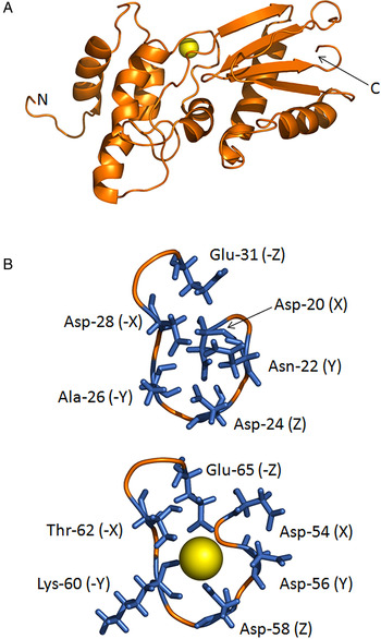
The molecular model of FhCaBP2 predicts a similar overall structure to FhCaBP3 and FhCaBP4 (Orr et al. Reference Orr, Kinkead, Newman, Anderson, Hoey, Trudgett and Timson2012; Banford et al. Reference Banford, Drysdale, Hoey, Trudgett and Timson2013) (Fig. 1A). The N-terminal domain contains two putative EF-hand sequences and the C-terminal domain is a largely β-sheet structure with similarities to DLCs. In contrast to FhCaBP3 and FhCaBP4, the linker between the two domains is shorter and, consequently, the two domains are predicted to be located closer to each other.
The predicted structure of FhCaBP2. (A) The predicted overall fold is shown in orange with a calcium ion (yellow) ball shown at the second EF-hand in the N-terminal domain. (B) Close-up views of the two EF-hands showing the potential ion-binding residues. Only the second EF-hand is shown to be interacting with a calcium ion, consistent with the data reported in this paper. (Colour version available online).

EF-hand sequences fold around calcium ions so that they provide six coordinating ligands for the ion. These ligands are arranged orthogonally and are referred to as the X, Y, Z, -X, -Y and -Z positions. Bioinformatics studies have revealed the preferred residues at each of these positions (Gifford et al. Reference Gifford, Walsh and Vogel2007). In FhCaBP2, the first EF-hand largely conforms to the consensus, except at the -Y position which is an alanine residue where threonine is preferred and alanine is not normally observed (Gifford et al. Reference Gifford, Walsh and Vogel2007) (Fig. 1B). However, this residue coordinates the ion using its backbone carbonyl group and, therefore, ion-binding cannot be ruled out by this substitution. In FhCaBP2's second EF-hand both the -Y (lysine) and -X (threonine) positions deviate from the most preferred residues (threonine and aspartate, respectively). However, both these residue types are ‘allowed’ (Gifford et al. Reference Gifford, Walsh and Vogel2007) and it should be noted the predicted structure of this EF-hand has the positive charge on the end of the lysine side chain directed away from the ion-binding site (Fig. 1B).
Expression and purification of FhCaBP2 and its domains
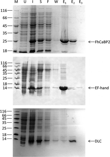
Recombinant FhCaBP2 could be readily expressed in, and purified from, E. coli using similar methods to those employed for FhCaBP3 and FhCaBP4 (Orr et al. Reference Orr, Kinkead, Newman, Anderson, Hoey, Trudgett and Timson2012; Banford et al. Reference Banford, Drysdale, Hoey, Trudgett and Timson2013) (Fig. 2A). Typical yields were 15 mg purified protein per litre of bacterial culture. In addition to the full length protein, fragments corresponding to the EF-hand domain and DLC-like domain (as estimated from the molecular model) were produced (Fig. 2B). Typical yields of these protein fragments were 16 and 7 mg L−1 of bacterial culture for the EF-hand and DLC-like domains, respectively. To probe the role of the second EF-hand, a point mutation which resulted in the alteration of Asp-58 to alanine, was introduced into the DNA coding sequence. The protein (FhCaBP2-D58A) and EF-hand domain fragment (EF-D58A) could also be expressed in, and purified from, E. coli in similar yields to the corresponding wild type proteins (data not shown).
Expression and purification of FhCaBP2 and its domains. Expression in, and purification of, recombinant proteins from E. coli was monitored by SDS-PAGE (10% for full length FhCaBP2 and 15% for the EF-hand and DLC-like domains). M, molecular mass markers (sizes shown to the left of the gel in kDa); U, extract from uninduced cells immediately prior to induction with IPTG; I, extract from induced cells immediately prior to harvesting; S, soluble material after sonication and centrifugation of cell extracts; F, material which flowed through the column; W, material removed from the column by washing; E1, E2 and E3, first, second and third elutions. For details of the purification methods, see Materials and Methods.

Ion-binding properties of FhCaBP2 and its domains
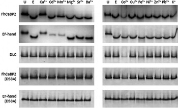
Full length, wild-type FhCaBP2 was resolved as a single band on native polyacrylamide gel electrophoresis (Fig. 3). Addition of EGTA increased the mobility of the protein in this gel system. This mobility could be restored by adding calcium chloride in a 2-fold molar excess over EGTA (Fig. 3). Therefore, it was concluded that FhCaBP2 interacts with calcium ions and that the recombinant protein, as purified from E. coli cells, is largely in the calcium-bound form. Therefore, further experiments to investigate ion-binding included EGTA to remove any calcium from the protein. A variety of other ions (cadmium, manganese, magnesium, strontium, barium, colbalt (II), copper (II), iron (II), nickel, zinc, lead and potassium) were tested for their ability to alter the mobility of FhCaBP2 in native gel electrophoresis. Of these, only manganese appeared to result in any change and this change was less than that observed with calcium (Fig. 3).
Ion-binding by FhCaBP2 and its domains as revealed by native gel electrophoresis. In each case protein (50 μ m) was resolved on 10% native polyacrylamide gels. The first lane contained the untreated protein (U) and the second protein incubated with EGTA (1 mm). The remaining lanes contained protein in the presence of EGTA (1 mm) and the indicated ion (2 mm).

A similar pattern was seen with the EF-hand domain. EGTA increased the mobility of the recombinant protein and this effect was revered by adding calcium ions. Manganese and, perhaps, cadmium ions, had a smaller effect, partially reversing the effect of EGTA. No other ions tested had any effect on the mobility of the EF-hand domain (Fig. 3). EGTA had no effect on the mobility of the DLC-like domain. Nor did calcium or any other of the ions tested here (Fig. 3). Thus, it was concluded that (as expected) that the EF-hand domain contains the calcium-binding site(s) in FhCaBP2.
This EF-hand domain has two EF-hands (Fig. 1). Of these, the second is closer to the consensus calcium-binding site and it was hypothesized that this is the main ion-binding site in FhCaBP2. To test this, a key residue in the second EF hand (Asp-58) was altered to alanine. The mobility of the resulting protein (FhCaBP2-D58A) was not affected by EGTA or any ion used in these experiments (Fig. 3). Similar results were observed with the EF-hand domain carrying the same alteration (EF-D58A, Fig. 3). The hypothesis that the second EF-hand in FhCaBP2 is the main calcium-binding site is supported by these experiments.
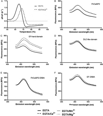
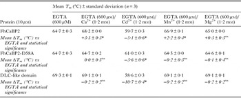
Calcium ions increased the thermal stability of FhCaBP2 (Fig. 4A; Table 1) consistent with them binding to the native, folded form of the protein (Matulis et al. Reference Matulis, Kranz, Salemme and Todd2005; Cummings et al. Reference Cummings, Farnum and Nelen2006). Manganese ions also increased the protein's thermal stability, but magnesium ions did not (Table 1). No ions stabilized FhCaBP2-D58A or the DLC-like domain (Table 1). Interestingly, cadmium ions radically destabilized wild type FhCaBP2, FhCaBP2-D58A and the DLC-like domain (Table 1), suggesting that this ion preferentially interacts with unfolded or partially folded forms of the protein (Cooper et al. Reference Cooper, Nutley, Wadood, Harding and Chowdhury2001). It was not possible to get reliable thermal stability measurements for the EF-hand domain or EF-D58A. Overall, these thermal stability data provide further support for the hypothesis that the second EF-hand in FhCaBP2 is the most important one in calcium (and manganese) binding. Since cadmium destabilized all forms of the protein tested (Table 1), it was concluded that its effects result not from specific interactions with an EF-hand, but from less specific interactions with multiple sites on the protein. It is not expected that these effects are physiologically important.
Ion-binding by FhCaBP2 and its domains as revealed by DSF and ANS fluorescence. (A) Representative first differential traces showing the thermal denaturation (‘melting’) of FhCaBP2 (10 μ m) in EGTA (0·6 mm) and EGTA (0·6 mm)/calcium chloride (1·2 mm). (B) ANS fluorescence spectra of FhCaBP2 (15 μ m). (C) ANS fluorescence spectra of the EF-hand domain (15 μ m). (D) ANS fluorescence spectra of the DLC-like domain (15 μ m). (E) ANS fluorescence spectra of FhCaBP2-D58A (15 μ m). (F) ANS fluorescence spectra of EF-D58A (15 μ m). In (b)–(f), spectra were obtained in the presence of ANS (30 μ m) and EGTA (1 mm, solid black line), EGTA (1 mm)/calcium chloride (2 mm; dashed black line), EGTA (1 mm)/magnesium chloride (2 mm; dotted black line) or EGTA (1 mm)/manganese chloride (2 mm; solid grey line).

Stabilization of FhCaBP2 by divalent ions as revealed by DSF

Statistical significance was determined using one-way ANOVA followed by Tukey's post hoc test for multiple comparisons.
nsNot significant (i.e. P > 0·5).
*P ⩽ 0·0001.
In calmodulin, and a number of other calcium-binding proteins, interaction with calcium results in an increase in the surface hydrophobicity of the protein (LaPorte et al. Reference LaPorte, Wierman and Storm1980). This increased hydrophobicity can be detected by probes such as anilinonaphthalene-8-sulfonate (ANS) whose fluorescence intensity increases when they interact in hydrophobic environments (Brand and Gohlke, Reference Brand and Gohlke1972). When FhCaBP2 was incubated with ANS in the presence of EGTA, the fluorescence intensity was lower than when the protein and probe were incubated in the presence of EGTA/calcium chloride (2:1 molar ratio, Fig. 4B). A similar result was observed with manganese, but not with magnesium ions (Fig. 4B). This suggests that, when FhCaBP2 binds to calcium or manganese ions, there is a conformational change resulting in a more hydrophobic surface. Similar results were seen with the EF-hand domain (Fig. 4C), but not with the DLC-like domain (Fig. 4D), FhCaBP2-D58A (Fig. 4E) or EF-D58A (Fig. 4F). Control experiments with human GALE (Accession number: NP_001121093; Timson, Reference Timson2005) showed no significant change with any of the ions studied here (data not shown). Once again, these results are consistent with the hypothesis that the second EF-hand is required for calcium-binding and also suggests that calcium-binding at this site is necessary to induce the conformational change detected by the increase in ANS fluorescence.
Dimerization of FhCaBP2 and its domains
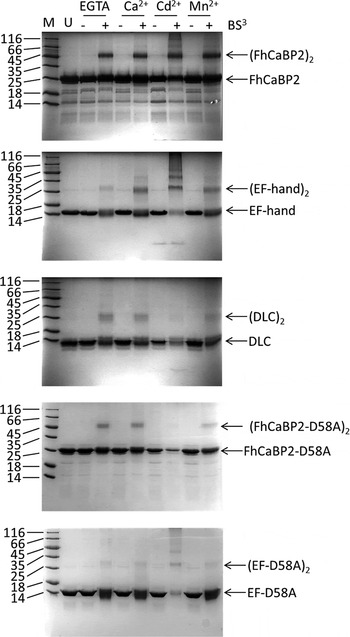
FhCaBP2 is a dimer as judged by protein–protein cross-linking experiments. The extent of dimerization was not affected by the presence or absence of calcium ions (Fig. 5). Both domains of the protein are involved in dimerization: the EF-hand domain and the DLC-like domain can both be cross-linked under similar conditions to the full length protein. The extent of cross-linking of the EF-hand domain was enhanced in the presence of calcium and manganese ions. However, cross-linking of the DLC-like domain was not greatly affected by the presence or absence of calcium or manganese ions (Fig. 5). FhCaBP2-D58A, which is unable to bind calcium ions (see above), could also be cross-linked and calcium or manganese ions did not affect the extent of cross-linking. Similar results were obtained with EF-D58A (Fig. 5). Cadmium ions appear to cause aggregation and multimerization of full length FhCaBP2 and the EF-hand domain (Fig. 5). Therefore, it can be concluded that FhCaBP2 forms homodimers which, overall, are insensitive to calcium ion concentrations. However, there is an alteration in the conformation of the EF-hand dimer on binding to calcium or manganese ions, which is reflected in the enhanced cross-linking. This may be the same change which was detected by ANS fluorescence. This contrasts with FhCaBP3 and FhCaBP4 which homodimerize in a calcium sensitive manner, but is similar to the S. mansoni proteins SmTAL1 and SmTAL2 which form calcium-insensitive homodimers (Orr et al. Reference Orr, Kinkead, Newman, Anderson, Hoey, Trudgett and Timson2012; Banford et al. Reference Banford, Drysdale, Hoey, Trudgett and Timson2013; Thomas et al. Reference Thomas, Fitzsimmons, Dunne and Timson2015). The structure of the homodimer is most likely a parallel one in which the two EF-hand domains interact and the two DLC-like domains also contact each other. In this arrangement the EF-hand domains alter conformation on binding calcium ions, but remain in contact in both the presence and absence of the ion.
Dimerization of FhCaBP2 revealed by cross-linking with BS3. FhCaBP2 and its domains (50 μ m) were incubated in the without (−) and with (+) of the cross-linking agent BS3 (800 μ m) in the presence of the ions indicated. M, molecular mass markers (sizes shown to the left of the gel in kDa); U, untreated protein. All the remaining lanes contained EGTA (1 mm) and, where indicated either calcium, cadmium or manganese chloride (2 mm).

Drug-binding properties of FhCaBP2 and its domains
Calmodulin (and related proteins) bind to a range of antagonists, some of which are important drugs used in the control of psychiatric conditions (Weiss et al. Reference Weiss, Prozialeck and Wallace1982). This suggests that calcium sensing signalling proteins are potential drug targets and that they may have potential in the control of liver fluke infections. Therefore, the interaction between FhCaBP2 and the calmodulin antagonists trifluoperazine (TFP), chlorpromazine (CPZ), N-(6-Aminohexyl)-5-chloro-1-naphthalenesulfonamide hydrochloride (W7) and thiamylal (ThA) was investigated. Since it has been previously reported that the antischistosomal drug praziquantel (PZQ) interacts with the calmodulin-like protein myosin regulatory light chain (Gnanasekar et al. Reference Gnanasekar, Salunkhe, Mallia, He and Kalyanasundaram2009), this drug was also included in these studies.
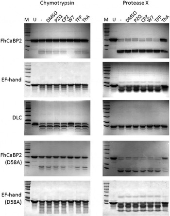
W7 protected wild type FhCaBP2 from limited proteolysis by chymotrypsin. This protection was less evident in FhCaBP2-D58A, in the isolated EF-hand domain and in EF-D58A. Some protection of the DLC-like domain was observed (Fig. 6, left column). Thiamylal protected FhCaBP2 from digestion with protease X and a similar result was observed with FhCaBP2-D58A, the EF-hand domain, EF-D58A but not with the DLC-like domain (Fig. 6, right column). None of these drugs had any effect on the digestion of a control protein, UDP-galactose 4′-epimerase, under the same conditions (data not shown).
Drug-binding by FhCaBP2 revealed by limited proteolysis. FhCaBP2 (50 μ m) was digested with either chymotrypsin (150 nm) or protease X (50 nm) as described in the Materials and Methods. M, molecular mass markers (sizes: 116, 66, 45, 35, 25, 18, 14 kDa); U, untreated protein; − protein treated with protease in the absence of drugs; DMSO, protein treated with protease in the presence of 1% (v/v) DMSO. In this experiment, all drugs were used at a concentration of 250 μ m.

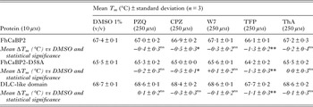
DSF demonstrated that, of the drugs tested only CPZ and TFP affected the melting temperature of the protein. Both caused a statistically significant reduction in the T m for the full length, wild-type protein (Table 2). This is consistent with these compounds binding to a partially folded form of FhCaBP2 (Cooper et al. Reference Cooper, Nutley, Wadood, Harding and Chowdhury2001). (Note that, like most proteins, FhCaBP2 most likely exists in equilibrium between folded and part-folded forms. Binding of a drug to a part-folded form will shift this equilibrium towards less stable, part-folded forms. Since an increased fraction of the molecules are now in this form, the overall T m is reduced). Both CPZ and TFP also bind to FhCaBP2-D58A and reduce this protein's stability by a similar amount to the wild-type (Table 2). This suggests that a functional calcium-binding site is not required for interaction with these drugs. TFP, but not CPZ, destabilizes the DLC-like domain (Table 2). This suggests that FhCaBP2's interaction with TFP is partly mediated by this domain. In contrast, CPZ is likely to interact entirely through the EF-hand domain.
Interaction of FhCaBP2 with drugs as revealed by DSF
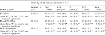
Statistical was significance determined using one-way ANOVA followed by Tukey's post hoc test for multiple comparisons.
nsNot significant (i.e. P > 0·5).
*P ⩽ 0·01.
**P ⩽ 0·0001.
DISCUSSION
Overall, these data demonstrate that FhCaBP2 is a calcium-binding protein and that this binding activity is primarily mediated by the second EF-hand in the N-terminal domain. The protein also interacts with manganese ions, a common feature with FhCaBP3 and FhCaBP4 and some members of the S. mansoni TAL family proteins (Orr et al. Reference Orr, Kinkead, Newman, Anderson, Hoey, Trudgett and Timson2012; Banford et al. Reference Banford, Drysdale, Hoey, Trudgett and Timson2013; Thomas et al. Reference Thomas, Fitzsimmons, Dunne and Timson2015). Interestingly, all these proteins have a lysine at the -Y position of the second EF-hand. In the Arabidopsis thaliana SOS3 protein, which has four EF-hands, manganese binds to the fourth EF-hand which is the only one with lysine at the -Y position (Sanchez-Barrena et al. Reference Sanchez-Barrena, Martinez-Ripoll, Zhu and Albert2005). In FhCaBP2, manganese ions are able to cause a similar conformational change to calcium ions which results in increased surface hydrophobicity (as judged from ANS fluorescence data; Fig. 3). This suggests that, in contrast to calmodulin where manganese ions stabilize the ‘closed’ state (Senguen and Grabarek, Reference Senguen and Grabarek2012), FhCaBP2 undergoes similar conformational changes with both manganese and calcium ions. Therefore, it is possible that both ions can induce signalling through this protein. However, to date, only limited functional information is available for these proteins and it is not possible to say whether or not manganese-binding is physiologically relevant. Cadmium (II) ions also interacted with FhCaBP2 and its domains. Very few cadmium (II) ion-binding proteins have been reported. Examples include carbonic anhydrases from marine diatoms and the cadmium sensing proteins of mycobacteria (Lane and Morel, Reference Lane and Morel2000; Cavet et al. Reference Cavet, Graham, Meng and Robinson2003). In both cases the metal ion is coordinated by structures which do not resemble EF-hands (Banci et al. Reference Banci, Bertini, Cantini, Ciofi-Baffoni, Cavet, Dennison, Graham, Harvie and Robinson2007; Xu et al. Reference Xu, Feng, Jeffrey, Shi and Morel2008). Calmodulin does bind to cadmium (II) through its EF-hands; this is not considered to be part of the normal functioning of this protein in vivo (Milos et al. Reference Milos, Comte, Schaer and Cox1989; Ouyang and Vogel, Reference Ouyang and Vogel1998). Indeed inappropriate cadmium-binding can lead to toxicity (Chmielowska-Bak et al. Reference Chmielowska-Bak, Izbianska and Deckert2013; Choong et al. Reference Choong, Liu and Templeton2014). In the case of FhCaBP2, cadmium (II)-binding is likely to be a non-physiological effect which results in partial unfolding of the protein's structure.
FhCaBP2's ability to homodimerize is unaffected by calcium ions. This is a different response to calcium-binding compared with FhCaBP3 and FhCaBP4 (Orr et al. Reference Orr, Kinkead, Newman, Anderson, Hoey, Trudgett and Timson2012; Banford et al. Reference Banford, Drysdale, Hoey, Trudgett and Timson2013). It is reasonable to assume that the oligomeric state of the protein affects its function. If this is the case, then these three proteins presumably have different cellular functions. In low calcium ion concentrations, FhCaBP2 and FhCaBP3 would be dimerized, but FhCaBP4 would be monomeric. However, in high calcium ion concentrations, FhCaBP2 would remain dimerized, FhCaBP4 would be a dimer and FhCaBP3 would be a monomer. Whether these proteins also have the capacity to heterodimerize is currently unknown.
FhCaBP2 interacts with a similar range of calmodulin antagonists to FhCaBP3, FhCaBP4 and the S. mansoni TAL proteins (Orr et al. Reference Orr, Kinkead, Newman, Anderson, Hoey, Trudgett and Timson2012; Banford et al. Reference Banford, Drysdale, Hoey, Trudgett and Timson2013; Thomas et al. Reference Thomas, Fitzsimmons, Dunne and Timson2015). CPZ and ThA interact with FhCaBP2, most likely through the EF-hand domain. These compounds have also previously been shown to interact with calmodulin (Tanaka et al. Reference Tanaka, Ohmura and Hidaka1983; Humar et al. Reference Humar, Pischke, Loop, Hoetzel, Schmidt, Klaas, Pahl, Geiger and Pannen2004) and so this result was not surprising. However, TFP and W7 were shown to interact (at least in part) with the DLC-like domain (by DSF and limited proteolysis, respectively). Previous work has demonstrated that these compounds interact with the EF-hand domains of calmodulin, preferentially interacting with hydrophobic clefts revealed as a consequence of calcium-binding (Cook et al. Reference Cook, Walter and Walter1994; Osawa et al. Reference Osawa, Swindells, Tanikawa, Tanaka, Mase, Furuya and Ikura1998). It will be interesting to see if other DLC-like structures can interact with TFP and W7.
In conclusion, FhCaBP2 has similar sequence and predicted structure to other members of this family of helminth proteins. It differs in its ion-binding properties and its response to ions to some other family members. Of the family members characterized to date, it is functionally most similar to FhCaBP3 (Orr et al. Reference Orr, Kinkead, Newman, Anderson, Hoey, Trudgett and Timson2012; Banford et al. Reference Banford, Drysdale, Hoey, Trudgett and Timson2013; Thomas et al. Reference Thomas, Fitzsimmons, Dunne and Timson2015). Its ability to reversibly bind calcium ions and alter its conformation in response suggests that the physiological role of the protein is in some form of calcium signalling process. The localization of a number of family members (including FgCaBP1, which has 85% similarity to FhCaBP2) to the tegument of the worm (Havercroft et al. Reference Havercroft, Huggins, Dunne and Taylor1990; Mohamed et al. Reference Mohamed, Shalaby, LoVerde and Karim1998; Vichasri-Grams et al. Reference Vichasri-Grams, Subpipattana, Sobhon, Viyanant and Grams2006; Kim et al. Reference Kim, Yoo, Lee, Kim, Lee, Kang, Na and Ju2012; Subpipattana et al. Reference Subpipattana, Grams and Vichasri-Grams2012), suggests that these proteins may play a key role in regulating this dynamic cellular structure. Localization studies on FhCaBP2 may reveal more about its physiological role. However, such studies may prove challenging due to the high level of similarity between FH22 and FhCaBP2 (78%) which is likely to make it difficult to generate specific antibodies. Stage specific expression studies may also be useful, especially if also carried out with other family members to enable comparisons. If FhCaBP2 and FH22 are located in the tegument, they may be good candidates for therapeutic intervention: antagonism of these calcium-binding proteins may result in dysregulation of the tegument which is a critical structure in parasite–host interaction (Wilson et al. Reference Wilson, Wright, de Castro-Borges, Parker-Manuel, Dowle, Ashton, Young, Gasser and Spithill2011).
SUPPLEMENTARY MATERIAL
To view supplementary material for this article, please visit http://dx.doi.org/S0031182015000736.
ACKNOWLEDGEMENTS
CMT is in receipt of a PhD studentship from the Department of Employment and Learning, Northern Ireland (DELNI). We thank Prof Aaron Maule (Institute for Global Food Security, Queen's University, Belfast) for access to qPCR machines which were used for the DSF experiments.