Severe acute respiratory syndrome-coronavirus-2 was initially regarded as a mild infection in children, but with the emergence of multisystem inflammatory syndrome in children (MIS-C), it has now been recognised as a virus with far more serious implications. Reference Belay, Abrams and Oster1,Reference Feldstein, Tenforde and Friedman2 Although MIS-C has now been described in more than 2000 children and half of the patients have biochemical signs of coagulopathy, thrombotic complications have only rarely been described with venous thromboembolisms in four children and cerebral artery thrombosis in one child. Reference Feldstein, Rose and Horwitz3 Further, two children who received extracorporeal membrane oxygenation developed right atrial thrombus and cerebral artery infarction. Reference Minen, Hands, Mustafa, Pienaar and Lillie4
Due to the novelty of MIS-C and resulting limited evidence, current treatment recommendations regarding antithrombotic prophylaxis are mainly founded on expert consensus. In patients with severe ventricular dysfunction anticoagulation prophylaxis is encouraged to reduce the risk of intra-cardiac thrombosis, a known but rare complication of severe non- coronarivus disease-related myocarditis. Reference Henderson, Canna and Friedman5,Reference Goldenberg, Sochet and Albisetti6
This case report describes the clinical and imaging features of a patient with MIS-C and left ventricular thrombus. To our knowledge, this is the only described case of left ventricular thrombus in a patient with MIS-C and illustrates the risk of severe intra-cardiac thrombotic complications in MIS-C.
History of presentation
A 17-year-old otherwise healthy Caucasian male adolescent fulfilled the case definition criteria for MIS-C by the Centers for Disease Control and Prevention and the World Health Organization. He presented to a paediatric emergency department in January 2021 with fever for 4 days, nausea, and abdominal pain. The physical examination revealed severe abdominal tenderness and a temperature of 39.7°C but no rash, mucous membrane change, or conjunctival injection. His initial blood pressure was 113/61 mmHg, heart rate was 104 beats/min, respiratory rate 18 breaths/min, and blood oxygen saturation by pulse oximetry was 99%.
Despite general supportive measures, his condition deteriorated in the following 48 hours with worsening epigastric pain, headache, emesis, and haemodynamic compromise (blood pressure 75/40, heart rate 125 beats/min, oliguria). On hospital day 3, he was transferred to a tertiary paediatric intensive care facility for further investigations and treatment.
Past medical history
There was no significant medical history.
Differential diagnosis
Presumed diagnosis was MIS-C due to previous severe acute respiratory syndrome-coronavirus-2 infection. Other considered differential diagnoses were bacterial sepsis, toxic shock syndrome, and Kawasaki disease.
Investigations
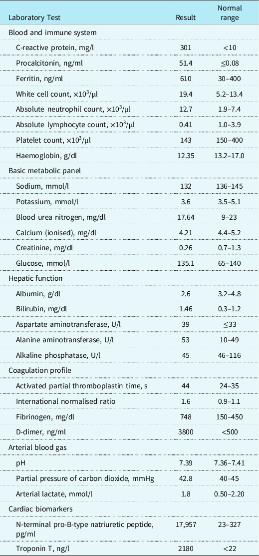
A 12-lead electrocardiogram showed flattening of T-waves and insignificant ascending ST-depression in the inferior extremity leads. Laboratory results showed elevated inflammatory markers and abnormal coagulation studies (Table 1). Serial chest radiographs were normal, and abdominal ultrasound indicated mesenteric lymphadenitis.
Clinical laboratory results, hospital day 3

A bedside focused transthoracic echocardiogram on hospital day 2 prior to haemodynamic shock showed left ventricular ejection fraction 55%, normal right ventricular systolic function, and no valvular disease.
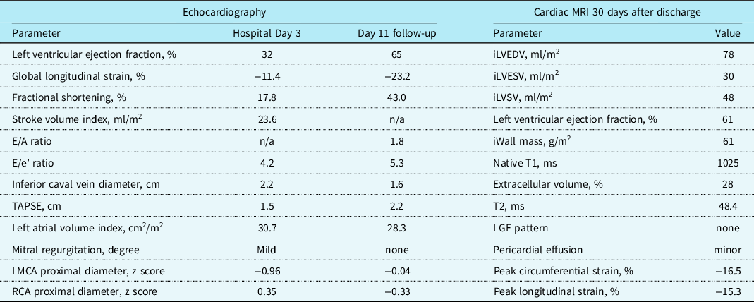
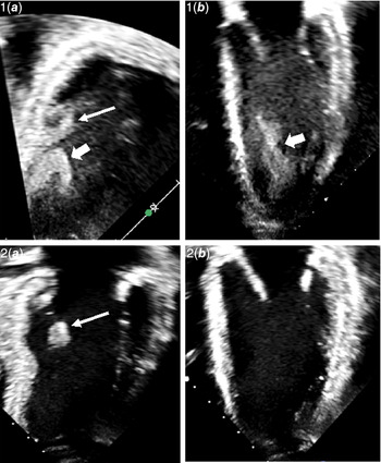
A transthoracic echocardiogram on hospital day 3 revealed global reduction of left ventricular ejection fraction, global longitudinal strain, and stroke volume index with normal diastolic function parameters (Table 2). Marked left ventricular spontaneous echo contrast was noted. In the 4- and 2-chamber views, a mural thrombus measuring 2.6 × 1.0 cm was identified near the posteromedial papillary muscle in the left ventricular apex (Fig 1, Online Video 1). The thrombus was more consolidated near the ventricular wall, while thinner strings of thrombus mass were swinging from its luminal head. There was a minor pericardial effusion. The dimension of the proximal coronary arteries was normal.
Cardiac imaging

iLVEDV = indexed left ventricular end-diastolic volume; iLVESV = indexed left ventricular end-systolic volume; iLVSV = indexed left ventricular stroke volume; LGE = late gadolinium enhancement; LMCA = left main coronary artery; n/a = not available; RCA = right coronary artery; TAPSE = tricuspid annular plane systolic excursion.
Echocardiogram images demonstrating left ventricular thrombus and subsequent dissolution. 1, Hospital Day 3. Reduced left ventricular function, notable spontaneous echo contrast and left ventricular thrombus near the posteromedial papillary muscle. 1A: 2-chamber view, 1B: 4-chamber view. 2, Hospital day 11. Normal left ventricular function, no spontaneous echo contrast, and absence of left ventricular thrombus with clear detection of endocardial border. 2A: 2-chamber view, 2B: 4-chamber view. Markers. Thin arrow = posteromedial papillary muscle. Wide arrow = Left ventricular thrombus.

Management
Immediate treatment on hospital day 1 was isotonic fluid, antibiotics, and prophylactic doses of tinzaparin. On hospital day 3, dopamine infusion at high concentrations was necessary to attain acceptable organ perfusion. Multisystem inflammatory syndrome in children treatment with intravenous immunoglobulin, anakinra, methylprednisolone, and acetylsalicylic acid was instituted. The left ventricular thrombus was treated with therapeutic doses of tinzaparin and monitored with daily echocardiograms. On hospital day 5, clinical status improved, left ventricular ejection fraction increased to 50%, and the mural thrombus was nearly dissolved. There were no findings suggestive of systemic thromboembolism from the left ventricular thrombus.
Follow-up
The patient was discharged on hospital day 9. Both inflammatory and cardiac biomarkers had decreased but were still moderately elevated. A transthoracic echocardiogram demonstrated normalisation of left ventricular function, complete absence of left ventricular spontaneous echo contrast, and no signs of intra-cardiac thrombus (Table 2, Fig 1). On follow-up 20 days after discharge, the patient and his parents reported a return to normal activities. Testing for inherited thrombophilia was negative.
Cardiac MRI was performed 30 days after discharge and showed normal left ventricular volumes, left ventricular ejection fraction, and stroke volume index but a reduction in both global longitudinal and circumferential strain and minor pericardial effusion. Dimensions of the proximal coronary arteries were normal. No late enhancement following gadolinium contrast and normal native T1, ECV, and T2 values was observed in all AHA segments, indicating no signs of myocardial injury or oedema (Table 2, Online Video 2).
Discussion
This represents the only reported case of left ventricular thrombus in a MIS-C patient and provides direct evidence of potential serious thrombotic complications despite prophylactic anticoagulant treatment. A previously healthy adolescent developed acute illness, cardiac dysfunction, and left ventricular thrombus. After treatment, he recovered and returned to normal activities without any signs of residual thrombus mass or arterial thromboembolic complications.
The pathogenesis of MIS-C remains poorly understood. The fact that the disease develops coinciding with the acquired immune response to severe acute respiratory syndrome-coronavirus-2 and most children have negative severe acute respiratory syndrome-coronavirus-2 RNA on presentation supports the hypothesis that MIS-C is caused by immune-mediated systemic inflammation. Given almost half of all children with MIS-C have elevated d-dimer and fibrinogen, Reference Belay, Abrams and Oster1,Reference Feldstein, Tenforde and Friedman2 inflammation-induced hypercoagulopathy could conceivably contribute to the multiple organ involvement in MIS-C.
In adult patients with primary severe acute respiratory syndrome-coronavirus-2 infection, procoagulant abnormalities have been associated with an increased risk of thrombotic complications, possibly due to a specific coronavirus disease 2019-associated coagulopathy. The incidence of venous thromboembolic events in critically ill severe acute respiratory syndrome-coronavirus-2 patients admitted to the ICU has been reported to be as high as 27% despite standard anticoagulant prophylaxis, and arterial vascular events up to 4%. Reference Connors and Levy7 Biochemical evidence of a similar hypercoagulable state in MIS-C patients has led to consensus-based guidelines recommending low-dose anticoagulation prophylaxis. Reference Henderson, Canna and Friedman5,Reference Goldenberg, Sochet and Albisetti6 Nevertheless, thrombotic events seem to be rare in patients with MIS-C. One study found that 4 of 186 children and adolescents had a venous thromboembolism, but the largest published MIS-C case series have not reported thrombotic complications. Reference Belay, Abrams and Oster1–Reference Feldstein, Rose and Horwitz3
In our patient, notable spontaneous echo contrast was noted on the transthoracic echocardiogram. This finding has been strongly correlated with intra-cardiac thrombus and is thought to represent erythrocyte aggregation under slow blood flow conditions in the left ventricle in patients with severe left ventricular dysfunction. Reference Black, Chesterman, Hopkins, Lee, Chong and Walsh8 Two previous echocardiographic studies in patients with MIS-C found median left ventricular ejection fraction was 55 to 57%. Reference Matsubara, Kauffman and Wang9,Reference Theocharis, Wong and Pushparajah10 In comparison, our patient had a more pronounced reduction in left ventricular ejection fraction from 55 to 32% in less than 48 hours perhaps resulting in particularly sluggish blood flow. It may be speculated that the risk of developing left ventricular mural thrombus is especially high in patients with MIS-C coagulopathy and large areas of poorly contracting left ventricular wall.
At follow-up, there was normalisation of echocardiographic parameters, while a mild reduction in global strain on cardiac MRI persisted. This is consistent with a previous study in 20 MIS-C patients in which all patients exhibited persisting abnormal strain patterns on cardiac MRI, possibly due to protracted but resolving myocardial inflammation. Reference Theocharis, Wong and Pushparajah10
In conclusion, this case demonstrates the genuine potential for intra-cardiac thrombus formation in MIS-C patients with coagulopathy and reduced left ventricular ejection fraction, even during treatment with prophylactic dose anticoagulants.
Learning objectives
-
To understand that the coagulopathy in multisystem inflammatory syndrome in children (MIS-C) may lead to intra-cardiac thrombotic complications in rare cases.
-
To be aware of the value of serial echocardiographic evaluations of possible thrombotic complications in MIS-C patients with haemodynamic shock.
Acknowledgements
The authors thank the patient and his parents for allowing us to share their story. They also thank all doctors and nursing staff at the Pediatric Emergency Department, Pediatric Intensive Care Unit, and Pediatric Cardiology Department for their efforts and care.
Financial support
This research received no specific grant from any funding agency, commercial, or not-for-profit sectors.
Conflicts of interest
None.
Supplementary material
To view supplementary material for this article, please visit https://doi.org/10.1017/S1047951121002456