Introduction
A replicating mammalian cell is estimated to suffer as many as 105 DNA lesions daily (Ref. Reference Hoeijmakers1). In order to protect the genomic integrity of the cell, complex biochemical pathways have been preserved to ensure the propagation of genomic material with a minimum of sustained damage. The DNA damage response and repair pathways are dependent on the signal transducers of the phosphoinositide 3-kinase (PI3 K)-related protein kinases (PIKKs) family, including ataxia-telangiectasia-mutated (ATM), ATM and RAD3-related (ATR) and DNA-dependent protein kinase (DNA-PK) (Ref. Reference Shiloh2).
Single-stranded DNA (ssDNA) is present during transcription and replication or is generated during the repair of DNA double-strand breaks (DSBs), stalled replication forks or damage triggering nucleotide excision repair (Refs Reference Hoeijmakers1, Reference Phillips3, Reference Lindahl and Barnes4). The signal emanating from ssDNA is relayed through a complex biochemical pathway that centres on the activity of ATR (Fig. 1). Activation and regulation of ATR ensures faithful replication as well as contributes to DNA damage signalling after DNA damage to enforce cell cycle arrest, maintenance of replication fork integrity and recruitment of DNA repair factors to lesions. As DNA lesions are repaired, the signal emanating from such sites dissipates and the cell is free to resume replication and cell division. ATR recruitment to sites of DNA lesions is dependent on the ATR-interacting protein (ATRIP) and its binding of replication protein A (RPA) coated ssDNA (Refs Reference Cortez5, Reference Zou and Elledge6). The activity of the ATR–ATRIP complex is in turn regulated by the evolutionary conserved Rad9–Rad1–Hus1 (Reference Mordes9–Reference Hoeijmakers1–Reference Hoeijmakers1) complex (Refs Reference Delacroix7, Reference Lee, Kumagai and Dunphy8), which upon being phosphorylated binds topoisomerase-binding protein 1 (TOPBP1) to activate the ATR-signalling cascade (Ref. Reference Mordes9). Recruitment of the 9–1–1 and ATR–ATRIP complexes occurs independently, possibly creating a built-in regulatory element of the pathway ensuring that activation will only occur when both complexes are present, minimising the chance of illicit firing of this regulatory pathway (Refs Reference Kondo10, Reference Melo, Cohen and Toczyski11). The recruitment and activation of ATR triggers the phosphorylation of a large number of target proteins. In fact, activation of ATR and ATM – responsible for signalling downstream of double-stranded DNA damage – regulates approximately 700 target proteins, spanning functional hubs, such as cell-cycle regulation, DNA replication and DNA repair (Ref. Reference Matsuoka12). Arguably the most important phosphorylation target of ATR is the p53 tumour suppressor. Ultraviolet (UV) irradiation induces ATR-dependent phosphorylation of p53Ser15, leading to p53 activation that, depending on cellular context, will either induce cell-cycle arrest, repair or apoptosis (Refs Reference Tibbetts13, Reference Dumaz and Meek14). The rationale for targeting ATR in cancer therapy comes from the evident engagement of this signalling hub following both radiation- and chemically induced DNA damage in cancer therapy, as well as the consistent and constitutive activation of ATR in fast proliferating cancers with aberrant replication stress (Ref. Reference López-Contreras and Fernandez-Capetillo15). The interest in ATR inhibitors (ATRi) is also linked with the vast interest in inhibitors of the CHK1 kinase, which is regulated downstream of ATR. Blocking of the downstream activation of repair and signalling factors by ATR inhibition would induce a more severe DNA damage phenotype of currently applied therapy. This in turn would cause a more efficient killing of cancer cells, enabling better disease control in the clinic. Indeed, by either genetically or chemically inhibiting ATR, several pre-clinical studies have shown the potential of applying this combination therapy to various cancer models (Ref. Reference Fokas16). The increased efficacy of ATR ablation in combination with chemotherapy has also been shown in cells with faulty checkpoint regulation, such as lack of p53, which is important for proper G2/M and G1 arrest following DNA damage. The inhibition of ATR causes a p53-independent death due to premature chromatin condensation and mitotic entry (Ref. Reference Nghiem17). Several inhibitors of ATR's main downstream effector, CHK1, have been published and reviewed elsewhere (Ref. Reference Dai and Grant18). A majority of clinical trials using CHK1 inhibitors are combination studies with different cytotoxic drugs, e.g. gemcitabine, showing positive clinical outcome (Ref. Reference Garrett and Collins19). This data uniformly support the use of ATRi's combined with DNA-damaging agents. Targeting ATR, a more central hub in the cell-cycle machinery and the direct upstream regulator of CHK1, will likely further increase the potential of these combination therapies.
ATR is recruited to sites of ssDNA damage. This in turn enables the recruitment of ATR–ATRIP and the 9–1–1 complex. Subsequent phosphorylation of 9–1–1 then enables TOPBP1 binding, starting the ATR-mediated phosphorylation cascade. ATR phosphorylates numerous proteins, including CHK1 and p53, to initiate DNA repair, cell-cycle regulation and control of replication origin firing.

The potential of using ATR inhibition, or by targeting its main downstream signal transducer CHK1, in a mono-therapy approach has also been evaluated in cancers with a high degree of replication stress or in those that carry an inherent defect in DNA repair, with positive results (Refs Reference Chen20, Reference Höglund21, Reference Cole22, Reference Schoppy23). Even though the efficacy of this approach have been questioned due to the lack of positive clinical outcome, the recent pre-clinical studies with encouraging data using model systems with inherent high degree of replication stress (N-MYC or C-MYC transformed cells) highlight the importance of patient stratification (Refs Reference Höglund21, Reference Cole22, Reference Schoppy23). Selecting disease or patients with, e.g. amplified MYC expression could potentially find a use for CHK1 and ATRi as monotherapeutic agents. Interestingly, the first clinical trials using ATRi's have been initiated by AstraZeneca, preferably in ATM-mutated chronic lymphocytic leukaemia (CLL), prolymphocytic leukaemia (PLL) or B-cell lymphoma patients (Ref. 24) and by Vertex Pharmaceuticals in advanced solid tumours (Ref. 25).
ATRi's published by academic groups
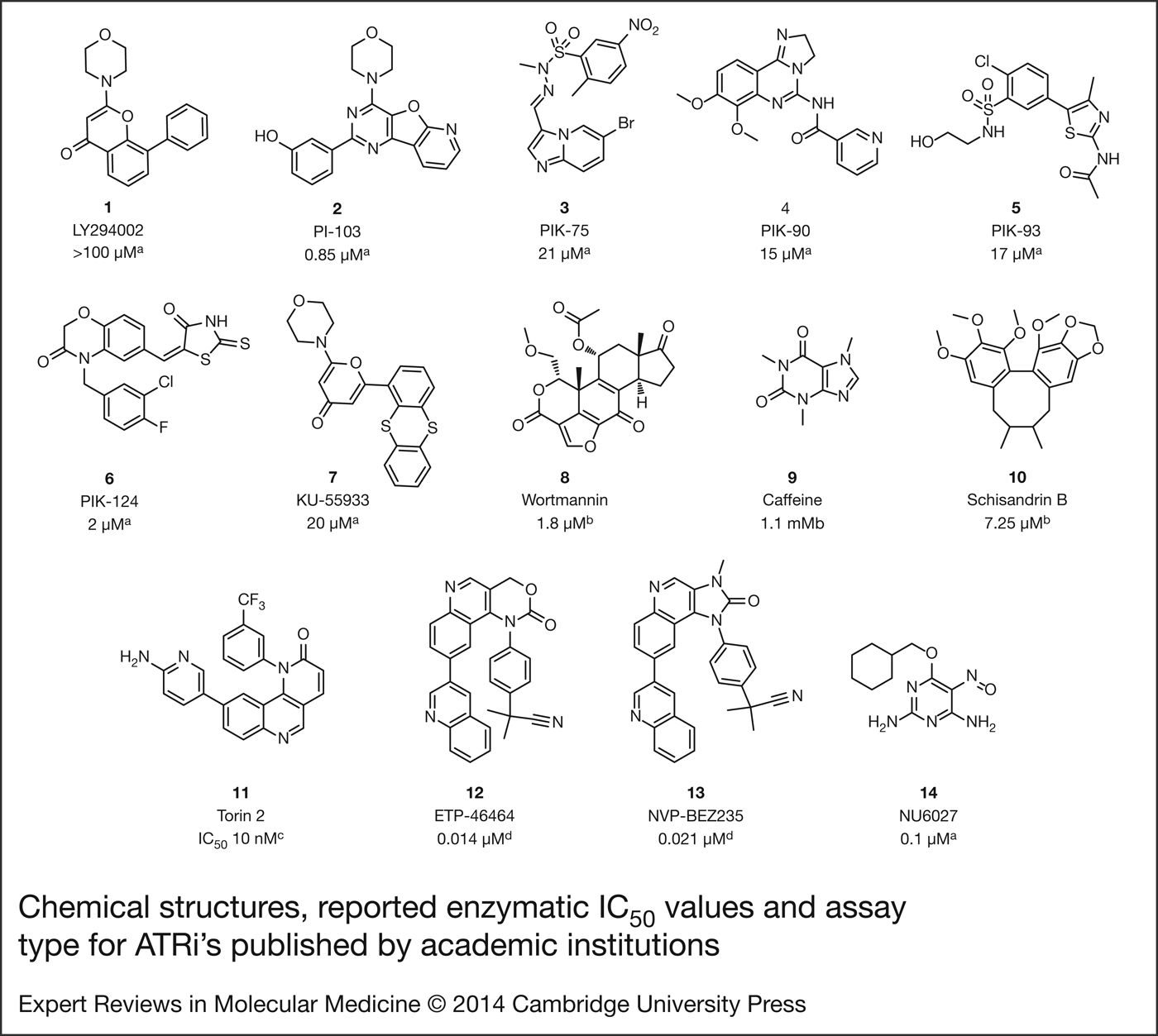
Research on ATRi's has been on-going for more than a decade and although the first small-molecule inhibitors were identified in the late 1990′s, most of the novel patented chemical matter appeared in 2010 and onwards. LY294002 1 (Fig. 2) was originally developed by Eli Lilly as a PI3 K inhibitor (Ref. Reference Vlahos26) but was later acknowledged as a pan-inhibitor of the PIKK family (Refs Reference Rosenzweig27, Reference Lee28). Although no enzymatic inhibition data were available from the initial publications, the ATR IC50 of 1 was determined to be >100 µm in a later publication focused on investigating the pharmacological roles of the PI3 K family (Ref. Reference Knight29). In this study, Knight et al. screened a chemically diverse set of PI3 K inhibitors in pre-clinical development against several lipid kinases and identified compounds 2, 3, 4, 5, 6 and 7 as moderate ATRi's in an enzymatic assay (ATR IC50 ≥ 0.85 µm but ≤21 µm, Fig. 2), with no selectivity over ATM or DNA-PK.
Chemical structures, reported enzymatic IC50 values and assay type for ATRi's published by academic institutions.aRadiolabelled assay; Rad17-derived peptide substrate; bRadiolabelled assay; PHAS-I substrate; cChemical proteomics, KiNativ ActivX Biosciences; dflag-IP/Radiolabelled assay, GST-p53 substrate.
The fungal metabolite wortmannin 8 (Fig. 2) has been characterised as an irreversible pan-inhibitor of the PIKK family (ATR IC50 = 1.8 µm, DNA-PK IC50 = 16 nm, ATM IC50 = 150 nm), suggesting that the radio-sensitising effect of 8 is probably due to the inhibition of DNA-PK kinase activity rather than ATR inhibition (Ref. Reference Sarkaria30). Caffeine 9 (Fig. 2) has also been identified as a weak pan-PIKK inhibitor (ATR IC50 = 1.1 mm, DNA-PK IC50 = 10 mm, ATM IC50 = 0.2 mm), but in this case the radio-sensitising effect of 9 was attributed to both ATM and ATR kinases (Ref. Reference Sarkaria31).
The first patent on the therapeutic use of ATR inhibition was filed in 2003 by the Universities of Rochester and Utah (Ref. Reference Planelles, Roshal and Zhu32). This filing claimed the use of ATR or Rad17 inhibitors to inhibit HIV replication but did not exemplify any novel chemical matter. Interestingly, in 2005 researchers at the Massachusetts General Hospital filed a US patent on the use of ATRi's to reduce UVB-induced skin damage (Ref. Reference Nghiem and Fujii33). The ATR-mediated checkpoint-signalling cascade is activated upon UV-induced DNA damage in replicating cells. Working under this hypothesis, topical application of an ATRi (1.2% solution of 9 in acetone) reduced UV-induced wrinkles in a murine model. No additional biological data were provided.
Nishida et al. filed in 2007 a patent on the first ATR-selective small-molecule inhibitors (Ref. Reference Nishida, Hamamori and Konishi34), the dibenzocyclooctadiene lignans schisandrins and gomisins and published a subsequent paper with detailed information on the mechanism of action of one of the compounds of interest, schisandrin B 10 (Fig. 2) (Ref. Reference Nishida35). 10 (30 µm) blocked the G2/M and S-phase checkpoint and reduced viability of UV-exposed A549 adenocarcinoma cells. In addition, 10 inhibited p53 and CHK1 ATR-dependent phosphorylation but did not prevent ATR-activating association to the ATRIP. 10 selectively inhibited ATR kinase activity in vitro with an IC50 of 7.25 µm (ATM IC50 = 1.74 mm, no inhibition detected for CHK1, PI3 K, DNA-PK and Mammalian target of rapamycin (mTOR). 10 (30 µm) blocked p53, CHK1, SMC1 and BRCA1 phosphorylation in ATM-deficient UV-exposed AT2KY fibroblasts and p53 and CHK1 phosphorylation in ATM-siRNA-depleted UV-exposed A549 cells, confirming the ATR-specificity of 10. Furthermore, 10 did not affect cell viability of UV-exposed ATR-deficient Seckel syndrome fibroblasts, in agreement with the other findings presented in this work.
Liu et al. at the Harvard Medical School characterised Torin 2 11 (Fig. 2) as a potent ATP-competitive inhibitor of mTOR, ATM and ATR (mTOR IC50 = <0.01 µm, ATM IC50 = <0.01 µm, ATR IC50 = <0.01 µm) using a KiNativ chemical proteomics assay and in cellular assays looking at phosphorylation status of downstream substrates (mTOR EC50 = 0.25 nm, ATM EC50 = 28 nm, ATR EC50 = 35 nm) (Ref. Reference Liu36). In the case of ATR, 11 inhibited the cellular activity of the kinase as assessed by phosphorylation status of CHK1S317 following exposure of HCT116 colon cancer cells to UV-induced DNA damage. 11 showed significant inhibition of tumour volume in a KRAS-driven murine model of lung cancer when administered in combination with the mitogen-activated protein/extracellular signal-regulated kinase (MEK) inhibitor AZD6244.
A group led by Dr Fernandez-Capetillo at the Spanish National Cancer Research Centre also screened a collection of PI3 K inhibitors with the aim of finding hits against the PIKK family, in particular ATR (Ref. Reference Toledo37). This phenotypic screen is very interesting as it exploit the fact that ATR is activated by binding the TOPBP1 protein (Ref. Reference Kumagai38). The team expressed the ATR-binding domain of TOPBP1 fused with ER which can enter the nucleus and activate ATR upon addition of 4-hydroxytamoxifen (4-OHT) in the absence of DNA damage (Ref. Reference Toledo39). Using this system the team carried out a high-throughput microscopy screen that quantified nuclear γH2AX, an early and reliable marker of double-stranded DNA damage, identified compound 12 (Fig. 2) as a promising hit. Compound 12 not only inhibited γH2AX, but also CHK1 phosphorylation, abolished the G2/M checkpoint and induced nuclei damage after ionising radiation (IR) exposure. 12 potently inhibited ATR in vitro and although it showed some selectivity over ATM (enzymatic ATR/ATM IC50's = 14 nm/545 nm) and 26 additional kinases, it strongly inhibited mTOR and DNA-PK (enzymatic mTOR/DNA-PK IC50's = 0.6 nm/36 nm). Given the complex network of ATR-binding/activating partners, using a flag-IP assay allowed the group to efficiently quantify ATR inhibition. 12 displayed outstanding ATR activity in the cellular assay quantifying γH2AX intensity in 4-OHT-treated U2OS cells (ATR IC50 = 25 nm). Upon co-treatment of U2OS cells with 12 and the DNA-damaging agent hydroxyurea, there was an increase in p53-binding protein 1 (53BP1) and RPA foci, together with a rise of ATM and Chk2 phosphorylation, indicating chromosomal damage. Cells treated with 12 stalled in G2 phase 16 h after removal of hydroxyurea, following a common pattern seen in selective ATR deficiency. 12 also showed synergy with the CHK1 inhibitor 7-hydroxystaurosporine. It is worth noting that staurosporine derivatives are well-known promiscuous compounds that inhibit a wide variety of protein kinases and this could blur the interpretation of these results (Ref. 40). Compound 13 (NVP-BEZ235), a dual PI3 K/mTOR inhibitor developed by Novartis (Ref. Reference Maira41), was also identified as a potent pan-PIKK inhibitor (ATR, ATM DNA-PK IC50's <21 nm) using the cell-free kinase assay and the ATR cell-based assay (ATR IC50 = 25 nm). 13 inhibited ionising radiation-induced ATM, CHK1, Chk2 and DNA-PK phosphorylation and affected ionising radiation-induced γH2AX formation, whereas 12 did not, in agreement with the less selective profile of 13. Compound 12 suffers from poor pharmacological properties in mice and has not been further developed, whereas compound 13 is currently undergoing clinical trials for solid tumours (Refs Reference Rosenzweig27, 42). The authors suggested that mTOR inhibition of 12 and 13 could be detrimental for the generation of replicative stress and such selectivity issue should be addressed in future generations of ATRi's. Furthermore, dual inhibition of PI3 K/mTOR did not show synthetic lethal interaction with cyclin E overexpression and/or p53 deficiency. This evidence, together with other data recently published (Refs Reference Konstantinidou43, Reference Gilad44), indicates that clinical effect of 13 could be due to potent inhibition of ATR rather than PIK3 or mTOR.
Prof Curtin's team at Newcastle University and colleagues at the Mayo Clinic identified the CDK2 inhibitor NU6027 14 (Fig. 2) as a moderate ATRi in a cell-free enzymatic assay (ATR IC50's = 0.1 µm; CDK2 IC50 = 2.2 µm) and using pCHK1S345 as a specific marker of ATR activity in GM847KD and human breast cancer MCF7 cells (IC50's = 2.8 and 6.7 µm, respectively), with good selectivity over related PIKK's, ATM and DNA-PK (Ref. Reference Peasland45). 14 was mildly cytotoxic in clonogenic assays when administered alone at 10 µm and sensitised GM847KD, MCF7 and A2780 cancer cell lines to the major classes of DNA-damaging agents by around 2-fold, but did not potentiate the cytotoxic effect of the mitotic inhibitor paclitaxel, indicating a specific chemo-sensitisation mechanism. 14 decreased DNA damage-induced G2/M arrest and inhibited RAD51 foci formation in cells treated with the poly(ADP-ribose) polymerase (PARP) inhibitor rucaparib. 14 was synergistic with rucaparib in MCF7 cells and synthetically lethal in XRCC1-defective EM9 cells, confirming that impaired DNA single-strand break repair is synthetically lethal with ATR inhibition.
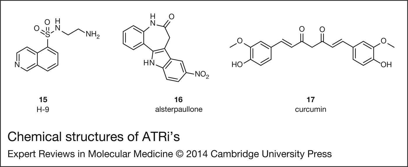
D'Andrea et al. at the Dana Farber Cancer Institute filed a patent on inhibitors of the Fanconi anaemia pathway and their use as inhibitors of DNA damage repair (Ref. Reference D'Andrea and Taniguchi46). During the screening campaign H-9 15, alsterpaullone 16 and curcumin 17 (Fig. 3) were found to inhibit mono-ubiquitination of the FANCD2 polypeptide. Further investigation indicated the compounds did not affect ATM-dependent phosphorylation of FANCD2 but did inhibit ATR-dependent phosphorylation of CHK1Ser345 in a concentration-dependent manner, suggesting that the compounds inhibit ATR either directly or indirectly, although no specific data were provided. This is in agreement with previous evidence suggesting that ATR, together with ATM and RPA, are upstream regulators of FANCD2 mono-ubiquitination (Refs Reference Andreassen, D'Andrea and Taniguchi47, Reference Ho48). Additionally, 15 and 16 sensitised the 2008 ovarian tumour cell line (deficient in FANCF protein) to cisplatin.
Chemical structures of ATRi's published by Dana Farber Cancer Institute.
ATRi's published by pharmaceutical companies
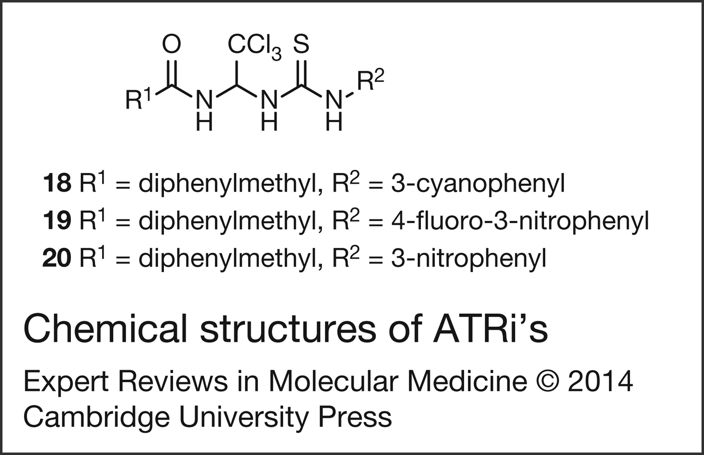
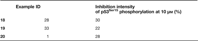
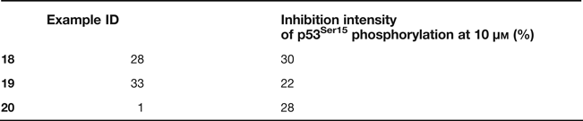
In 2007 CGK Co. Ltd. filed a patent on a series of urea derivatives as ATM/ATR inhibitors (Fig. 4) (Ref. Reference Kim, Won and Yi49). The compounds claimed blocked ATM and ATR-mediated phosphorylation of p53Ser15 in a concentration-dependent manner, both in an enzymatic assay and in RKO and GM847 cells, although no IC50 values were provided in this filing. The compounds inhibited the ATM and ATR-mediated phosphorylation selectively over a panel of known p53-targeting kinases (Table 1). Compound 18 showed synergistic effect with the cytotoxic agents cisplatin, etoposide and/or doxorubicin in Va-13, HeLa and/or MCF-7 cells. Additionally, co-treatment of RKO cells with 19 and doxorubicin-inhibited cell cycle at the G2/M phase. When BJ cells in replicative senescence were treated with compound 20, the cell division was increased and the process showed to be reversible. Compounds 19 and 20 were well tolerated in mice acute toxicity tests. It is worth noting that the same research group published data on compound 19 elsewhere (Ref. Reference Won50) but subsequently retracted the paper after an investigation (Ref. Reference Won51).
Chemical structures of ATRi's published by CGK Co.
Biological data of ATRi's published by CGK Co.
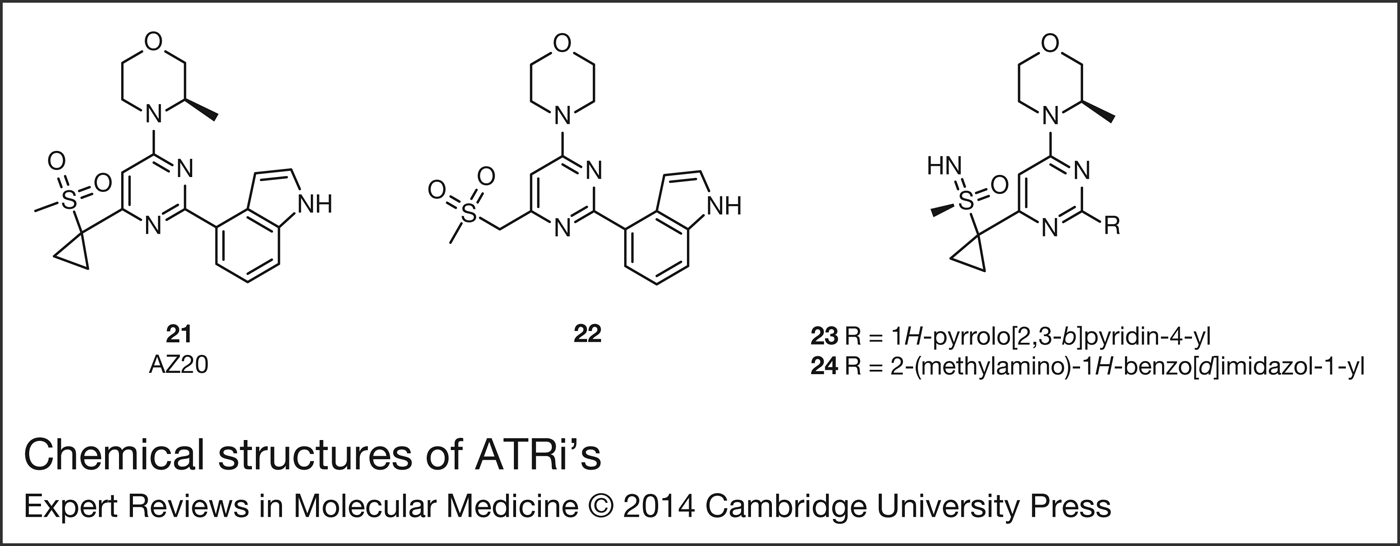
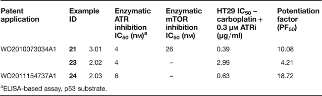
The first patents on potent and selective small-molecule inhibitors of ATR were filed in 2010 by AstraZeneca and Vertex Pharmaceuticals. These compounds were claimed for the treatment of cancer, as single agents or in combination with other anti-cancer drugs (Refs Reference Foote and Nissink52, Reference Foote, Nissink and Turner53, Reference Charrier54, Reference Charrier55, Reference Charrier56, Reference Charrier, Durrant and Knegtel57, Reference Charrier58, Reference Charrier59, Reference Charrier60, Reference Charrier61, Reference Charrier, Durrant and Shaw62, Reference Charrier63, Reference Charrier and Kay64, Reference Charrier65, Reference Charrier66, Reference Charrier67, Reference Charrier68, Reference Charrier69, Reference Everitt70, Reference MacCormick71, Reference Pollard and Reaper72, Reference Storck73). AstraZeneca presented compounds with ATR and/or mTOR IC50 values and IC50's for HT29 colon cancer cell growth, alone and in combination with the DNA-damaging agent carboplatin (IC50 = 3.97 µg/ml in HT29 cells) (Table 2) (Refs Reference Foote and Nissink52, Reference Foote, Nissink and Turner53). The synergism of the compounds was assessed by the potentiation factor 50 (PF50) which is calculated as a ratio of the IC50 of control cell growth in the presence of carboplatin, divided by the IC50 of cell growth in the presence of this agent and the ATRi. Compounds were compared using the PF50 value at concentrations of ATRi that showed minimal growth inhibition on their own in a sulforhodamine B (SRB) assay after 5 days of culture. In accordance with Toledo et al. (Ref. Reference Toledo37), the AstraZeneca group suggested that compounds with reduced mTOR inhibition are preferred since they may ameliorate off-target effects. A follow-up publication by the same group reported on the development of compound 21 (Fig. 5) from the 2010 patent (Ref. Reference Foote74). Initial hit 22 (Fig. 5) was identified as a potent ATRi from an mTOR screening campaign (enzymatic/cellular ATR IC50's = 30 nm/1.1 nm; enzymatic/cellular mTOR IC50's = 0.33 µm/5.9 µm) with high selectivity against other PIKK and PI3 K kinases. The group determined cell-free ATR inhibition using an ELISA-based assay for the detection of p53Ser15 phosphorylation whilst cellular ATR inhibition was measured in HT29 cells using pCHK1Ser345 phosphorylation as a marker. Medicinal chemistry optimisation of 22 lead to the discovery of 21 (enzymatic/cellular ATR IC50's = 5 nm/50 nm). 21 showed weak potency in a mTOR cell-based pAKT assay and displayed good selectivity against PI3Kα, ATM and DNA-PK, amongst a panel of 442 kinases, and no inhibition of the hERG channel, but suffered from time-dependent inhibition of CYP450 3A4. Compound 21 inhibited cell growth in LoVo colorectal adenocarcinoma cells (GI50 = 0.2 µm), as measured by an MTS assay after 72 h. Despite poor aqueous solubility (10 µm), 21 showed high Caco-2 permeability and good stability in rat hepatocytes, which led to respectable bioavailability and plasma exposure in a rat pharmacokinetic (PK) study. Female nude mice grafted with LoVo cells and treated with 21 alone (25 mg/kg twice a day or 50 mg/kg once a day) for 13 days presented a significant decrease in tumour growth and the treatment was generally well tolerated by the animals (Ref. Reference Foote74). No mention was made in the paper to the fact that 21 also exhibited good synergy with carboplatin in HT29 colon adenocarcinoma cell growth inhibition (Table 2) (Ref. Reference Foote74). Sulfonimides 23 and 24 (Fig. 5) from a subsequent patent were also potent ATRi's and compound 24 strongly increased the cytostatic effect of carboplatin (Table 2) (Ref. Reference Foote, Nissink and Turner53). The company has recently announced a clinical trial in patients with 11q-deleted relapsed/refractory CLL, PLL or B-cell lymphomas to determine the safety, tolerability, PKs and biological activity after oral administration of the ATRi AZD6738 alone (undisclosed structure). The study will examine biomarkers of ATR inhibition/status such as and γH2AX, pCHK, pATR and will identify patients with ATM-deficient status, determine ATM mutation load and correlate this with clinical efficacy (Ref. 24).
Chemical structures of ATRi's published by AstraZeneca.
Biological data of ATRi's published by AstraZeneca.
aELISA-based assay, p53 substrate.
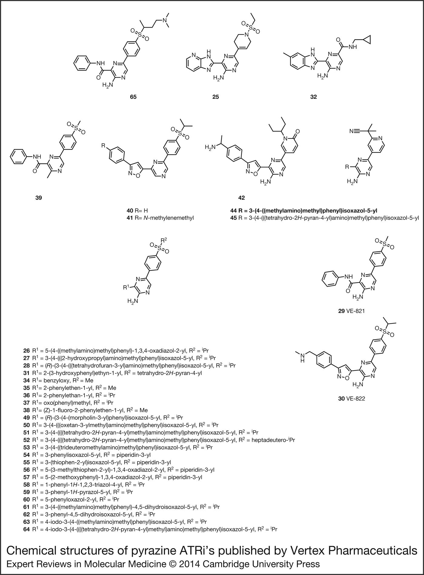
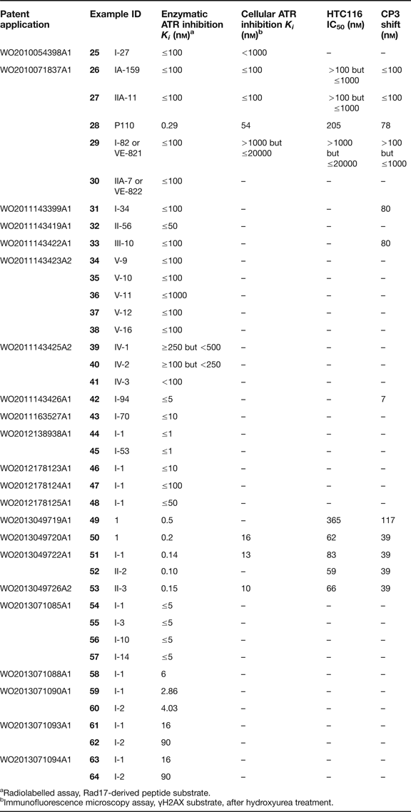
In 2010, researchers at Vertex Pharmaceuticals filed two patents covering a broad number of pyrazines as ATRi's (Fig. 6) (Refs Reference Charrier54, Reference Charrier56). The initial filings were followed-up by a number of patents providing detailed information on selected compounds, including enzymatic/cellular inhibition of ATR kinase and viability values for HCT116 cell growth, as measured by an MTS assay after 96 h, alone and in combination with the DNA-damaging agent cisplatin (Table 3). Many of the examples listed in these filings inhibited the full-length ATR with K i <100 nm. Vertex expressed the synergy level of the inhibitors using the ‘CP3 shift’, which is the concentration of compound required to reduce the IC50 of cisplatin alone by at least 3-fold. This metric does not take into account the cytotoxicity displayed by the inhibitors alone in contrast to AstraZeneca's measure of synergism.
Chemical structures of pyrazine ATRi's published by Vertex Pharmaceuticals.
Biological data of ATRi's published by Vertex Pharmaceuticals.
aRadiolabelled assay, Rad17-derived peptide substrate.
bImmunofluorescence microscopy assay, γH2AX substrate, after hydroxyurea treatment.
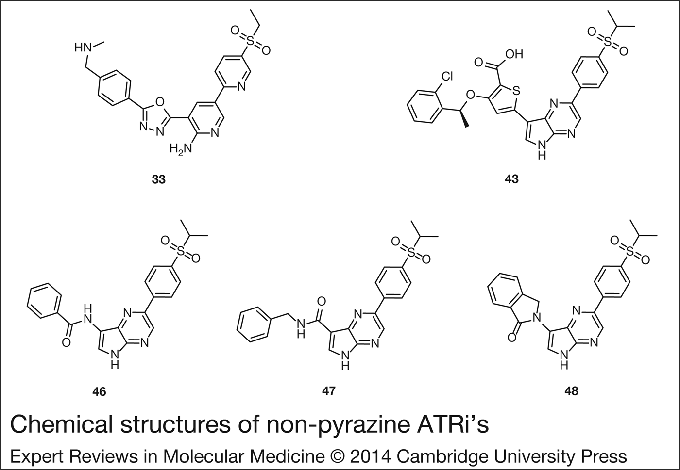
The compounds claimed generally feature a central 2-aminopyrazine core, but 2-aminopyridine (33) or pyrrolopyrazine (43, 46, 47, 48) are also described (Fig. 7) (Refs Reference Charrier55, Reference Charrier59, Reference Charrier61, Reference Charrier63, Reference Charrier66, Reference Everitt70, Reference MacCormick71, Reference Storck73). Examples lacking the 2-amino group such as 39, 40 and 41 are less frequent and result in higher enzymatic ATR K i ranges (Ref. Reference Charrier61). The pyrazine ring is often flanked at the 3-position by an amide (29) or a 5-membered heterocyclic linker, most commonly isoxazole (30) but also oxadiazole (26) (Refs Reference Charrier56, Reference Charrier, Durrant and Knegtel57), pyrazole (59) (Ref. Reference Charrier67), oxazole (60) (Ref. Reference Charrier67), triazole (58) (Ref. Reference Charrier, Durrant and Shaw62) and dihydroisoxazole (61) amongst others (Ref. Reference Charrier and Kay64). Iodinated isoxazole rings (63, 64) maintain acceptable enzymatic activity and present an attractive option to access triterated isotopes of ATRi, useful as tool compounds (Ref. Reference Charrier and Kay64). Other linkers such as ether (34) (Ref. Reference Charrier63), ketone (37) (Ref. Reference Charrier63), alkane (36) (Ref. Reference Charrier63), alkene (35) (Ref. Reference Charrier63), alkyne (31) (Ref. Reference Charrier59) or bicyclic systems (25, 32) (Refs Reference Charrier54, Reference Charrier58) are generally mentioned with higher K i ranges and to a lesser extent. Structural motifs that allow for intramolecular hydrogen bonding with the exocyclic amino group at the 2-position of the pyrazine ring usually display good enzymatic potency. The above-mentioned linker usually leads to an aromatic ring, mainly benzene (29) but also thiophene (55) (Ref. Reference Charrier, Durrant and Knegtel57), often containing an aliphatic amine substituent (30).
Chemical structures of non-pyrazine ATRi's published by Vertex Pharmaceuticals.
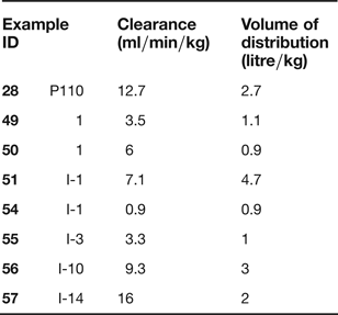
The central heterocyclic core is usually substituted on the 5-position with an appropriate spacer leading to a hydrogen bond acceptor placed within a ‘sphere’ located 6 Å away from the pyrazine's C5 on a line defined by the C5–C2 axis (Fig. 8) (Ref. Reference Charrier54). This hydrogen bond acceptor is usually presented as an aliphatic sulfone, but occasionally a pyridone or a cyanopyridine can also assume this role, as seen with examples 42 and 44 (Refs Reference Charrier55, Reference Charrier65, Reference Charrier68). It is worth noting that 3-piperidine sulfones (54, 55, 56, 57) confer good PK properties when compared with similar 4-piperidine compounds (Table 3) (Ref. Reference Charrier, Durrant and Knegtel57).
Schematic representation of the position of the hydrogen bond acceptor.
During 2013 additional patents providing biological data on very potent ATRi's were published. Compounds 49 (Ref. Reference Charrier69) and 50 (Ref. Reference Charrier, Durrant and Shaw62) are sub-nanomolar inhibitors with encouraging synergy in combination with cisplatin and good PK profiles (Tables 3 and 4). Compound 51 was characterised as various solid forms and several deuterated analogues such as 52 were described (Ref. Reference Charrier, Durrant and Knegtel57). Deuterium bioisostere 52 showed the same biological efficacy as the parent compound 51. The tetrahydropyran ring in 51 and 52 led to an improvement in comparison with the tetrahydrofuran derivative 28 included in a 2010 filing (Ref. Reference Charrier56), not only in terms of cytotoxic effect but also in the PK profile (Tables 3 and 4). Compound 51 was tested in two mouse models: colon adenocarcinoma Colo205, 51 (40 mg/kg), in combination with irinotecan (20 mg/kg) and primary non-small cell lung cancer (NSCLC), 51 (30 mg/kg), in combination with cisplatin (3 mg/kg). In both cases there was a marked tumour regression in comparison with the control groups (<98% reduction in tumour size in the case of cisplatin combination). Equivalent doses of a compound with a favourable in vitro profile such as 28, did not show sensitisation in similar in vivo experiments. The greater effectiveness of 51 versus 28 in the animal models can be explained by the improved in vitro and PK properties of 51.
PK data of ATRi's published by Vertex Pharmaceuticals.
The Vertex group expanded on their findings with two follow-up publications. The first communication presented extensive biological characterisation of the synthetic lethal interaction between ATR and the ATM-p53 tumour suppressor pathway in cells responding to DNA damage (Ref. Reference Reaper75). Compound 29 is a potent ATP-competitive ATRi (enzymatic K i = 13 nm) with good selectivity over a panel of kinases including ATM, DNA-PK, mTOR and PI3Kγ (K i of 16 µm, 2.2 µm, >1 µm and 3.9 µm, respectively), although Rsk3 was highly inhibited at 2 µm. 29 showed synergy with a range of genotoxic agents in HCT116 cells, as measured by an MTS assay, in particular with the DNA cross-linking drugs cisplatin and carboplatin, nevertheless this effect was not observed in normal cells or ATR-defective cell lines. HCT116 cells express normal levels of p53 but are ATM-deficient, indicating that these cells are more reliant on ATR for survival from DNA damage. This hypothesis was confirmed with ATM-null cells from an individual with ataxia telangiectasia (AT1BR cells) and with cells from a healthy donor (161BR cells). In both cases 29 showed clear synergism with cisplatin. Apoptosis levels were studied in a p53-mutant cancer line (H23); 29 in combination with cisplatin did not induce cell death after 24 h and apoptosis only became apparent at the 96 h time point. When administered alone, 29 reversibly limited cell cycle progression in 161BR normal cells without inducing significant cell death or long-term detrimental effects but induced substantial cell death in AT1BR cancer cells. The group claimed that ATR inhibition promotes DSB formation at sites of DNA replication stress which in normal conditions activates an ATM-dependant checkpoint that stops DNA replication, decreasing further DNA damage and promoting cell survival. In ATM pathway-deficient cancer cells, S phase arrest does not occur, leading to cytotoxic levels of DNA damage accumulation. The second paper expanded on the structure–activity relationships (SAR) of some pyrazine ATRi's and introduced an ATR homology model based on the structure of the related kinase PI3Kγ (Ref. Reference Charrier76). Although the sequence identity between the kinase domain of the two enzymes is low, the group identified key amino acid residues and discussed the predicted interactions. Compound 65 was highlighted as a potent ATP-competitive inhibitor of ATR (enzymatic K i = 12 nm, cellular ATR IC50 = 420 nm) with excellent selectivity over a panel of more than 50 kinases, including ATM and DNA-PK (IC50's >8 µm), good aqueous solubility as well as cell permeability and minimal efflux in a Caco-2 assay. 65 affected ATM-defective HCT116 cancer cells, both alone (IC50 = 1.1 µm) and in combination with cisplatin or ionising radiation (IR), but showed no effect in HFL1 non-cancerous cells, in agreement with the results observed when cancer cells are treated with other ATR-selective inhibitors in combination with DNA cross-linking agents.
Compounds 29 and 30 have also been specifically claimed for the treatment of pancreatic cancer and NSCLC (Ref. Reference Pollard and Reaper72). Both compounds sensitised pancreatic cancer cells, but not normal cells, to radiation therapy. 30 synergised well with cisplatin and gemcitabine in pancreatic cancer cells and enhanced not only their anti-tumour effect, but also the effect of IR in pancreatic cancer xenograft mouse models. Additionally 30 synergised with cisplatin, oxaliplatin, etoposide, gemcitabine and irinotecan in several lung cancer cell lines. Compound 30 has been characterised as various solid forms and deuterated analogues such as 53 have also been described (Ref. Reference Charrier68). Compound 30 (also known as VX-970) has entered a clinical trial to evaluate the safety, tolerability and response after intravenous administration, alone and in different combinations with gemcitabine, cisplatin or etoposide in subjects with advanced solid tumours, squamous NSCLC), relapsed or refractory small cell lung cancer (SCLC) and metastatic oestrogen receptor, progesterone receptor, and HER2 negative (triple negative) breast cancer (TNBC) with and without BRCA1/BRCA2 mutation(s) (Ref. 25).
Summary/Outlook
The efforts towards the development of ATRi's have undergone a remarkable upswing in the past years. From classical phenotypic approaches based on complex natural products (Refs Reference Sarkaria30, Reference Sarkaria31, Reference Nishida35) or simple small molecules (Ref. Reference Peasland45), through rational discoveries associated with investigations of the PIKK family and/or related lipid kinases (Refs Reference Knight29, Reference Toledo37) to industrial high-throughput screening campaigns (Refs Reference Foote74, Reference Charrier76), the ATR field has benefited from the work of both academic groups and pharmaceutical companies. Although the first ATR-selective inhibitor schizandrin B 10 was published in 2007 (Ref. Reference Nishida, Hamamori and Konishi34), the weak enzymatic potency and low microsomal stability in mice has limited its preclinical development (Ref. Reference Cao77). NVP-BEZ235 13 entered clinical trials as a dual PI3 K/mTOR inhibitor but recent data published by Toledo et al. questions the mechanism of action of this clinical candidate and starts a debate on the true molecular target behind the observed therapeutic effect (Ref. Reference Toledo37). There is a growing body of opinion suggesting that in the oncology therapy, and especially within the kinase field, a ‘selective polypharmacology’ approach might be required to overcome cancer resistance (Refs Reference Morphy78, Reference Knight, Lin and Shokat79, Reference Dar80). The early clinical success of the Novartis pan-PIKK inhibitor NVP-BEZ235 13 in mono-therapy could be attributed to a favourable ‘non-selective’ profile as seen with other marketed kinase inhibitors such as imatinib or sorafenib (Ref. Reference Morphy78). On the other hand, AstraZeneca and Vertex Pharmaceuticals entered the ATR arena with selective small-molecule inhibitors which have been progressed into clinical evaluation. The AstraZeneca clinical trial will help to establish the safety and tolerability of AZD6738, as well as giving insight on ATR inhibition and ATM functional status, which will aid to determine what patients will benefit the most from the treatment. The AstraZeneca inhibitors slow cancer cell growth when used alone, as seen with 21, indicating a potential use in mono-therapy. However, the compounds show a remarkable synergistic effect when combined with carboplatin, in accordance with the literature: ATRi's kill cancer cells more effectively when combined with DNA-damaging agents such as antimetabolites, alkylating agents, topoisomerase inhibitors or platinating compounds, being the later most commonly used, translating to marked tumour regression in cancer mouse models (Ref. Reference Wagner and Kaufmann81). In contrast to the mono-therapy trial sponsored by AstraZeneca, Vertex Pharmaceuticals has applied the pre-clinical information available on synergistic combinations with ATRi's for the rational design of the on-going clinical trial with VX-970 30. The scientific community will follow with interest the outcome of the NVP-BEZ235, AZD6738 and VX-970 clinical trials, which will provide invaluable information on the therapeutic validity of DNA-damage enhancing agents.
Acknowledgements
The authors would like to thank Oliver Mortusewicz, Martin Scobie and Ulrika Warpman-Berglund for insightful comments.
Financial support
We would like to acknowledge the Knut and Alice Wallenberg Foundation, Vinnova, the Swedish Research Council, Swedish Cancer Society, the Swedish Pain Relief Foundation and the Torsten and Ragnar Söderberg Foundation for funding.
Conflicts of interest
None.