Introduction
Glandular fever (also known as infectious mononucleosis; caused by the Epstein–Barr virus) is a very common illness with a wide range of described neurological and other complications. True nerve compression occurring as a direct result of cervical lymphadenopathy has not previously been described. This case report describes such a complication.
Case report
A 35-year-old female doctor self-presented to the ENT department with an 18-hour history of a unilateral Horner syndrome. Two weeks previously, she had become unwell with symptoms of lethargy, malaise, anorexia, swinging pyrexia and tender cervical lymphadenopathy.
She had presented to her general practitioner one week following symptom onset, and a diagnosis of glandular fever had been confirmed by blood tests (a positive monospot test, lymphocytosis with atypical mononucleocytes, a raised erythrocyte sedimentation rate (60 mm/hour)) and deranged liver function tests (alanine aminotransferase 280 U/l, alkaline phosphatase 680 U/l). During the second week, the swinging pyrexia and anorexia subsided; however, cervical lymphadenopathy increased in volume, with associated neck pain, and she developed left upper quadrant pain. At day 13, she developed partial left Horner syndrome, but this was not associated with increased neck pain, headache or other neurological symptoms.
Examination at the ENT department identified left upper lid ptosis (blepharoptosis) and meiosis but no clear anhydrosis. She had extensive bilateral lymphadenitis, more notable on the left side (Figure 1).
Photograph showing unilateral left Horner syndrome.

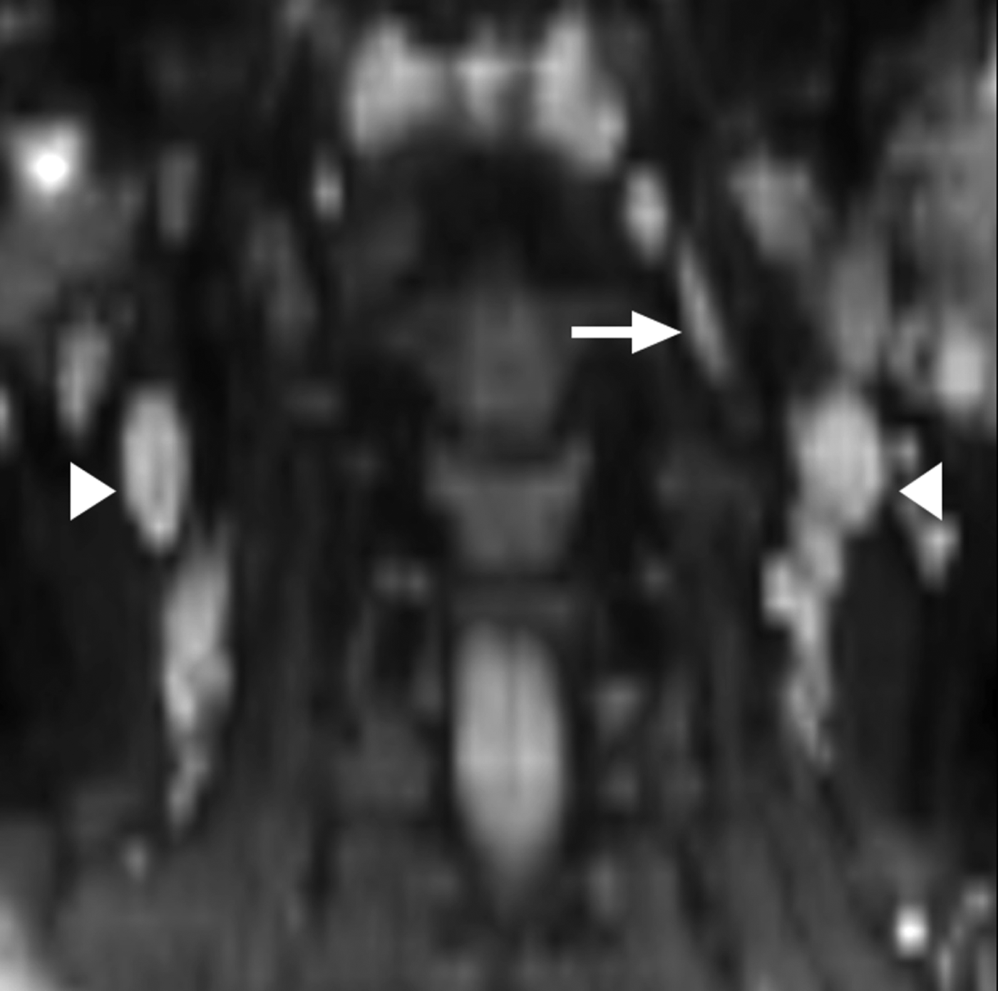
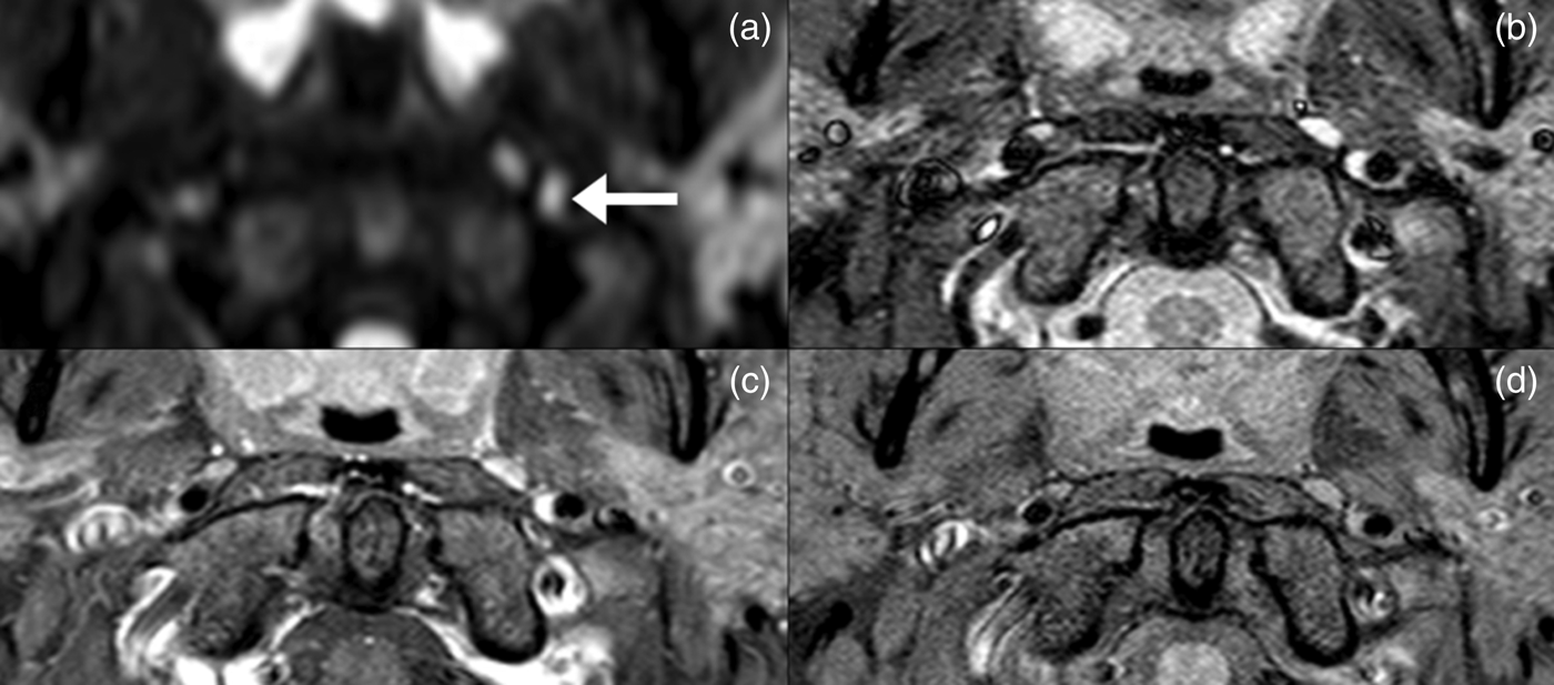
Repeated baseline blood tests confirmed the diagnosis of glandular fever. A chest X-ray was unremarkable and she underwent magnetic resonance imaging (MRI) of the neck. The MRI demonstrated bilateral cervical lymphadenopathy, which was particularly extensive along the carotid sheath and at levels IIa and III. There was also a clear abnormality within the carotid sheath superior to the left carotid bifurcation. Contrast-enhanced magnetic resonance angiography (MRA) of the neck was also performed, which confirmed this to be an intra-carotid sheath lymph node, and excluded a small internal carotid artery (ICA) dissection as the causative pathology (see Figures 2 and 3).
Coronal reformatted diffusion-weighted magnetic resonance imaging (b-value 700 seconds/mm2) demonstrating bilateral reactive cervical lymphadenopathy (arrowheads). There are also bilateral retropharyngeal nodes, with an asymmetrically large left-sided node within the posteromedial carotid space (arrow).
(a) Axial (b-value 700 seconds/mm2), (b) short tau inversion recovery (STIR), (c) fat-saturated (FS) T1-weighted post gadolinium and (d) FS T1-weighted magnetic resonance imaging of the intra-carotid sheath node (arrow on image a), which resembles a small carotid dissection on image b.
The patient was managed expectantly and the Horner syndrome symptoms resolved within six days of onset. This coincided with a visible reduction in the volume of cervical lymph nodes. She continued to improve and was able to return to work after four weeks.
Glandular fever is most commonly caused by infection with Epstein–Barr virus (EBV), a herpes-type DNA virus. Over 95 per cent of adults worldwide have been exposed to EBV, usually during the first two decades of life.Reference Luzuriaga and Sullivan1 The disease is classically characterised by the triad of pharyngitis, pyrexia and cervical lymphadenopathy, although other common symptoms include profound fatigue, malaise, anorexia and splenic enlargement.Reference Connelly and DeWitt2, Reference Odumade, Hogquist and Balfour3 It is usually self-limiting, with symptoms resolving within a mean of 19 days, and almost all within 4 weeks. Whilst cervical lymphadenopathy and fatigue may resolve more slowly, most patients return to normal activities within two to three months with no long-term sequelae. Complications of acute EBV infection, such as splenic rupture, haematological sequelae or neurological involvement, are rare.
The sympathetic nerve supply to the eye, head and neck occurs via a three-neuron pathway; disruption at any point in this chain may result in Horner syndrome. The first-order neuron arises in the hypothalamus, descends through the midbrain and terminates in the ciliospinal centre between C8 and T2. The second-order neuron exits the spinal cord at T1 and joins the para-vertebral sympathetic chain, running over the lung apex to the superior cervical ganglion which lies within the posterior medial carotid space at the level of C2–C3. Third-order neurons leave the ganglion and form a plexus within the adventitia of both the ICA (oculosympathetic branches) and external carotid artery (ECA); these nerve fibres are responsible for facial sweating.Reference Kedar, Biousse and Newman4–Reference Kong, Wright, Pesudovs, O'Day, Wainer and Weisinger6
Disruption of the cervical sympathetic nerves leading to Horner syndrome in the context of known EBV infection could result from a number of pathological processes. However, we believe the most likely cause in this patient to be physical compression of the sympathetic chain. This has been well described at different levels as a result of apical lung tumours, goitre and tuberculous lymphadenopathy.Reference Wilhelm, Ochsner, Kopycziok, Trauzettel-Klosinski, Schiefer and Zrenner7, Reference Ismail, Hodkinson and Tuling8 Clinical and MRI findings show that the compression may be caused by either the large number of nodes surrounding the carotid sheath at levels 3 and 4 or the intra-carotid sheath lymphoid tissue.
The patient demonstrated incomplete or partial Horner syndrome with meiosis and ptosis, but no anhidrosis. This suggested involvement at the level of or distal to the ICA because the nerve fibres innervating the sweat glands, which run alongside the ECA, were preserved. In Figure 3, MRI short tau inversion recovery images showed an abnormality in the carotid sheath, but cannot differentiate between an intra-carotid sheath node and a small carotid artery dissection. Pre-contrast fat-saturated MRI images showed that the node did not demonstrate a high T1 signal, which would have been typical of a mural haematoma. Diffusion and post contrast enhanced MRI images showed that the area of abnormality mirrors the signal of the other cervical nodes, confirming lymphoid tissue. Taken in combination with the clinical picture, these findings suggest that this intra-carotid sheath lymph node is probably the site of sympathetic nerve compression.
Interestingly, neurological complications of EBV infection are well recognised, occurring at a rate of 0.37–7.3 per cent.Reference Connelly and DeWitt2, Reference Mohanaruban and Fisher9, Reference Gavin, Langan and Hutchinson10 These may occur at the central (lymphocytic meningitis or encephalitis), cord (transverse myelitis) or peripheral (mono- or polyneuritis) levels. Involvement of almost all cranial nervesReference Flanagan, Hawkins and Bryars11–Reference Salazar, Martinez and Sotelo13 and a number of peripheral neuropathiesReference Mohanaruban and Fisher9, Reference Gavin, Langan and Hutchinson10, Reference Vucic, Palmer and Cros14, Reference Dodig, Ngo, Bailey and Bril15 have been reported, but none involve the autonomic nervous system, specifically not the cervical sympathetic nerves. The pathophysiology of nerve involvement previously described has not been clearly delineated, but is thought to be caused by immune complex deposition, direct viral invasion or an EBV immune-mediated process.Reference Van Baalen, Petersen and Stephani12 Physical nerve compression has not previously been suggested in any of these cases.
Direct ocular involvement can occur with EBV infection, affecting the central neuro-ophthalmological apparatus or causing direct inflammation of the eye and its adnexa.Reference Librach16 However, direct involvement of the sympathetic apparatus (either alone or in conjunction with other nerves) has not been described. In this case, in which there was a good alternative diagnosis, it seems unlikely.
• Glandular fever is a common viral illness that presents to ENT surgeons
• Neurological complications occur in up to 7 per cent of patients
• Horner syndrome may result from sympathetic nerve disruption at any level from the brain to the eye
• A common cause of Horner syndrome in young, fit patients is an internal carotid artery dissection
• Patients should be investigated by magnetic resonance imaging and magnetic resonance angiography
An important and relatively common cause of a partial Horner syndrome is an ICA dissection, which classically presents with unilateral head and neck pain, focal cerebral ischaemic symptoms and sympathetic nerve disruption.Reference Flaherty and Flynn17–Reference Chan, Paine and O'Day19 This was the main differential diagnosis in our patient, but was successfully ruled out by MRA, the accepted imaging modality of choice in diagnosing an ICA dissection and investigating Horner syndrome.Reference Kong, Wright, Pesudovs, O'Day, Wainer and Weisinger6, Reference Flaherty and Flynn17–Reference Yoshiura, Wu and Sorensen25
Conclusion
Whilst the neurological complications of Epstein–Barr virus infection are well recognised, Horner syndrome has not been previously described. We believe the most likely cause in this patient to be physical compression of the internal carotid artery sympathetic neurons by the enlarged intra-carotid sheath lymphoid tissue. This was demonstrated clearly upon MRI and magnetic resonance angiography and fits both the partial nature of Horner syndrome symptoms and their rapid resolution coincident with the reduction in lymph node bulk.