Congenital posterolateral diaphragmatic hernia – Bochdalek hernia – is a life-threatening condition frequently associated with various congenital cardiac diseases, which make the prognosis of congenital diaphragmatic hernia even worse.Reference Graziano1, Reference Cohen, Rychik and Bush2 Repair of congenital diaphragmatic hernia has been known to improve ventilation and perfusion of the once hypoplastic lung; however, parenchymal abnormality and ventilation-perfusion mismatch of the ipsilateral lung may persist as patients with repaired congenital diaphragmatic hernia grow up.Reference Hayward, Kharasch and Sheils3 In congenital diaphragmatic hernia associated with tetralogy of Fallot, central pulmonary artery size of the affected side thus may not reflect the capacitance of peripheral pulmonary vascular beds. In this regard, decision to close ventricular septal defect upon tetralogy of Fallot repair merely based on pulmonary artery size may be misleading, even though the affected lung appears to have grown adequately and the ipsilateral pulmonary artery looks sizeable. We report a case of congenital diaphragmatic hernia associated with tetralogy of Fallot, which was repaired after assessing the adequacy of the pulmonary vascular beds by intra-operative pulmonary blood flow study.Reference Honjo, Al-Radi and MacDonald4, Reference Reddy, Petrossian, McElhinney, Moore, Teitel and Hanley5
Case report
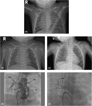
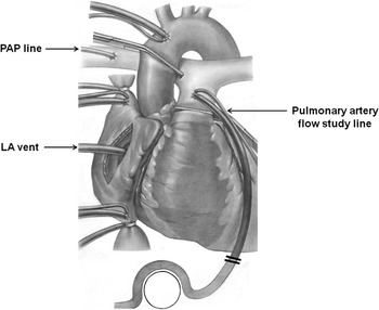
A male baby was born at 34 weeks of gestation and 2280 grams of birth weight. He had respiratory distress and cyanosis, and simple chest X-ray showed bowel gas at the left hemithorax (Fig 1a). He was diagnosed as having congenital diaphragmatic hernia associated with tetralogy of Fallot, and hence underwent repair of congenital diaphragmatic hernia on postnatal day 1. Owing to the fact that the patient was adequately saturated and his body size was small, surgical intervention for tetralogy of Fallot was deferred until postnatal day 39 when the baby weighed 3130 grams and a systemic–pulmonary shunt using a 3.5-millimetre polytetrafluoroethylene vascular graft was placed. Post-operative course was complicated by mechanical ileus from previous hernia surgery, and he underwent emergency small bowel resection and anastomosis. During post-operative follow-up, left lung volume gradually increased (Fig 1b and c), and pulmonary angiogram at 13 months after birth both showed sizeable pulmonary arteries (Fig 1d), which encouraged us to contemplate performing definitive repair of tetralogy of Fallot. Visualisation of the left pulmonary veins in the late phase of pulmonary angiogram was not evident compared with the right side (Fig 1e), and thus we speculated that left pulmonary artery size might not signify the proper growth of the peripheral pulmonary vascular beds in the left side. Thus, we elected to conduct intra-operative pulmonary blood flow study. After cardiopulmonary bypass was initiated and aorta was cross-clamped, an additional 8-French straight aortic cannula (Medtronic, Minneapolis, Minnesota, United States of America) was inserted into the main pulmonary artery through the divided end of the previous shunt (Fig 2). A pressure line was inserted into the right pulmonary artery for the continuous measurement of mean pulmonary artery pressure. The pulmonary vasculature was perfused with an incrementally increasing flow to a target rate of 2.5 litres per metre square per minute, whereas the left atrium was fully vented and the lungs were adequately ventilated. We decided to close the ventricular septal defect without fenestration because the mean pulmonary artery pressure obtained at the target flow rate was only 20 millimetres of mercury. With respect to the right ventricular outflow tract reconstruction, pulmonary valve annulus was preserved and the infundibular patch was placed after extensive infundibulectomy. After the discontinuation of cardiopulmonary bypass, systemic systolic blood pressure and right ventricular systolic pressure were 63 and 43 millimetres of mercury, respectively. The post-operative course was uneventful, and post-operative echocardiography showed right ventricular outflow tract pressure gradient of 25 millimetres of mercury and tricuspid regurgitation jet velocity of 2.7 metres per second. The patient was discharged home on post-operative day 8, and is currently in good clinical condition.
(a) Simple chest X-ray before hernia repair showing bowel gas in the left hemithorax and mediastinal shifting towards the right side. (b) Simple chest X-ray after the repair of congenital diaphragmatic hernia showing scanty lung parenchymal shadow in the left hemithorax. (c) Simple chest X-ray before the total correction of tetralogy of Fallot showing balanced ventilation and decreased perfusion of the left lung. (d) Pulmonary angiography before the total correction of tetralogy of Fallot showing adequately grown both pulmonary arteries. Ao = aorta; CS = conal septum; LPA = left pulmonary artery; RPA = right pulmonary artery. (e) Late phase of pulmonary angiogram showing faint visualisation of the left pulmonary veins. RPV = right pulmonary vein.

Intra-operative flow study. The ascending aorta is cross-clamped, and 8-French straight aortic cannula is inserted in the main pulmonary artery distal to the clamp. A pressure line is in the right pulmonary artery to measure the mean pulmonary artery pressure, and the left atrium is vigorously vented. LA = left atrium; PAP = pulmonary artery pressure.

Discussion
Congenital diaphragmatic hernia is a life-threatening anomaly with a mortality rate of approximately 40–50%. From the “Congenital Diaphragmatic Hernia Study” encompassing 82 centres and enrolling 2636 patients, the association of significant cardiovascular malformation was found in approximately 10% of the whole congenital diaphragmatic hernia cohort.Reference Graziano1 Cardiovascular malformation association of congenital diaphragmatic hernia has been reported to increase the risk of death by up to three times higher than congenital diaphragmatic hernia without cardiovascular malformation, mainly because of the detrimental effect of unilateral pulmonary hypoplasia, which would be poorly tolerated by the severely malformed heart.Reference Cohen, Rychik and Bush2 Furthermore, altered lung structure – that is, reduced number and increased size of alveoliReference Peetsold, Heij, Kneepkens, Nagelkerke, Huisman and Gemke6 – pulmonary vascular abnormality – that is, decreased number of small arteries and increased wall thicknessReference Peetsold, Heij, Kneepkens, Nagelkerke, Huisman and Gemke6 – and ventilation–perfusion mismatchReference Hayward, Kharasch and Sheils3 in the affected lung may hinder the correction of certain cardiovascular malformation, such as tetralogy of Fallot, in which the adequacy of the pulmonary vascular bed to accommodate systemic venous return is a critical prerequisite for successful outcome. There are a variety of methods to measure pulmonary vascular compliance, including analysis of pulmonary arterial diastolic pressure curve obtained from cardiac catheterisation;Reference Huez, Brimioulle, Naeije and Vachiery7 analysis of pulsed wave Doppler pattern;Reference Weinberg, Hertzberg and Ivy8 assumption of pulmonary vascular compliance from electrical circuit analogy;Reference Senzaki, Isoda, Ishzawa and Hishi9 intra-operative aortic/pulmonary flow measurement;Reference Kitagawa, Hori and Chikugo10 and intra-operative flow study. Intra-operative flow study excels others in that this technique allows direct assessment of flow/pressure relationship of the pulmonary vascular bed without any assumption. In this case, ventilation and perfusion of the left lung appeared to develop adequately before the repair of tetralogy of Fallot. Given the possibility of peripheral vascular abnormality in the left lung, however, we intended to ascertain the capacitance of the pulmonary vascular beds employing intra-operative pulmonary blood flow study, which was proposed to determine the physiological tolerance of ventricular septal defect closure in pulmonary atresia with ventricular septal defect and major aortopulmonary collateral arteries.Reference Honjo, Al-Radi and MacDonald4, Reference Reddy, Petrossian, McElhinney, Moore, Teitel and Hanley5