Introduction
Osteolysis of the temporal bone may be associated with cholesteatoma, Langerhans cell histiocytosis, osteoradionecrosis, benign tumours such as paragangliomas, and malignant and metastatic disease. Osteolysis can also occur as part of systemic bone resorption seen in hyperparathyroidism, and in connective tissue disorders. Gorham–Stout disease of the skull is a rare entity. It presents with gradual bone resorption, and proliferation of lymphoid and vascular channels within the bony matrix. Gorham–Stout disease is identified under a number of other names, including vanishing, disappearing or phantom bone disease. It was first described in 1838 by Jackson, and was described as a disease much later in 1954 by Gorham and Stout.Reference Gorham and Stout 1 It causes progressive loss of normal bone, through the process of osteolysis being replaced by proliferative vascular and lymphatic tissue.
The precise aetiology of Gorham's disease remains poorly understood and largely unknown. There is no evidence of a malignant, neuropathic, autoimmune, connective tissue error or infectious component involved in the causation of this disease. The reason for the angiogenic proliferation and the mechanism of bone resorption is also unclear. The diagnosis is normally one of exclusion with the benefits of histological and radiological investigations.Reference Ruggieri, Montalti, Angelina, Alberghini and Mercuri 2 The most commonly affected areas include the spine, shoulder, clavicle, ribs, pelvis, skull and mandible. Gorham–Stout disease is rarely ever fatal. However, because of its progressive nature, it can cause functional loss of the affected area, or even lead to fracture with low impact secondary to osteopenia.
We report a case of Gorham–Stout disease of the temporal bone that initially presented as a diagnostic dilemma and was confirmed on serial imaging.
Case report
A 51-year-old male presented with a gradually progressive painless swelling in his left mastoid bone and left temporomandibular joint (TMJ) for over 20 years, with a history of recurrent TMJ dislocation. He generally experienced approximately five episodes of TMJ dislocation a year, which seemed to be triggered by opening his jaw wide. He had discomfort on wearing spectacles, which pressed on his progressive mastoid swelling.
For many years, he had fluctuating left-sided hearing loss associated with deep-seated otalgia. At presentation, his hearing had reduced significantly on the left. He had no past history of middle-ear cleft disease. He had no tinnitus, vertigo or cranial neuropathy. There was no history of head trauma, or cranial, ear or TMJ surgical procedures. There was no family history of hearing loss, ototoxic drug use or loud noise exposure. He had no similar complaints in his right ear or right TMJ. He was systemically well, with no history of bone disease elsewhere in his body.
Clinical examination revealed normal tympanic membranes. The mastoid swelling was diffuse, with firm to hard areas. The swelling was non-compressible, non-pulsatile and non-tender, with no signs of inflammation. The contour of his TMJ appeared distorted, but there was no arthralgia and his mouth opening was adequate. He had no signs of infection in the skin over his mastoid or TMJ. There were no palpable cervical lymph nodes. Vestibular and cranial nerve examination findings were normal except for left-sided sensorineural hearing loss, confirmed on tuning fork tests. A pure tone audiogram showed near-normal hearing with slight sensorineural hearing loss in the higher frequencies on the right hand side, and a moderate to severe sensorineural loss on the left (Figure 1).
Audiogram of patient (a = right ear, b = left ear), showing left ear severe hearing loss. ○ = air conduction unmasked (right ear); △ = bone conduction unmasked (right ear); × = air conduction unmasked (left ear);] = bone conduction masked (left ear)

Initial scans of the temporal bone showed osteolytic changes in the left temporal bone region involving the TMJ. A thorough systemic metabolic bone disease investigation did not indicate any known condition. There was no similar bony pathology in any other bones in the patient's body.
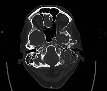
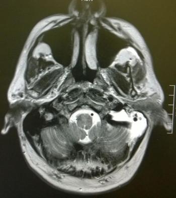
Two subsequent scans demonstrated a similar but progressive picture. There was evidence of further bone density loss in the temporal bone region and TMJ. The integrity of the middle-ear cleft and ossicles was preserved, but there were degenerative changes in the TMJ, resulting in recurrent dislocation. There was no evidence to suggest any affliction of vital structures in the lateral skull base. There was no radiological suspicion that this may be a malignant process. Hence, no biopsy of this lesion was recommended. The most recent computed tomography and magnetic resonance imaging scans (Figures 2 and 3 respectively) were compared with the previous scans and discussed in the skull base multidisciplinary team (MDT) meeting. The diagnosis of vanishing bone was confirmed, with the recommendation of continued observation and conservative management of symptoms.
Axial computed tomography scan showing vanishing left temporal bone replaced with lymphovascular soft tissues. R = right; L = left; A = anterior

Axial magnetic resonance imaging scan showing enhancement of the lymphovascular soft tissues within the temporal bone.

According to the MDT consensus of opinion, the clinical picture was in keeping with a benign, slowly progressive process, with no impending complications expected. Hence, the patient is currently being monitored with annual imaging in the otology clinic at our institute, and is being treated by the maxillofacial surgeons to improve TMJ stability. The future plan is to manage the symptoms conservatively, as this condition has been reported in the literature to become self-limiting over time and does not create life-threatening complications.
Discussion
Gorham–Stout disease is a very rare bone disease, with less than 200 cases having been reported across all body systems. There have been more reports from orthopaedic departments than from other specialties. It is a non-hereditary disease, with equal gender distribution, with onset typically in children or young adults. The random growth of abnormal venous and lymphatic tissues within the bone matrix causes destruction of bone integrity and contour. The clinical course is generally protracted but rarely fatal, with eventual stabilisation of the affected bone being the most common sequelae. Chylous, pericardial and pleural effusions may occur due to mediastinal extension of the disease process from involved vertebra, scapula, ribs or sternum.Reference Ruggieri, Montalti, Angelina, Alberghini and Mercuri 2 The disease may also lead to blood, lymph and cerebral spinal fluid leaks if involving the skull base suture lines communicating with the dural sheath of the central nervous system.Reference Morimoto, Ogiwara, Miyazaki, Kitamuara, Nishina and Nakazawa 3 , Reference Cushing, Ishak, Perkins and Rubinstein 4 There can be neurological deficits if the disease involves the craniocervical junction causing instability of spine.Reference Morimoto, Ogiwara, Miyazaki, Kitamuara, Nishina and Nakazawa 3 , Reference Plontke, Koitschev, Ernemann, Pressler, Zimmermann and Plasswilm 5
There have been a few case reports in the world literature that describe this rare entity involving the temporal bone.Reference Morimoto, Ogiwara, Miyazaki, Kitamuara, Nishina and Nakazawa 3 , Reference Plontke, Koitschev, Ernemann, Pressler, Zimmermann and Plasswilm 5 – Reference Mowry and Canalis 7 However, it is notable that only one such case has been associated with sensorineural hearing loss, similar to this report.Reference Nozawa, Ozeki, Kuze, Asano, Matsuoka and Fukao 6 There are no cases reported on Gorham–Stout disease isolated to the TMJ, although the mandible cortex and zygoma-maxilla complex are known to be involved.Reference Oujilal, Lazrak, Benhalima, Boulaich, Amarti and Saidi 8 There is a spectrum of presentations because of the varied osseous deformities associated with Gorham–Stout disease, but the disease is mostly expected to be self-limiting over time. Two reports have also mentioned spontaneous regression of the disease, but this is uncommon.Reference Mowry and Canalis 7 , Reference Choma, Biscotti, Bauer, Mehta and Licata 9
-
• Gorham–Stout disease can affect any head and neck bony structures
-
• Symptoms vary depending on the bone affected, and include hearing loss and temporomandibular joint dislocation
-
• This disease should be considered when imaging shows osteolytic involvement
-
• Treatment includes conservative, medical, radiotherapy or surgical approaches, depending on disease progression severity
If the disease progresses to become symptomatic, medical treatment is indicated. Bisphosphonates, propranolol, clodronate and alpha-2b interferon have been found to be useful given their anti-osteoclastic and anti-angiogenic effects.Reference Hagberg, Lamberg and Aström 10 Radiotherapy and surgery are alternative modalities if the disease is severe or needs reconstruction; moderate doses of radiotherapy have good results by inducing scarring of the lymphovascular channels within the bone, without causing significant morbidity.Reference Dunbar, Rosenberg, Mankin, Rosenthal and Suit 11
Conclusion
Gorham–Stout disease is a very rare, gradually progressive, but self-limiting osteolytic bone disease, which has the potential for spontaneous regression. The disease may affect many parts of the body, including the skull and mandible, as in this report, giving rise to symptoms of progressive hearing loss and recurrent TMJ dislocation. A conservative approach should be the first line of management; however, medical and radiotherapy treatment options are available if this disease should progress. In cases of complications such as cerebrospinal fluid leak, cranial neuropathy or TMJ dislocation, appropriate surgical remedy needs to be explored.