Introduction
Fasciolosis/liver fluke disease is caused by the trematode parasites Fasciola gigantica (tropical liver fluke) and F. hepatica (temperate liver fluke). It is considered to be one of the most serious pathogens causing ‘liver rot’ in ruminants and it inflicts a significant economic loss to the livestock sector, estimated at US$3.2 billion per annum worldwide (Spithill et al., Reference Spithill, Smooker, Sexton, Bozas, Morrision, Creaney, Parsons and Dalton1999). Moreover, fasciolosis is increasingly recognized as causing a significant human disease, with 2.4 million people infected and about 6 million people at the risk of infection (Mas-Coma et al., Reference Mas-Coma, Bargues and Valero2005; Moghadami & Mardani, Reference Moghadami and Mardani2008). The Ministry of Agriculture, Government of India, in its annual report for the year 1996–1997, has recorded fasciolosis as a disease of prime economic concern.
Control of this disease has been a challenging task for several decades and a subject of interest for researchers. The strategic control measures against fasciolosis mainly rely upon epizootiology-based forecasting and judicious use of antifluke drugs (Knox, Reference Knox1994). Other conventional methods for the control of fasciolosis include elimination of snails using molluscicides or by applying biological methods (Armour, Reference Armour1975). Application of molluscicides is a costly proposition and it has adverse effects on the non-target flora and fauna in the habitat (Amin & Fenwick, Reference Amin and Fenwick1977).
Very few antifluke drugs, such as triclabendazole, are efficient against both adult and immature flukes (Boray et al., Reference Boray, Crowfoot, Strong, Allison, Schellenbaum, Von Orelli and Sarasin1983). However, chemotherapy has limitations on account of development of resistance to anthelmintics (Overend & Bowen, Reference Overend and Bowen1995) and the cost of treatment impedes application of chemotherapy within the rural areas of developing countries. Alternative control measures, such as development of vaccines, are therefore warranted. The development of a suitable immunoprophylactic strategy involves evaluation of various defined antigens of F. gigantica for their potential in inducing immunoprotection in susceptible animals, but to date no successful vaccine has been developed against this disease.
In order to identify the novel immunogen conferring significant protection against fasciolosis, research has been focused on the defined antigens of the fluke. These include glutathione S-transferase, fatty acid binding protein, cathepsin-L, fluke haemoglobin, paramyosin, myosin, leucine amino peptidase and F. hepatica-Kunitz Type molecule. These molecules are involved in various metabolic processes that are crucial to the development and survival of the parasite. One of the most promising candidates has been glutathione S-transferase (GST) (Morrison et al., Reference Morrison, Colin, Sexton, Bowen, Wicker, Friedel and Spithill1996). These molecules have been tried as likely vaccine candidates against Schistosoma spp. and Fasciola spp. (Mitchell, Reference Mitchell1989; Preyavichyapugdee et al., Reference Preyavichyapugdee, Somphong, Riengrojpitak, Grams, Viyanant and Sobhon2008). In the various immunization trials in cattle, buffaloes and sheep, GST has been evaluated for its protective efficacy and has given promising results of 49–69% protection in these animals (Sexton et al., Reference Sexton, Milner, Thompson, Wilson, Spithill, Mitchell and Campbell1990; Morrison et al., Reference Morrison, Colin, Sexton, Bowen, Wicker, Friedel and Spithill1996). Recently, GST of S. japonicum has found application in the serodiagnosis of Schistosoma japonicum infection in water buffaloes and cattle (Zhu, Reference Zhu2005; Cheng et al., Reference Cheng, Tsaihong and Lee2007).
The aim of the present study was to clone the essential molecule, glutathione S-transferase of F. gigantica for generating a recombinant protein for its future exploitation in the immunodiagnosis and immunoprophylaxis against tropical liver fluke disease.
Materials and methods
Parasite
Adult F. gigantica flukes were recovered from the biliary system/gall bladder of the bubaline liver, at necropsy of animals slaughtered at Bareilly abattoir (Uttar Pradesh, India). The flukes were thoroughly washed several times with chilled phosphate-buffered saline (PBS), pH 7.2, until they were free from adhering host tissues and host secretion (bile), and were thereafter processed for the isolation of total RNA for cDNA synthesis.
RNA isolation
Isolation of the total RNA from these live parasites was carried out using an RNA isolation kit (RNeasy kit: Qiagen, GmbH, Germany). Briefly, 50 mg of tissue from a single fluke was triturated with a sterile pestle and mortar in lysis buffer and further steps for the isolation of RNA were followed as per the protocol recommended by the manufacturer.
Reverse transcriptase-PCR (RT-PCR)
Complementary DNA (cDNA) from the total RNA was synthesized by reverse transcription of the gene signal. One microlitre of template RNA (1 μg), 1 μl (0.5 μg/μl) of oligo (dT) primer and water added to a total volume of 12 μl were incubated at 70°C for 10 min. Following incubation, the reaction mixture was chilled on ice and 1 × reaction buffer, 1 m dNTP, RNase inhibitor (20 U) and 200 U of RevertAid M-MLV reverse transcriptase (MBI Fermentas, Latvia) were added and incubated at 42°C for 60 min in a thermocycler (MJ Research, Waltham, Massachusetts, USA), followed by incubation at 70°C for 10 min. The cDNA thus synthesized was preserved at − 20°C until further use.
PCR amplification of cDNA
The cDNA was used as a template for polymerase chain reaction (PCR) amplification, using FgGST-F (5′-ATG CCA GCC AAA CTC GGA TAC-3′) and FgGST-R (5′-TCA AGC CGG TGC AGC GTC TC-3′) primers. Gene amplification was performed with 0.2 mm dNTPs, 1.5 mm MgCl2, Taq DNA polymerase buffer (1 × ), 2.5 units of Taq DNA polymerase (Promega Corporation, Madison, Wisconsin, USA) and 1 μl of template cDNA, with initial denaturation for 5 min at 94°C, followed by annealing for 45 s at 52°C and elongation for 1 min at 72°C for 35 cycles. The PCR product was analysed by electrophoresis in 1% agarose gel.
The purified PCR product was cloned in pDrive (T-A) cloning vector (Qiagen). Recombinant colonies with the GST gene were confirmed by colony PCR and restriction digestion of the clones. The nucleotide sequence of the cDNA clones was determined by custom sequencing. The 657 bp GST gene was sub-cloned at BamH1 and HindIII restriction sites of the prokaryotic expression vector pPROEXHTb (Invitrogen, Carlsbad, California, USA) in frame with a histidine tag as a fusion partner.
Expression and purification of the recombinant protein
Two positive clones harbouring the desired GST gene were induced for protein expression by 1 mm isopropyl thiogalactoside (IPTG) and analysed on 10% SDS-PAGE (Laemmli, 1970) under reducing conditions.
One hundred millilitres of the IPTG-induced recombinant Escherichia coli DH5α culture were pelleted and the bacterial cells lysed in the lysis buffer (pH 8.0) for 1 h at room temperature (RT) with intermittent vortexing. The supernatant was mixed with 600 μl of nickel nitrilotriacetic acid (Ni-NTA) agarose (Qiagen), supplemented with 10 mm imidazole. Binding of the recombinant protein with Ni-NTA resin was carried out at RT for 1 h with gentle shaking. The lysate–resin mixture was washed with 15 ml of wash buffer (pH 7.0) and finally eluted with 4 × 500 μl of elution buffer (pH 4.5). All the three buffer concentrations were 10 mm Tris and 100 mm NaH2PO4 with 8 m urea. The eluted protein was dialysed overnight (O/N) against PBS (pH 7.2) or Tris–saline–EDTA, pH 7.0 (Tris 25 mm, NaCl 25 mm, EDTA 2 mm), with a step gradient of 6 m to 0 m urea.
The recombinant protein was also purified from the bacterial lysate under non-denaturing conditions. Bacterial cells were treated with lysozyme (1 mg/ml on ice for 1 h), sonicated and the soluble fraction of the bacterial lysate was subjected to Ni-NTA affinity chromatography purification. Glutathione S-transferase was purified in its native conformation by using 10 mm imidazole in the binding and wash buffers (pH 8.0), respectively, and 250 mm imidazole in the elution buffer (pH 8.0). Buffer concentrations were 10 mm Tris, 100 mm NaH2PO4 and 300 mm NaCl for all the steps. The eluted protein was dialysed against PBS (pH 7.2) and the purified protein stored at − 20°C.
Dot-ELISA
Dot-ELISA (enzyme-linked immunosorbent assay) with soluble GST protein was carried out with the antigen concentrations of 1 μg, 500 ng, 200 ng and 100 ng, respectively, blotted to the nitrocellulose membrane (Biorad, Hercules, California, USA). The antigen was reconstituted in PBS (pH 7.2) and dots of 1 μl volume of the antigen were applied to the nitrocellulose strips. The antigen spots were air dried, blocked with 5% skimmed milk and washed with PBS-Tween 20 (wash buffer). Fasciola gigantica infected sera of buffaloes, collected from a local abattoir, were screened for anti-Fasciola antibodies at serial dilutions of 1:50–1:400. Immuno-reactivity of the F. gigantica positive sera was probed with rabbit anti-bovine IgG–horse radish peroxidase (HRP) conjugate (Sigma, St. Louis, Missouri, USA) at 1:2000 dilution. ELISA dots were developed with diaminobenzedine substrate (Sigma). Sera positive for F. gigantica infection showed purple dots on the membrane.
Results
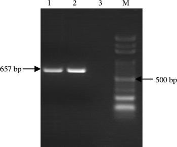
RT-PCR carried out using cDNA as a template resulted in an amplification product of 657 bp GST (fig. 1). Cloning of the gene was confirmed by colony PCR and restriction digestion of the recombinant clones confirmed the correct insert size of 657 bp. Sequence analysis of the isolate revealed 99.1% homology with F. gigantica GST of cattle origin (accession no. AF112567), with six nucleotide changes at base pairs 72, 74, 423, 513, 549 and 627 observed in the present isolate. The mutation at 74 bp caused a change of Asp to Gly, at 423 bp a change from Gln to His, at 513 bp from Asn to Lys, and at 549 bp caused a change of Ile to Met, respectively. However, mutations at 72 bp and 627 bp did not lead to any change of amino acids. Mutations at six nucleotides caused an overall change of four amino acids in the protein. The GST gene sequence of this Indian isolate of F. gigantica (buffalo origin) has been accorded the NCBI GenBank accession number FJ 024084.
RT-PCR amplification of the GST gene. Lanes: 1 and 2, 657 bp PCR product; 3, negative control; M, 100 bp DNA ladder marker.

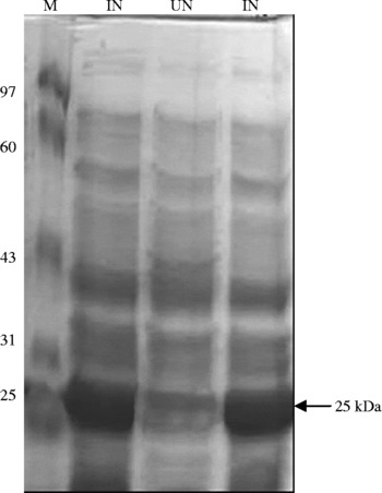
Glutathione S-transferase protein was expressed in E. coli DH5α using a prokaryotic expression vector pPROEXHTb. A time course study for optimizing the level of protein expression showed that 8–12 h growth of bacteria post-IPTG induction was sufficient for optimal expression of the recombinant protein (fig. 2). Likewise, 1 mm IPTG induced optimal protein expression, when 0.25 mm, 0.5 mm, 0.75 mm and 1 mm IPTG concentration were checked for the protein induction.
Expression of the recombinant GST protein in E. coli. SDS-PAGE fractionation (15%) of the recombinant E. coli lysate. Lanes: UN, un-induced control; IN, shows expression of 25 kDa GST induced with 1 mm IPTG; M, protein marker.

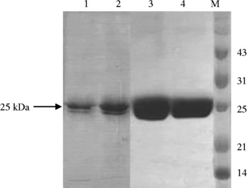
The recombinant protein (rGST) was purified under denaturing conditions using Ni-NTA metal chelating affinity chromatography. Certain modifications, such as incorporation of 10 mm imidazole in the binding buffer and washing with wash buffer without imidazole at pH 6.3, improved the purity of the recombinant protein. Elution of the recombinant protein with elution buffer, pH 5.9, eliminated the contaminating bacterial proteins. Purification of the enzyme yielded a single polypeptide at 25 kDa (fig. 3).
SDS-PAGE showing purified recombinant protein under denaturing (urea treatment) and non-denaturing conditions. Lanes: 1 and 2, purified rGST protein under denaturing conditions; 3 and 4, purified rGST protein under non-denaturing conditions; M, protein marker.

The purified protein eluted in a denaturation buffer on dialysis against PBS pH 7.2, or Tris–saline–EDTA pH 7.0, resulted in the precipitation of over 80% of the recombinant protein as an insoluble product and only 20% of the recombinant protein was retrieved as a soluble fraction. Protein yield was ~1 mg/l of bacterial culture.
Purification of the recombinant protein from the bacterial lysate under non-denaturing conditions led to the purification of the recombinant protein to its complete homogeneity in its native conformation (fig. 3). The protein yield was 4–5 mg/l of bacterial culture.
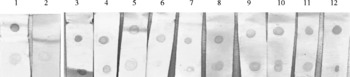
Recombinant GST protein detected F. gigantica field infection in buffaloes by dot-ELISA. Sixty buffalo sera samples collected from a local abattoir, including those from F. gigantica infected animals, were screened in this assay and F. gigantica infected animals were detected as positive, showing no reactivity with the sera from F. gigantica non-infected animals (fig. 4). A concentration of 100–200 ng of the antigen was found to be optimal for use in dot-ELISA assay.
Dot-ELISA showing immunoreactivity of the recombinant GST with F. gigantica positive field sera. Strips: 1, positive control serum; 2, negative control serum; 3–12, field sera positive for F. gigantica infection.

Discussion
Glutathione S-transferases are a family of enzymes that are involved in the cellular detoxification process, and are found in a wide spectrum of organisms, ranging from bacteria to mammals. They function primarily by catalysing the conjugation of the tripeptide glutathione (GSH, γ-Glu–Cys–Gly) to a wide variety of electrophilic toxic substrates, rendering the toxin more water soluble, less toxic and more readily eliminated by the host. GSTs are dimeric molecules of approximately 52 kDa that generate their substrate diversity, in part, by existing in multiple forms, making the GST family well suited to the cellular detoxification process (Rossjohn et al., Reference Rossjohn, Feil, Wilee, Sexton, Spithill and Parker1997). Glutathione S-transferases have been characterized from a number of parasitic helminths, including cestodes, trematodes and nematodes (Brophy et al., Reference Brophy, Crowley and Barrett1990; Brophy & Pritchard, Reference Brophy and Pritchard1994). The interest in helminth GSTs stems from the fact that they represent the major detoxification enzymes in these organisms and consequently represent chemotherapeutic and immunological targets (Sexton et al., Reference Sexton, Wilce, Colin, Wijffels, Salvatore, Feil, Parker, Spithill and Morrison1994; McTigue et al., Reference McTigue, Williams and Tainer1995; Morrison et al., Reference Morrison, Colin, Sexton, Bowen, Wicker, Friedel and Spithill1996).
The GST of the bubaline isolate of F. gigantica was analysed for sequence variation in the nucleotides and amino acids in comparison to an isolate of cattle origin (Thailand isolate). A variation of six nucleotides caused a change of four amino acids: Asp to Gly, Gln to His, Asn to Lys and Ile to Met, respectively, but further characterization of this recombinant protein is required to assign the protein to the proper isoform of the glutathione S-transferases and to ascertain whether these changes in four amino acids have any effect on the biological activity of the enzyme.
A non-denaturation protocol for the purification of the recombinant protein resulted in a significantly higher yield (P < 0.05), with 4–5 mg of protein/litre of bacterial culture, when compared with the denaturation protocol (1 mg protein/litre of bacterial culture). Expression of the protein in its native conformation will be an advantage for its subsequent biological characterization and in immunoprophylactic studies.
The role of GSTs in the immunoprophylaxis of schistosomosis and fasciolosis in various ruminants has been found to be encouraging, with reports of worm reduction, anti-fecundity effect, reduced pathological signs of the host and improvement in the body weight gain of the host in response to vaccination with parasite GST (Sexton et al., Reference Sexton, Wilce, Colin, Wijffels, Salvatore, Feil, Parker, Spithill and Morrison1994; Morrison et al., Reference Morrison, Colin, Sexton, Bowen, Wicker, Friedel and Spithill1996; Estuningsih et al., Reference Estuningsih, Smooker, Wiedosear, Widjante, Vaiano, Partoutoms and Spithill1997; Muro et al., Reference Muro, Casanueva, Lopez-Aban, Ramajo, Martinez-Fernandez and Hillyer2007; Preyavichyapugdee et al., Reference Preyavichyapugdee, Somphong, Riengrojpitak, Grams, Viyanant and Sobhon2008) but with varying results. A protection level of 19–69% or even more has been reported by the above authors in their different immunization schedules with different adjuvant formulations in sheep, cattle and buffaloes. Studies for exploiting the full potential of these molecules are under way in different hosts.
Serological assays will be the key players in the diagnosis of fasciolosis in near future, and serological tests based on several antigens have been commercialized for the diagnosis of F. hepatica infection. The diagnostic potential of cysteine proteinase for the detection of human and animal fasciolosis has been the most evaluated, and the protein has shown promise in the specific diagnosis of this infection (O'Neill et al., Reference O'Neill, Parkinson, Strauss, Angles and Dalton1998; Córdova et al., Reference Córdova, Reátegui and Espinoza1999; Cornelissen et al., Reference Cornelissen, Gaasenbeek, Borgsteede, Holland, Harmsen and Boersma2001; Raina et al., Reference Raina, Yadav, Sriveny and Gupta2006). Other defined antigens, such as glutathione S-transferase, fatty acid binding proteins, parasite haemoglobin, paramyosin, etc. have been exploited in the immunoprophylaxis of the disease but not in its serodiagnosis. However, utility of GST in the serodiagnosis of schistosomosis has been shown by Chinese workers by using peptide antigen of rSJ26 GST (Zhu, Reference Zhu2005) and a recombinant S. japonicum 26 GST in water buffaloes by ELISA and Western blotting (Cheng et al., Reference Cheng, Tsaihong and Lee2007); but this protein has not been tested in the diagnosis of fasciolosis in animals.
In the present preliminary study, the recombinant GST was probed in the dipstick-ELISA for the detection of an antibody response in buffaloes infected with F. gigantica. The antigen could differentiate between the F. gigantica positive and negative sera, as the antigen showed positive reactivity with animals infected with F. gigantica. Helminth mu-class GSTs, such as those seen in F. hepatica, are essentially internal, cytosolic proteins (Pemble et al., Reference Pemble, Wardle and Taylor1996; Hoarau et al., Reference Hoarau, Garello, Gnassia-Barelli, Romeo and Girard2002). The release of GST into the host for antibody stimulation is most likely via tegumental shedding or degradation following death. This could be a possible means of stimulating the host immune system for antibody production. No cross-reactivity studies on this recombinant protein with other helminths parasitizing buffaloes were conducted. This needs to be undertaken for evaluating the specificity of the antigen in the serological diagnosis of F. gigantica infection in ruminants.
Acknowledgements
The authors are thankful to the Director, Indian Veterinary Research Institute for providing facilities to carry out the present investigation.