The ability to mark organisms individually is vital to ecological studies linking individual traits to performance and evolutionary studies of selection on individual phenotypes. The relationship between individual traits and performance has been central to understanding evolutionary processes and intraspecific trait variation has also been increasingly recognised as an important factor affecting processes and patterns in population and community ecology (Bolnick et al. Reference Bolnick, Svanback, Fordyce, Yang, Davis and Hulsey2003, Reference Bolnick, Amarasekare, Araújo, Bürger, Levine and Novak2011). In addition to relating individual level variation to performance, effective marking techniques can facilitate comparisons of organisms collected from different sources (e.g., different ponds or clutches), making it feasible to address questions about the effects of source environment on performance. However, tagging the juvenile stages of many insects is complicated by moulting, which can result in tag loss and because external marks that are visible to other organisms may change the outcome of the process under study. For example, predators may be differentially able to see certain colours (Kusche and Meyer Reference Kusche and Meyer2014) making colour-coded marks unsuitable for predation studies.
We examined the potential for using an internal marking approach in dragonfly larvae (Odonata: Anisoptera). Odonata are a useful model system in ecology and evolution (Córdoba-Aguilar Reference Cordoba-Aguilar2008) and there are well-established techniques for individually marking adults (e.g., McCauley Reference McCauley2010). However, marking the larval stages is more difficult and has created barriers to studies requiring individual or group identification of larval odonates. Previous approaches to marking larvae have included removal of tarsi and scarring the eye with a needle (Johnson et al. Reference Johnson, Martin, Mahato, Crowder and Crowley1995) but these approaches are limited in the number of distinctive marks they provide and they have potential drawbacks including processing time that make them unsuitable for many studies. We tested the use of coded-wire tags (from Northwest Marine Technology Inc., Shaw Island, Washington, United States of America, hereafter: CWT) as a potential tool to enable individual identifications of odonate larvae. CWT are an injectable, internal tagging method with an individual code etched into each tag which can be identified when tags are recovered.
Coded-wire tags have been used extensively in fisheries studies (Bumgarner et al. Reference Bumgarner, Schuck and Blankenship2009) and have also been used in a variety of other animals including amphibian larvae (Martin Reference Martin2011) and several malacostracan crustaceans (e.g., lobster, McMahan et al. Reference McMahan, Cowan, Sherwood, Grabowski and Chen2012; crayfish, de Graaf Reference de Graaf2007; crabs, Davis et al. Reference Davis, Young-Williams, Hines and Zmora2004; and shrimp, Kneib and Huggler Reference Kneib and Huggler2001). Tests of this tagging approach in larval insects have been limited but they have been demonstrated to be an effective tagging approach with larval Coleoptera (Tenebrionidae) with a relatively high rate of retention through metamorphosis to the adult stage (Schaffler and Isely Reference Schaffler and Isely2001). These studies suggest that tag retention may be relatively high in organisms such as odonate larvae that undergo 10–12 moults and emergence when there is an increased risk of tag loss. However, the potential use of CWT as a means of tagging and individually identifying odonate larvae has not been addressed in the literature.
To evaluate the potential use of CWT as a tool for individually marking odonate larvae, we conducted two experiments comparing survival of marked and unmarked larvae and the retention rates of these tags. We also assessed the effects of injecting tags on growth in these experiments.
For each experiment, we collected larvae by dip-netting from a pond at the Koffler Scientific Reserve at Jokers Hill (hereafter: KSR), in Ontario, Canada (44°01'48''N, 79°32'01''W). Larvae were transported to the University of Toronto Mississauga (Ontario, Canada) where they were transferred into 473 mL plastic cups containing a strip of plastic mesh (for use as substrate by larvae) and de-chlorinated tap water.
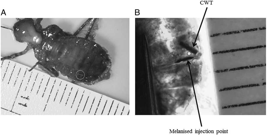
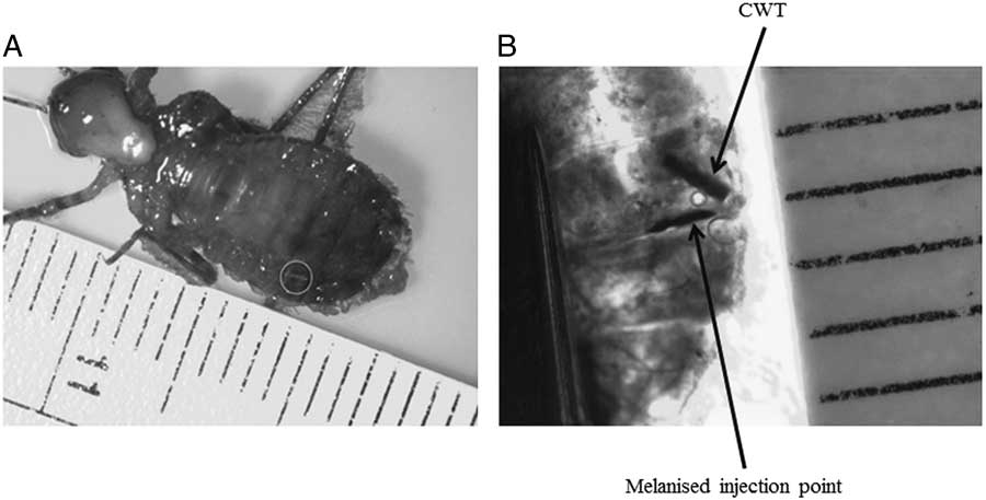
Larvae in both experiments were randomly assigned to either the tagged or control treatment (n=24 for each treatment). Head width (mm) of each individual was measured using digital calipers (±0.0003 mm). Larvae were marked using a single-shot injector (Northwest Marine Technologies Inc., Shaw Island, Washington, United States of America) to inject a pre-cut magnetic CWT into the lateral-dorsal suture of segment eight (S8) of each larva in the tagged treatment of both experiments (Fig. 1). Each CWT was 1.1 mm in length and 0.25 mm in diameter.
Coded-wire tags are frequently visible in live larvae through the larval exoskeleton. (A) A ventral view of a preserved Epitheca canis showing CWT (circled); and (B) a dorsal view of preserved Epitheca canis showing the melanised point of injection and the coded-wire tag at lateral-dorsal suture of S8.
In the first experiment, larval Epitheca canis McLachlan (Odonata: Corduliidae) were housed individually in 473 mL cups filled with dechlorinated tap water in the laboratory for 22 days. Larvae (initial head-width, mean±1 SD: 4.51±0.51 mm) were maintained under 24 hour lighting and fed a standard 5 mL volume of zooplankton concentrated in dechlorinated water twice per week. Water in each cup was changed twice per week. During water changes, each cup was inspected for a lost tag using a circular magnet (25 mm in diameter) that was moved along the bottom and sides of the cup in order to attract the tag if it had been lost from the larva. When a tagged individual moulted, the shed exoskeleton was removed from the cup. The cup and exoskeleton were then inspected for a lost tag both visually and using the magnet. Growth was estimated based on the number of larvae that moulted during these experiments. We compared the proportion of larvae that moulted in each treatment using a Pearson χ2 test. Increases in body size occur at moulting and differences in the frequency of moulting would indicate differences in growth rates between treatments.
In the second experiment, larval Leucorrhinia intacta Hagen (Odonata: Libellulidae) (initial head-width, mean±1 SD: 4.39±0.39 mm) were housed and maintained in the same way as in experiment 1 for 30 days. At day 30 larvae were transferred from plastic cups to larger plastic tubs (30×43 cm) filled with dechlorinated tap water to encourage larval growth. Larvae were held individually in this way for an additional 30 days. In the tubs the feeding schedule remained the same as in the cups, each larva was fed a standard 5 mL of zooplankton solution at each feeding and fed twice per week. Water changes in the tubs were performed once per week as the extra volume of water allowed the water to remain cleaner for longer than in the smaller cups. Tubs received a total of eight water changes over the experimental period. During each water change, larvae were removed and all water in each tub was replaced with dechlorinated water and larvae were returned to tubs. Larvae were fed at the time of the water change.
Larval head widths were re-measured at the end of this experiment and larval growth rates were calculated using head width measurements taken pre-experiment and post-experiment as ([ln(ending head width) – ln(starting head width)]/total experimental period). Head widths were natural log transformed to linearise these data and facilitate comparisons. An independent samples t-test was used to compare larval growth rates in control and experimental treatments of each experiment.
To assess tag retention rates, larvae in the tagged treatments of experiments 1 and 2 were preserved in 70% ethanol and 95% ethanol, respectively. Tags were recovered by making a small incision through the tag injection site (visible as a melanised line in the exoskeleton) and using forceps to remove the tag. This process was facilitated by the magnetic attraction between the forceps and the CWT.
Under magnification, we found that CWT were visible within both preserved and live larvae. Both the CWT and melanisation line were apparent when handling the larvae (Fig. 1). The CWT typically held their initial positions in the lateral-dorsal S8 sites, which aided in their detection. Two tagged individuals had their tags shift slightly anterior of the S8 area. However, this shift in position did not inhibit tag recovery from either larva during dissection.
In experiment 1, all larvae from the tagged treatment survived the experimental period, and a single untagged larva died on the final day of this experiment (a 100% and 98% survivorship rate for tagged and untagged larvae, respectively). Tags from 22 of 24 tagged larvae were successfully recovered (a 92% retention rate). Two larvae lost their tags, one of which had moulted during the experiment. Neither lost tag was detected during inspection of the cups throughout the experiment. A total of eight tagged larvae and 12 untagged larvae each underwent a single moult during the experiment and there was no difference in the proportion of larvae that molted in each treatment (χ2=1.37, df=1, n=48, P=0.242).
In experiment 2, survival in both tagged and untagged larvae was 100% and all tags were recovered from the 24 tagged larvae (a 100% retention rate). Only one larva moulted during this experiment, a single tagged individual. Tags within the abdomen of four individuals were observed to have shifted distally into the anterior segments of the abdomen (location at dissection approximated to be between S4 and S6 for each individual), though these shifts in position did not induce mortality, nor prevent tag recovery. We did not observe a difference in growth rate between tagged and untagged larvae (t=0.952, df=46, P=0.346).
All odonate larvae injected with CWT survived across monitoring periods that ranged from 22 to 60 days and retention rates of tags were very high (92–100%). This indicates that injected CWT can be a useful tagging method in these animals despite the relative invasiveness of the approach. While limited, our data do not suggest that CWT affect odonate larval growth. This conclusion is based on finding no difference in molting frequency data in experiment 1 and no growth rate differences in experiment 2 when few larvae moulted.
Once the experimenter had gained experience with handling the injector and larvae, the tagging process was rapid and injecting a single tag could be completed in approximately 10 seconds. Taken together these results indicate that injecting CWT is an effective, time-efficient, and inexpensive approach to individually identifying dragonfly larvae. There are limits to the use of this tagging approach, particularly in the body size of larvae that can be safely injected with a tag. The smallest larva tagged had a head width of 3.5 mm. Larval Cordulliidae and Libellulidae larvae have relatively wide abdomens compared to Zygoptera larvae and these wide abdomens are likely to be essential for the safe injection of these tags. Further work is necessary to assess the feasibility of tagging other groups of odonate larvae but it is likely that only the largest Zygoptera larvae could be tagged with this approach.
This study provides a foundation for the use of CWT in larval dragonflies. Future studies should assess how tagging affects larval behaviour and whether retention rates remain high during adult emergence. This would provide an opportunity to track individual characteristics associated with changes from larval to adult form. The ability to individually identify odonate larvae using CWT without increasing mortality, and in a time and cost-effective manner, will facilitate addressing numerous questions in the ecology and evolution of these animals.
Acknowledgements
Helpful feedback on this project and manuscript were provided by T. Duong, D. Frances, S. French, C. Searcy, and two anonymous reviewers. The Koffler Scientific Reserve provided research support and access to collection sites. This work was funded by The University of Toronto Mississauga and a Natural Sciences and Engineering Research Council of Canada Discovery Grant (RGPIN 435614-13) to S.J.M.