




No CrossRef data available.
Article contents
Purification and characterization of a metacestode cysteine proteinase from Taenia solium involved in the breakdown of human IgG
Published online by Cambridge University Press: 11 May 2005
Abstract
Infection of the central nervous system by Taenia solium cysticerci is the cause of human neurocysticercosis, a major neurological infection in the Third World and an emerging infectious disease in the United States. We previously isolated a cysteine proteinase from cysticerci of Taenia crassiceps and demonstrated that it degrades human IgG in vitro. We have now isolated a 48 kDa thiol-dependent proteinase from T. solium. The T. solium enzyme also degrades human IgG, but does not significantly degrade albumin. IgG degradation was inhibited by cysteine proteinase inhibitors, but not significantly by inhibitors of aspartic, serine, or metalloproteinases. The peptide substrate specificity and pH optimum resemble cathepsin L. The Km for the peptide substrate Z-Phe-Arg-AFC was calculated to be 7·0×10−6 M, the Kcat was 1·98×105 s−1, and the Kcat/Km 2·84×109 M−1 s−1, a value which is within the diffusion control limit for highly catalytic enzymes. We propose that immunoglobulin degradation by the T. solium cysteine proteinase may play a key role in the host-parasite interface and could be employed as a target for chemotherapy.
Information
- Type
- Research Article
- Information
- Copyright
- © 2005 Cambridge University Press
INTRODUCTION
The metacestode (larval cyst) stage of the tapeworm, Taenia solium infects over 50 million persons world-wide and causes the neurological disorder neurocysticercosis (White, 2000; Garcia et al. 2003). Pigs are the natural host for the metacestode stage and are the source of human infection with the adult tapeworm stage of the parasite (taeniasis). Neurocysticercosis is characterized by an incubation period of several years (Dixon & Lipscomb, 1961). During this period, the parasite survives in the human brain without causing symptoms. Thus, the parasitic cysts are able to survive and evade destruction by the host immune response (White, Robinson and Kuhn, 1997).
The survival of Taenia cysts for many years in asymptomatic intermediate hosts requires that the Taenia cysts evade host immune responses and acquire essential nutrients including amino acids (Hayunga, Sumner & Letonja, 1989; Ambrosio, Merchant & Laclette, 1994). We and others have hypothesized that the parasites may use proteases (White et al. 1992; Molinari et al. 2000) and degrade host immune molecules to satisfy both of these requirements (Hayunga et al. 1989; Ambrosio et al. 1994; Molinari et al. 2000). Taenia cysts actively bind and take up host IgG (Kalinna & McManus, 1993). Degraded IgG has been observed in Taenia cysts (Hayunga et al. 1989; Ambrosio et al. 1994). Ambrosio and colleagues (Ambrosio et al. 1994) noted that the IgG concentration in human serum is sufficient to allow saturated uptake of IgG in Taenia cysts. We identified and purified a cysteine proteinase from T. crassiceps cyst walls, that rapidly degrades human IgG at acidic pH (White, Baig & Chappell, 1997). We demonstrated localized cysteine protease activity in the cyst wall (Khalil, Burns & White, 1998). Others found degraded IgG incorporated into Taenia cysticerci (Hayunga et al. 1989; Ambrosio et al. 1994). Based upon these data, the parasite may be able to evade and exploit a host immune response through the mechanism of IgG degradation.
MATERIALS AND METHODS
Proteinase assays
Enzyme activity was assessed through the cleavage of peptide substrates coupled to 7-amino-4-trifluoromethyl-coumarin (AFC) (Enzyme Systems Products, Livermore, CA) as previously described (White, Robinson & Kuhn, 1997). Following incubation of enzyme preparations overnight (18 h, 37 °C) with peptide in assay buffer (0·2 M citrate, pH 4·9, supplemented with 0·01 M cysteine), free AFC concentrations were measured fluorometrically (excitation 405 nm, emission 505 nm) and converted into moles based upon a standard curve.
Enzyme purification
Active fractions in chromatographic steps were identified by their cleavage of the substrate Z-Phe-Arg-AFC. T. solium cysts were obtained from 6 to 12-month-old cysticercotic pigs from an endemic area of porcine cysticercosis in Mexico. Cysts were carefully dissected from the parasitized tissues, washed 3 times with PBS, and lyophilized for storage and subsequent fractionation. Parasite extracts were prepared by suspending 0·1 g of dry frozen cysts in 10 ml of extraction buffer (0·4 M citrate, pH 4·9). The cysts were vortexed for 3×5-min intervals with intermittent incubations (5 min) at 4 °C. Particulate material was removed by centrifugation (20000 g, 120 min). The soluble fraction was filtered and loaded onto a 10 ml collagen-agarose column equilibrated with 10 mM Tris, pH 7·6, in order to remove paramyosin, which is the major protein within the T. solium cyst wall (Laclette et al. 1990). Proteinase activity that was detected in the unbound material was further fractionated by gel filtration (AcA 54 column, bed volume 480 ml, 2·5×86 cm, equilibrated in 10 mM Tris, pH 7·6). Subsequently, the protein was further eluted by FPLC anion-exchange chromatography (Mono Q HR 5/5 column, Pharmacia Biotech Inc., Piscataway, NJ) using a gradient of 10 mM Tris, pH 7·6 with or without 1 M NaCl (solution B) at a flow rate of 1 ml per min as follows: 0–11 min 0% B, 11–14 min 0–10% B, 14–44 min 10–30% B, 44–47 min 30–100% B, 47–50 min 100% B. Active fractions from the Mono Q column were pooled and subsequently characterized. Enzyme purity was assessed by silver staining on SDS-PAGE gels using the method of Wray et al. (1981).
IgG degradation
IgG digestion was detected by incubating 20 μl of T. solium purified enzyme or T. solium extract with 1 μg of human IgG (Sigma, St Louis, MO) in a final volume of 100 μl of assay buffer. Exogenous thiol dependence was assessed by incubating the assay in the presence or absence of 10 mM L-cysteine. The catalytic class of the IgG proteinase was determined by pre-incubating the enzyme for 30 min with the following proteinase inhibitors: L-trans-epoxysuccinyl-leucylamide-(4-guanidino)-butane (E64) (10−6 M, cysteine proteinases), phenylmethylsulfonyl fluoride (PMSF) (10−3 M, serine proteinases), pepstatin A (10−6 M, aspartic proteinases), and 1,10 phenanthroline (10−3 M, metalloproteinases). After addition of IgG, the assay was incubated overnight (37 °C, 18 h). Subsequently, 100 μl of the mixture were boiled in sodium dodecyl sulfate polyacrylamide gel electrophoresis (SDS-PAGE) sample buffer with 2% 2-mercaptoethanol, separated by 12·5% SDS-PAGE, and blotted onto polyvinylidene difluoride. Tris-buffered saline containing 0·1% bovine serum albumin and 0·05% Tween 20 (TBS-BSA) was used to block non-specific binding. The blot was washed 3 times with TBS containing 0·05% Tween (TBS-Tween), and incubated with biotinylated anti-human heavy and light chain antibody diluted 1[ratio ]1000 in TBS-BSA (Boehringer-Mannheim, Indianapolis, IN) followed by incubation with a peroxidase-conjugated strepavidin-avidin complex. Bound immunoglobulin was detected by chemiluminescence and autoradiography (ECL Kit, Amersham International, Sweden).
Biochemical characterization
Determination of the percentage of active enzyme was based upon the observation that E-64 binds irreversibly in a 1[ratio ]1 molar ratio to cysteine proteinases (Barrett et al. 1982). In order to determine the percentage of enzyme in active form, the purified cysteine proteinase was titrated with E-64 inhibitor at 10-fold dilutions with final concentrations from 10−3 M to 10−21 M in a final volume of 500 μl of assay buffer. Each dilution was performed in triplicate and the inhibitor was pre-incubated for 30 min with the enzyme prior to addition of substrate. The liberation of free AFC after overnight incubation was determined fluorometrically as described above. Percentage calculations were performed by dividing the actual molar yield of active enzyme by the theoretical molar yield of purified enzyme protein (protein concentration divided by molecular weight).
Substrate specificity profiles were obtained by incubating the enzyme with 1 μg of various peptide substrates in a final volume of 500 μl of assay buffer. The final concentration of substrate (1·92×10−5 M) was at least 50 times the Km, such that the substrate was provided for in saturating quantities. The optimal pH for purified enzyme was determined by incubating purified enzyme with Z-Phe-Arg-AFC in 0·1 M citrate-phosphate buffer at a pH range from 2·5 to 8·5. Inhibitor profiles were obtained by pre-incubating inhibitors of various catalytic classes and effective concentrations for 30 min with the purified enzyme before adding Z-Phe-Arg-AFC.
Km and Vm were assessed by adding increasing concentration of Z-Phe-Arg-AFC to triplicate tubes of the purified enzyme into a final buffer volume of 500 μl. Kinetic calculations were derived from the Lineweaver-Burke plot of substrate concentration (mol) versus proteinase velocity (mol/h). Kcat was calculated from the Vmax. Kinetic values were assessed along the linear segment of the reaction. To assess the effect of E-64 on the kinetic parameters, enzyme was pre-incubated with 10−6 M E-64 for 30 min prior to the addition of Z-Phe-Arg-AFC.
RESULTS
When the parasite extract was fractionated on a collagen column, 30–40% of the proteinase activity was removed. Subsequent fractionation by gel filtration resulted in a single peak of enzyme activity with an approximate molecular weight of 50 kDa (mean±2 kDa) (Fig. 1). Further separation by FPLC anion-exchange chromatography resulted in a single peak of enzyme activity (Fig. 2). This purification scheme resulted in a 690-fold overall purification (Table 1). Based upon titration with E-64, approximately 43% of the purified enzyme was determined to be active with half of the maximal proteolytic activity occurring at 5×10⁻¹⁰ moles of E-64. A single band (Mr 48 kDa) on silver-stained gels confirmed the purity of the enzyme (Fig. 3).

Fig. 1. Gel filtration chromatography of acid extracts of Taenia solium cysts. The extract was loaded onto an AcA54 column (2·5×86 cm), which was equilibrated and eluted with 10 M Tris buffer, pH 7·6. Absorbance was monitored at 280 nM (A) and for proteinase activity that was detected by cleavage of Z-Phe-Arg-AFC in assay buffer (0·2 M citrate buffer, pH 4·9, supplemented with 10 mM L-cysteine) (B). Proteinase activity eluted at a molecular weight of 50 kDa±2 kDa based upon a standard curve produced for the column.

Fig. 2. FPLC anion-exchange chromatography of active fractions from AcA 54 gel-filtration separations of Taenia solium. Active fractions from the AcA 54 gel filtration column were fractionated further by FPLC anion-exchange chromatography using a Mono Q column. Enzymes were eluted with a gradient (dashed line) of 10 mM Tris, pH 7·6 (solution A) and 10 mM Tris buffer, pH 7·6 plus 1 M NaCl (solution B). One ml fractions were collected and assayed with Z-Phe-Arg-AFC substrate (closed circles) in assay buffer (0·2 M citrate, pH 4·9, supplemented with 10 mM L-cysteine). Absorbance was monitored at 280 nm (solid line).


Fig. 3. Silver-stained SDS-polyacrylamide gel showing the purified Taenia solium cysteine proteinase. Active pool from the MonoQ FPLC column treated immediately with E64. The molecular weight of the cysteine proteinase was determined to be 48 kDa based upon a standard curve from various molecular weight standards.
When human IgG was incubated with a homogenate of T. solium cysts, both heavy and light chain bands decreased in intensity. Degradation was eliminated by pre-incubation of the homogenate with E-64. Similarly, incubation of human IgG with the purified cysteine proteinase resulted in a loss of both the heavy and light chain bands. This degradation was reversed by the addition of E-64, but not by inhibitors of other mechanistic proteinase classes (Fig. 4).

Fig. 4. Degradation of IgG heavy chain by cysteine protease of Taenia solium. Human IgG was incubated for 18 h with T. solium purified cysteine protease (CP). Incubated tubes were subsequently fractionated by SDS-PAGE under reducing conditions, blotted onto PVDF membrane, and visualized using biotinylated anti-IgG (heavy chain/light chain), biotin-peroxidase conjugated-streptavidin complex and enhanced chemiluminescence. Only the heavy chain is shown. (A) Human IgG alone, (B) human IgG with CP minus cysteine, (C) human IgG with CP with cysteine, (D) human IgG with CP with E64.
To determine substrate specificity, a series of synthetic peptide substrates was incubated with the purified enzyme. Z-Phe-Arg-AFC was found to be maximally cleaved, with other substrates cleaved 14% or less in comparison (Fig. 5). Based upon the substrate cleavage data, Z-Phe-Arg-AFC was chosen as the primary substrate for subsequent characterizations. The pH profile of the purified proteinase, measured by incubating the enzyme with Z-Phe-Arg-AFC over a broad pH range in a 0·1 citrate-phosphate buffer system, was found to be bell-shaped with optimal activity at pH 4·7.

Fig. 5. Substrate specificity of purified Taenia solium cysteine protease assessed by cleavage of a panel of peptide substrates. The final concentration of substrate was 1·92×10−5 M.
Inhibitor studies demonstrated that enzyme activity was inhibited by a variety of cysteine proteinase inhibitors, including E-64, Ep459, Ep475, iodoacetate, and HgCl2. Enzyme activity was also inhibited by the chloromethyl ketones TLCK and TPCK. Partial inhibition was recorded for 1,10 orthophenanthroline, but not for EDTA, PMSF, or pepstatin (Table 2). A Lineweaver-Burke plot was employed to ascertain kinetic parameters. Km for Z-Phe-Arg-AFC was calculated to be 7·0×10−6 M and Kcat was 1·98×105 s−1. Kcat/Km was calculated to be 2·84×109 M−1 s−1, a value which is within the diffusion control limit for highly catalytic enzymes (108 to 109 M−1 s−1) (Albery & Knowles, 1977).

DISCUSSION
We have identified and purified an acid cysteine proteinase from the cyst wall of T. solium metacestodes. We isolated the major endopeptidase activity by acid extraction, followed by 3 chromatographic steps. Collectively, these chromatographic procedures resulted in an overall purification of 690-fold from the original acid extract. The purified enzyme appeared as a single 48 kDa band on silver-stained gels and was approximately 50 kDa in size, based upon gel filtration chromatography. The acidic pH optimum, requirement for exogenous thiols, total inhibition by the specific cysteine protease inhibitors E-64 and iodoacetate and dose inhibition by HgCl2 demonstrated that the enzyme is a cysteine protease. Although the enzyme was inhibited by TPCK and TLCK (which inhibit both serine and cysteine proteinases), limited inhibition by the serine protease inhibitor PMSF also indicated that the enzyme is a cysteine protease. Partial inhibition by 1,10 phenanthroline suggested that the enzyme may be characterized by some partial dependence on a metal co-factor at the active site, as has been noted in the inhibitor profiles of cysteine proteinases in other parasites including Plasmodium (Salas et al. 1995) and Diplostomum (Moczon, 1994). Alternatively, the enzyme may be synthesized as a pro-enzyme requiring cleavage by a metalloproteinase.
The purified proteinase was marked by an unusually high Kcat/Km ratio on the synthetic peptide substrate, Z-Phe-Arg-AFC, equivalent to 2·2×109 m−1 s−1, which approximates the enzymatic diffusion control limit which some investigators have likened to ‘catalytically perfect’ enzymes (Rickard, 1978). Thus, the impressive efficacy of the Taenia cysteine proteinase in turning over substrates suggests a critical role in the survival of the organism. This capability is most likely related to an active site highly evolved for turning over the substrate.
The T. solium cysteine proteinase cleaves human IgG in vitro optimally at the pH and reducing environment typical of lysosomes. This enzyme was predominantly responsible for complete human IgG digestion, in comparison to other proteinases present in the original acid extract of the purification protocol. The observation that both the heavy chain and light chains were degraded, leaving only fragments at the lower end of the gel, suggests that the chains were reduced to peptides or amino acids. Dose-response experiments showed that the purified cysteine proteinase completely degraded both heavy and light chains of human IgG within a period of 15 min, and was completely inhibited by the cysteine proteinase inhibitor, E64. These results are consistent with the nanomolar Ki value of the time-dependent irreversible inhibition of the cleavage of Z-Phe-Arg-AFC by E64. In a previous study (Khalil et al. 1998), we demonstrated that E64 suppresses cysteine proteinase activity in the Taenia cyst wall in situ, suggesting that specifically designed cysteine proteinase inhibitors would also be expected to suppress IgG degradation by the cysteine proteinase.
We believe that human IgG may serve as a selective biological target for the Taenia cysteine proteinase. For example, the T. solium cyst wall cysteine proteinase does not degrade the human blood protein albumin, at similar proteinase concentrations. Thus, this evidence suggests that the Taenia proteinase is somewhat selective for IgG. The homologous T. crassiceps cysteine proteinase localizes to sites (Khalil et al. 1998) where IgG is degraded (e.g. cyst lysosomes) (Ambrosio et al. 1994). The acidic pH optimum and reducing condition optimum for the Taenia proteinase's activity is also consistent with a lysosomal location. Moreover, the substrate specificity profile of the purified enzyme most closely resembles cathepsin L-like cysteine proteinases, which are mammalian lysosomal enzymes that have also been linked to IgG degradation (Carmona et al. 1993). Taken together, these observations suggest that the Taenia cyst wall cysteine proteinase may be a significant agent responsible for IgG degradation in Taenia, and may actually be specialized for this process.
The cysteine proteinase may help the parasite to exploit the host immune response (Damian, 1987). Antibody may provide a readily available source of amino acids for the parasite in vivo, as has been suggested (White, 1997; White et al. 1997). The growth of Taenia cysts in the presence of cysticercosis derived antibody-enriched immune serum, further supports this contention (Bojalil et al. 1993). The correlation between increased cyst growth and increased cysteine proteinase activity in these smaller cysts is consistent with the suggestion that the enzyme may play a key role in breaking down proteins, like IgG, for parasite nutrition (Khalil et al. 1998).
This proposal suggests a potential dual mode of action for a specific proteinase inhibitor that would be designed against the Taenia cysteine proteinase. First, specific inhibition of the cysteine proteinase may allow host immunoglobulins to recognize cysts thereby ‘unmasking’ them to the immune system. A second mode may be that inhibition of the cysteine proteinase may weaken the cyst by depriving it of IgG derived amino acids, thus impairing it metabolically. In support of this, preliminary experiments have demonstrated that mice were protected against a Taenia challenge by cysteine proteinase inhibitors.
The high kinetic turnover ratio of Taenia cyst wall cysteine proteinase, its potentially key role in parasite nutrition and host immune evasion, its accessible location within the cyst wall as demonstrated by localization studies in T. crassiceps, and the observation that its activity can be reduced by cysteine proteinase inhibitors in situ (Khalil et al. 1998) suggest that this enzyme may be a useful target for anti-parasitic chemotherapy.
We thank Tiffany Paul for help in technical portions of this work. This work was supported in part by a grant from the National Institutes of Health Pre-doctoral Training Grant in Molecular Parasitology and a grant from the US/Mexico Foundation.
References
REFERENCES
Albery, W. J. and
Knowles, J. R. (
1977).
Efficiency and evolution of enzyme catalysis.
Angewandte Chemie International Edition in English
16,
285–293.CrossRefGoogle Scholar
Ambrosio, J.,
Landa, A.,
Merchant, M. T. and
Laclette, J. P. (
1994).
Protein uptake by cysticerci of Taenia crassiceps.
Archives of Medical Research
25,
3235–3330.Google Scholar
Barrett, A. J.,
Kembhavi, A. A.,
Brown, M. A.,
Kirschke, H.,
Knight, C. G.,
Tamai, M. and
Hanada, K. (
1982).
L-trans-Epoxysuccinyl-leucylamido (gaunidino) butane (E-64) and its analogues as inhibitors of cysteine proteinases including cathepsins B, H, and L.
The Biochemical Journal
201,
189–198.CrossRefGoogle Scholar
Bojalil, R.,
Terrazas, L. I.,
Govezensky, T.,
Sciutto, E. and
Larralde, C. (
1993).
Thymus-related cellular immune mechanisms in sex-associated resistance to experimental murine cysticercosis (Taenia crassiceps).
Journal of Parasitology
79,
384–389.CrossRefGoogle Scholar
Carmona, C.,
Dowd, A. J.,
Smith, A. M. and
Dalton, J. P. (
1993).
Cathepsin L proteinase secreted by Fasciola hepatica in vitro prevents antibody-mediated eosinophil attachment to newly excysted juveniles.
Molecular and Biochemical Parasitology
62,
9–17.CrossRefGoogle Scholar
Damian, R. T. (
1987).
The exploitation of host immune responses by parasites.
Journal of Parasitology
73,
3–13.Google Scholar
Dixon, H. B. F. and
Lipscomb, F. M. (
1961).
Cysticercosis: an analysis and follow-up of 450 cases.
Medical Research Council Special Report Series, vol. 299,
Her Majesty's Stationary Service,
London, pp.
1–58.CrossRef
Garcia, H. H.,
Gonzalez, A. E.,
Evans, C. A. and
Gilman, R. H. (
2003).
Taenia solium cysticercosis.
Lancet
362,
547–556.CrossRefGoogle Scholar
Hayunga, E. G.,
Sumner, M. P. and
Letonja, T. (
1989).
Evidence for selective incorporation of host immunoglobulin by strobilocerci of Taenia taeniaeformis.
Journal of Parasitology
75,
638–642.CrossRefGoogle Scholar
Kalinna, B. and
McManus, D. P. (
1993).
An IgG (Fc gamma)-binding protein of Taenia crassiceps (Cestoda) exhibits sequence homology and antigenic similarity with schistosome paramyosin.
Parasitology
106,
289–296.CrossRefGoogle Scholar
Khalil, A. I.,
Burns, A. R. and
White, A. C., Jr. (
1998).
Demonstration of Taenia crassiceps cysteine proteinase activity in tegumentary lysosome-like vesicles.
Journal of Parasitology
84,
513–515.CrossRefGoogle Scholar
Laclette, J. P.,
Alagon, A.,
Willms, K. and
Torre-Blanco, A. (
1990).
Purification of antigen B from Taenia solium cysticerci by affinity to mammalian collagen.
Journal of Parasitology
76,
273–275.CrossRefGoogle Scholar
Moczon, T. (
1994).
A cysteine proteinase in the cercariae of Diplostomum pseudospathaceum (Trematoda, Diplostomatidae).
Parasitology Research
80,
680–683.CrossRefGoogle Scholar
Molinari, J. L.,
Mejia, H.,
White, A. C., Jr.,
Garrido, E.,
Borgonio, V. M.,
Baig, S. and
Tato, P. (
2000)
Taenia solium: a cysteine protease secreted by metacestodes depletes human CD4 lymphocytes in vitro.
Experimental Parasitology
94,
133–142.CrossRefGoogle Scholar
Rickard, J. (
1978).
Generalized microscopic reversability, kinetic co-operativity of enzymes and evolution.
The Biochemical Journal
175,
779–791.CrossRefGoogle Scholar
Salas, F.,
Fichmann, J.,
Lee, G. K.,
Scott, M. D. and
Rosenthal, P. J. (
1995).
Functional expression of falcipain, a Plasmodium falciparum cysteine proteinase, supports its role as a malarial hemoglobinase.
Infection and Immunity
63,
2120–2125.Google Scholar
White, A. C., Jr. (
1997).
Neurocysticercosis: a major cause of neurological disease worldwide.
Clinical Infectious Diseases
24,
101–113.; quiz
114–115.CrossRefGoogle Scholar
White, A. C., Jr. (
2000).
Neurocysticercosis: updates on epidemiology, pathogenesis, diagnosis, and management.
Annual Review of Medicine
51,
187–206.CrossRefGoogle Scholar
White, A. C., Jr.,
Baig, S. and
Chappell, C. L. (
1997).
Characterization of a cysteine proteinase from Taenia crassiceps cysts.
Molecular and Biochemical Parasitology
85,
243–253.CrossRefGoogle Scholar
White, A. C., Jr.,
Molinari, J. L.,
Pillai, A. V. and
Rege, A. A. (
1992).
Detection and preliminary characterization of Taenia solium metacestode proteases.
Journal of Parasitology
78,
281–287.CrossRefGoogle Scholar
White, A. C., Jr.,
Robinson, P. and
Kuhn, R. (
1997).
Taenia solium cysticercosis: host-parasite interactions and the immune response.
Chemical Immunology
66,
209–230.CrossRefGoogle Scholar
Wray, W.,
Boulikas, T.,
Wray, V. P. and
Hancock, R. (
1981).
Silver staining of proteins in polyacrylamide gels.
Annals of Biochemistry
118,
197–203.CrossRefGoogle Scholar

Fig. 1. Gel filtration chromatography of acid extracts of Taenia solium cysts. The extract was loaded onto an AcA54 column (2·5×86 cm), which was equilibrated and eluted with 10 M Tris buffer, pH 7·6. Absorbance was monitored at 280 nM (A) and for proteinase activity that was detected by cleavage of Z-Phe-Arg-AFC in assay buffer (0·2 M citrate buffer, pH 4·9, supplemented with 10 mM L-cysteine) (B). Proteinase activity eluted at a molecular weight of 50 kDa±2 kDa based upon a standard curve produced for the column.

Fig. 2. FPLC anion-exchange chromatography of active fractions from AcA 54 gel-filtration separations of Taenia solium. Active fractions from the AcA 54 gel filtration column were fractionated further by FPLC anion-exchange chromatography using a Mono Q column. Enzymes were eluted with a gradient (dashed line) of 10 mM Tris, pH 7·6 (solution A) and 10 mM Tris buffer, pH 7·6 plus 1 M NaCl (solution B). One ml fractions were collected and assayed with Z-Phe-Arg-AFC substrate (closed circles) in assay buffer (0·2 M citrate, pH 4·9, supplemented with 10 mM L-cysteine). Absorbance was monitored at 280 nm (solid line).

Table 1. Purification of the Taenia solium cysteine protease

Fig. 3. Silver-stained SDS-polyacrylamide gel showing the purified Taenia solium cysteine proteinase. Active pool from the MonoQ FPLC column treated immediately with E64. The molecular weight of the cysteine proteinase was determined to be 48 kDa based upon a standard curve from various molecular weight standards.

Fig. 4. Degradation of IgG heavy chain by cysteine protease of Taenia solium. Human IgG was incubated for 18 h with T. solium purified cysteine protease (CP). Incubated tubes were subsequently fractionated by SDS-PAGE under reducing conditions, blotted onto PVDF membrane, and visualized using biotinylated anti-IgG (heavy chain/light chain), biotin-peroxidase conjugated-streptavidin complex and enhanced chemiluminescence. Only the heavy chain is shown. (A) Human IgG alone, (B) human IgG with CP minus cysteine, (C) human IgG with CP with cysteine, (D) human IgG with CP with E64.

Fig. 5. Substrate specificity of purified Taenia solium cysteine protease assessed by cleavage of a panel of peptide substrates. The final concentration of substrate was 1·92×10−5 M.
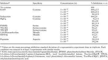
Table 2. Sensitivity of Taenia solium cysteine protease to inhibition by a panel of inhibitors when Z-Phe-Arg-AFC is used as the substrate, in assay buffer (0·2 M citrate, pH 4·9, supplemented with 10 mM L-cysteine)
 You have
Access
- 27
- Cited by