Introduction
In the modern biosphere, the green macroalga Codium Stackhouse, Reference Stackhouse1797, known by a variety of common names, including dead man's fingers, stag seaweed, and oyster thief, has a cosmopolitan distribution in shallow, low- to mid-latitude seas, locally occurring in numbers large enough to reach nuisance levels (Mathieson et al., Reference Mathieson, Dawes, Harris and Hehre2003). Because it does not calcify, the fossilization potential of this taxon is undoubtedly quite low. Nonetheless, over a century ago, White (Reference White1901) noted strong similarities between Codium and the nonbiomineralized taxa Buthotrephis divaricata White, Reference White1901, B. newlini White, Reference White1901, and B. lesquereuxi Grote and Pitt, Reference Grote and Pitt1876 from the upper Silurian of the Great Lakes area. Beyond being comparable in terms of external form, White (Reference White1901) observed that all three species shared key aspects of internal structure with Codium. Little more than a decade after publication of that report, however, Ruedemann (Reference Ruedemann1916) transferred Buthotrephis lesquereuxi to the genus Inocaulis Hall, Reference Hall1852 and redescribed it as a graptolite. No in-depth studies focusing on the affinities of the three taxa described as Codium-like by White (Reference White1901) have been published since that time.
The present study has two objectives. The first is to thoroughly reexamine the morphology, taxonomy, and paleoecology of Codium-like taxa from the Silurian of North America. This encompasses all three species investigated by White (Reference White1901) but also focuses on lower Silurian taxa, including forms from New York and Ontario and Thalassocystis striata Taggart and Parker, Reference Taggart and Parker1976 from northern Michigan, material of which is formally described herein from a new occurrence. The second objective is to use the assembled data to guide an in-depth assessment of the phylogenetic affinity of these taxa, including reconsideration of the algal interpretation proposed by White (Reference White1901). Application of scanning electron microscopy (SEM) and stable carbon isotope analysis provided new insights benefitting both parts of the study.
Materials and methods
Study materials included specimens from the Bertie Group at Buffalo, New York, and near Fort Erie, Ontario, the Kokomo Limestone Member of the Wabash Formation at Kokomo, Indiana, the Eramosa Formation near Wiarton in the Bruce Peninsula of Ontario, the Goat Island Formation near Gasport, New York, the Rochester Shale at Middleport, New York, and the Schoolcraft Formation in the southern part of the Upper Peninsula of Michigan. All descriptions provided herein were developed from first-hand examination of the study materials.
Reflected light images were obtained using a Canon Rebel digital camera fitted with a macrolens. To enhance contrast, specimens in some cases were photographed immersed in alcohol (if small) or water (if large). Further gains in contrast were obtained using polarizing filters. SEM study of the material was conducted using a Hitachi S-3200N and a JEOL JSM-7800F at the Electron Microbeam Analysis Laboratory (EMAL), Department of Earth and Environmental Science, University of Michigan. All specimens were imaged uncoated, and backscattered electron (BSE) images were obtained using an accelerating voltage of 25 keV. High-resolution morphometric data were collected from digital images using the open source program ImageJ (Schneider et al., Reference Schneider, Rasband and Eliceiri2012). Attempts to obtain novel morphological information using acetate peels proved unsuccessful.
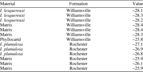
For stable carbon isotope analyses, samples were prepared according to the method outlined by LoDuca and Pratt (Reference LoDuca and Pratt2002). Briefly, fossil material (carbonaceous compressions) was carefully separated from the rock matrix using a razor knife under stereoscopic magnification, and ~10 mg of material was collected for each specimen. A separate sample of the bulk rock matrix for each specimen was also collected by micro-drilling fossil-free areas with a rotary tool equipped with a diamond-tipped bit. The separated fossil material and the bulk matrix powders were then placed in separate centrifuge tubes (15 mL), treated with an excess of 2M HCl (typically 5–10 mL), and allowed to react for a minimum of 24 hours to remove all carbonate components. These solutions and insoluble residues were then diluted with doubly deionized water and centrifuged, and the supernatant liquid was carefully decanted. This process was repeated until the solutions achieved a neutral pH of 7 (usually three times). Once neutralized, samples were decanted and allowed to dry completely in an oven at 45°C. When dry, the final sample residues were homogenized using an agate mortar and pestle and stored in glass vials pending analysis. To obtain isotopic measurements, between 0.1 and 1 mg of the homogenized sample powders were loaded into tin capsules and combusted at 950°C in an Elementar Vario Cube elemental analyzer connected to an Isoprime 100 continuous-flow gas-source isotope-ratio mass spectrometer (CF-GS-IRMS) in the stable isotope lab in the Department of Geosciences at Virginia Tech. Samples were run with international and commercial isotopic (CH-6, CH-7, Elemental Microanalysis wheat flour) and elemental standards (acetanilide, wheat flour) for calibration to the Vienna Pee Dee Belemnite (VPDB) scale and to monitor in-run and inter-run accuracy and precision. The standard deviation (1σ) of the isotopic standards was < ± 0.1‰ for all analyses.
Morphological terminology applied herein is that for algae. Terminology specific to bryopsidalean algae follows Taylor (Reference Taylor1960).
Repositories and institutional abbreviations
Types, figured, and other specimens examined in this study are deposited in the following institutions: D. M. Fisk Museum of Natural History (DMF), Hillsdale, Michigan, USA; Smithsonian Institution National Museum of Natural History (USNM), Washington, D.C., USA; University of Michigan Invertebrate Paleontology Collection (UMMP), Ann Arbor, Michigan, USA; Field Museum of Natural History (FMNH), Chicago, Illinois, USA; Indiana State Museum, Indianapolis, Indiana, USA (INSM); Indiana University Paleontology Collections, Bloomington, Indiana, USA (IUPC); New York State Museum (NYSM), Albany, New York, USA; Buffalo Museum of Science (BSNS), Buffalo, New York, USA; Royal Ontario Museum (ROMIP), Toronto, Canada.
Systematic paleontology
Division Chlorophyta Reichenbach, Reference Reichenbach1828
Class Ulvophyceae Mattox and Stewart, Reference Mattox, Stewart, Irvine and John1984
Order Bryopsidales Schaffner, Reference Schaffner1922
Genus Inocladus new genus
Type species
Buthotrephis lesqueruexi Grote and Pitt, Reference Grote and Pitt1876, as designated herein.
Other species
Buthotrephis newlini White, Reference White1901, B. divaricata White, Reference White1901, Chondrites verus Ruedemann, Reference Ruedemann1925.
Diagnosis
Erect nonbiomineralized unsegmented thallus comprising dichotomously branched elongate axes; branching mainly isotomous, occasionally sub-isotomous. Axes consist of a dense array of medullary and cortical tubes; medulla consisting of numerous longitudinally arranged isodiametric tubes; cortical tubes closely packed, oriented at an angle of about 90° to the supporting medullary tube and arrayed along it in a single row, unbranched, with closed endings, apex obtuse to truncate. External reproductive structures not present. Holdfast small, consisting of a loose bundle of thin filaments.
Occurrence
Bertie Group (Pridolian) in western New York and southern Ontario; Kokomo Limestone Member of the Wabash Formation (latest Ludlovian to Pridolian) at Kokomo, Indiana; Goat Island Formation (Wenlockian) in western New York; Eramosa Formation (Wenlockian) in the Bruce Peninsula, Ontario.
Etymology
From the Greek ino, meaning fibrous, and klados, denoting branch.
Remarks
The genus Buthotrephis was erected by Hall (Reference Hall1847) to accommodate forms that were then regarded as fossil seaweeds with thalli consisting of cylindrical, dichotomously branched axes. It has since been determined that many of the species assigned to this taxon instead represent trace fossils (Simpson, Reference Simpson1957; Johnson and Koneshi, Reference Johnson and Konishi1959; Osgood, Reference Osgood1970; Häntzschel, Reference Häntzschel and Teichert1975; Schweitzer, Reference Schweitzer1983), and none assigned to it in the same publication that established the genus show complex internal structure. This requires, then, that a new genus be erected to accommodate body fossils previously assigned to Buthotrephis that show an internal morphology consisting of tubes divided into medullary and cortical regions and that cannot be accommodated by existing taxa. Accordingly, the new genus Inocladus n. gen. is proposed, with Inocladus lesquereuxi (Grote and Pitt, Reference Grote and Pitt1876) n. comb. as the type species (Figs. 1–6).
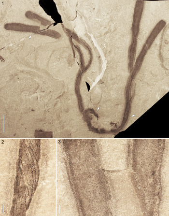
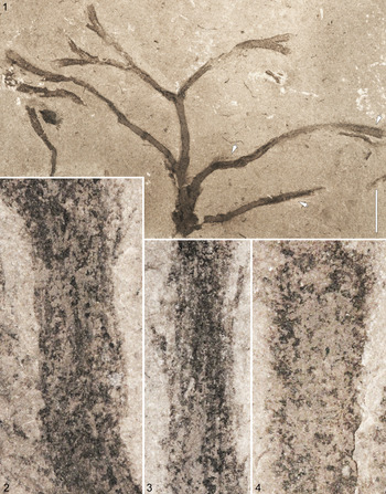
Inocladus lesquereuxi (Grote and Pitt, Reference Grote and Pitt1876) from the Bertie Group, Pridolian, Ridgemount Quarry, Fort Erie, Ontario, ROMIP 56896: (1) entire specimen, arrow at center marks holdfast; (2) enlargement of area at right arrow in (1) showing details of axis, including medullary tubes arranged in spiral fashion; (3) enlargement of area at left arrow in (1) showing papillate texture on axis surfaces. Specimen photographed dry with polarized light. (1) Scale bar = 20 mm; (2, 3) scale bars = 2 mm.

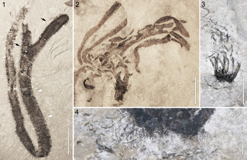
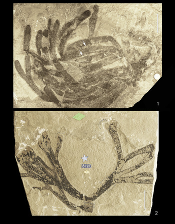
Inocladus lesquereuxi (Grote and Pitt, Reference Grote and Pitt1876) from the Bertie Group, Pridolian, Ridgemount Quarry, Fort Erie, Ontario: (1) entire specimen, ROMIP 50692; (2) entire specimen, ROMIP 63971; (3) small specimen, ROMIP 65539 (image courtesy of Jean-Bernard Caron); (4) detail of lower part of specimen in (3) showing holdfast (image courtesy of Jean-Bernard Caron). All specimens photographed immersed in water with polarized light. (1–3) Scale bars = 20 mm; (4) scale bar = 2 mm.

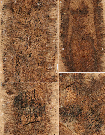
Details of axial structure for Inocladus lesquereuxi (Grote and Pitt, Reference Grote and Pitt1876), ROMIP 50692, Bertie Group, Ridgemount Quarry, Fort Erie, Ontario: (1) detail of area at lower white arrow in 2.1 showing medullary and cortical tubes; (2) detail of area at upper black arrow in 2.1 showing medullary and cortical tubes near tip of axis; (3) detail of area at upper white arrow in 2.1 showing medullary and cortical tubes at various levels within axis; (4) detail of area at lower black arrow in 2.1 showing medullary and cortical tubes at bifurcation. Specimen photographed immersed in alcohol with polarized light. m = medullary region; c = cortical region. Scale bar = 1 mm for all.

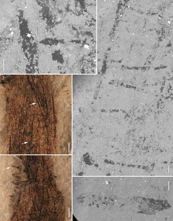
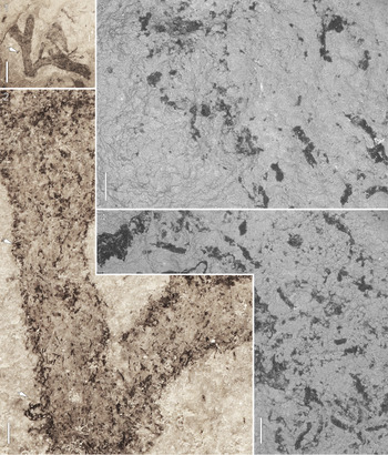
Details of axial structure for Inocladus lesquereuxi (Grote and Pitt, Reference Grote and Pitt1876), ROMIP 63966, Bertie Group, Ridgemount Quarry, Fort Erie, Ontario: (1) SEM-BSE mosaic image of area at upper arrow in (3) showing medullary tube (m) bearing cortical tube (c); (2) SEM-BSE mosaic image of area at lower arrow in (3) showing medullary tube (m) bearing cortical tubes (c) along one side; (3) enlargement of a portion of an axis showing twisted medullary tubes bearing cortical tubes; (4) enlargement of a portion of an axis showing twisted medullary tubes bearing cortical tubes; (5) SEM-BSE mosaic image of area at arrow in (4) showing distal portion of a cortical tube with closed ending at left. Specimens in non-SEM images photographed immersed in alcohol with polarized light. (1, 2, 5) Scale bars = 0.1 mm; (3, 4) scale bars = 1 mm.

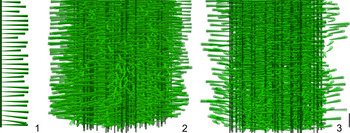
Computer-generated 3D models of axial structure for Inocladus lesquereuxi (Grote and Pitt, Reference Grote and Pitt1876) (see text for details): (1) isolated medullary tube bearing cortical tubes in a single row; (2) axis composed of a bundle of elements shown in (1); (3) longitudinal cross section of the axis shown in (2). Scale bar = 1 mm for all.

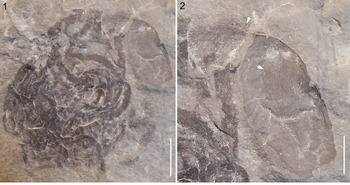
Inocladus lesquereuxi (Grote and Pitt, Reference Grote and Pitt1876), Bertie Group, Pridolian, Ridgemount Quarry, Fort Erie, Ontario, UMMP 75068: (1) complete specimen attached to phyllocarid carapace; (2) detail of upper right part of specimen in (1) showing thin filaments of holdfast at attachment point (upper arrow) and scale-like pattern on phyllocarid carapace (lower arrow). Specimen photographed dry. (1) Scale bar = 20 mm; (2) scale bar = 10 mm.

Among extant genera, Inocladus is most similar to Codium. In particular, many of the ~145 Codium species, as with all assigned herein to Inocladus, are characterized by erect thalli that reach a height of 10 cm or more and consist of elongate cylindrical to clavate axes with dichotomous branching. More crucially, the axes of species that make up both genera show an array of thin tubes divided into medullary and cortical regions. In this regard, the range in average medullary tube width reported by Oliveira-Carvalho et al. (Reference Oliveira-Carvalho, Pereira and Pedroche2010) for various Codium species (0.023–0.075 mm) overlaps with that observed herein for specimens assigned to Inocladus (0.05–0.19 mm). Key differences between Inocladus and Codium concern the arrangement of the medullary tubes, these being strongly longitudinal in the former versus an entangled mesh in the latter, and the development in Codium of cylindrical to fusiform reproductive structures (gametangia) along the sides of the cortical tubes (utricles), which have not been observed among any material of Inocladus. These differences establish Inocladus as a genus distinct from Codium. The cortical tubes of many Codium species also differ from those of Inocladus in being greatly expanded relative to the supporting medullary tube, often by a factor of six or more, but this is not always the case. For example, in Codium spongiosum Harvey, Reference Harvey1855, the cortical tubes are only about three times as wide as the medullary tubes (Oliveira-Carvalho et al., Reference Oliveira-Carvalho, Pereira and Pedroche2010, table 1), and the widths of both the cortical tubes and medullary tubes compare closely to those of Inocladus lesquereuxi. Inocladus also shows some similarity to Pseudocodium Weber-van Bosse, Reference Weber-van Bosse1896. In particular, the medullary tubes in this case have a longitudinal arrangement not unlike that of Inocladus. Pseudocodium, however, differs markedly in that the cortical tubes of all currently accepted species within the genus (see De Clerck et al., Reference De Clerck, Verbruggen, Huisman, Faye, Leliaert, Schils and Coppejans2008) are much wider than the supporting medullary tubes and have a rather bulbous form. Additionally, in Pseudocodium, external reproductive structures are developed along the sides of the siphons and the cortical tubes cohere strongly laterally (De Clerck et al., Reference De Clerck, Verbruggen, Huisman, Faye, Leliaert, Schils and Coppejans2008). Collectively, these differences establish Inocladus as a genus distinct from Pseudocodium.
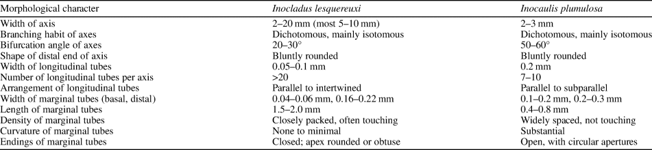
Morphological comparison between the respective type species of Inocladus n. gen. and Inocaulis Hall, Reference Hall1852.

Among fossil taxa, Inocladus shares much in common with Shonabellia Retallack, Reference Retallack1983, a monospecific noncalcified algal taxon erected on the basis of material from the Triassic of New Zealand. Thalli of both taxa are characterized by cylindrical axes with dichotomous branching and internally comprise numerous tubes divided into medullary and cortical regions. The cortical tubes of Shonabellia, however, branch dichotomously, and the medullary region consists of a “main thread and a loose mesh of narrow longitudinal filaments” (Retallack, Reference Retallack1983, p. 130), whereas in Inocladus the cortical tubes are unbranched, and all of the medullary tubes are of similar aspect. Noncalcified algal material designated ‘codiacean (?) sp. A’ and ‘codiacean (?) sp. B’ by Nitecki and Spjeldnaes (Reference Nitecki, Spjeldnaes, Barattolo, DeCastro and Parente1993) from the Silurian of Gotland (Lower Visby Marl and Hemse Group, respectively) also shows a structure similar to that of Inocladus, with longitudinally arranged medullary tubes giving rise to cortical tubes. In this case, all of the medullary tubes within a given axis have the same diameter (0.15–0.2 mm), and the cortical tubes are unbranched. Despite these similarities, this material is herein excluded from Inocladus because the fragmentary axes do not show definitive bifurcation although, as noted by Nitecki and Spjeldnaes (Reference Nitecki, Spjeldnaes, Barattolo, DeCastro and Parente1993), the possibility of branching is suggested by the fact that some axes increase in diameter toward the distal end. Indeed, this material agrees so closely with Inocladus that should further specimens reveal unequivocal bifurcation, it could be readily accommodated within the genus, in which case it would be maintained as a distinct species on the basis of the markedly bulbous (guttiform) shape of the cortical tubes. Other fossil taxa characterized by elongate cylindrical axes comprising narrow tubes divided into medullary and cortical regions include the early Paleozoic genera Palaeoporella Stolley, Reference Stolley1893, Dimorphosiphon Høeg, Reference Høeg1927, and Maslovina Obrhel, Reference Obrhel1968 and the late Paleozoic genus Anchicodium Johnson, Reference Johnson1946 (Kozłowski and Kaźmierczak, Reference Kozłowski and Kaźmierczak1968; Torres and Baars, Reference Torres and Baars1992; Boyd, Reference Boyd2007). All of these algal taxa differ significantly from Inocladus in being heavily calcified. In addition, in Palaeoporella, both the medullary and cortical regions are more complex than in Inocladus (see Kozłowski and Kaźmierczak, Reference Kozłowski and Kaźmierczak1968, fig. 3). Powysia Edwards, Reference Edwards1977, a monospecific noncalcified algal taxon erected on the basis of material from the Silurian (Ludlovian) of Wales, shows dichotomous branching of elongate axes that contain narrow longitudinally aligned tubes, but thallus branching is highly irregular, and crucially, no cortical tubes are developed from the longitudinal tubes.
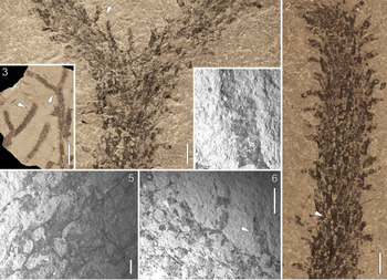
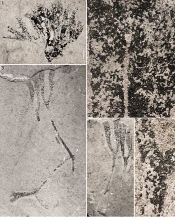
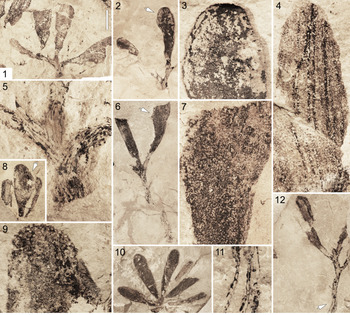
Inocladus is also similar to Inocaulis Hall, Reference Hall1852, a nonbiomineralized taxon that has been reported from uppermost Cambrian through lowermost Devonian strata (summarized by Ruedemann, Reference Ruedemann1947). The type species of the latter is Inocaulis plumulosa Hall, Reference Hall1852 (Fig. 7.1–7.6). This species was originally described on the basis of material from the Silurian Rochester Shale of New York and has also been recorded from the slightly younger Goat Island Formation of New York and Ontario (Ruedemann, Reference Ruedemann1947). It has been variously interpreted as a graptolite (Hall, Reference Hall1865; Ruedemann, Reference Ruedemann1908, Reference Ruedemann1947; Muir et al., Reference Muir, Zhang and Lin2013) or hydroid (Mierzejewski, Reference Mierzejewski1986); the Paleobiology Database currently lists it as belonging to the latter. The respective type species of these genera are similar in that both consist of elongate, largely isodiametric cylindrical axes that develop isotomous to subisotomous dichotomous branching and have an internal structure consisting of bundles of longitudinal tubes that bear, at more or less regular intervals, marginal tubular processes, the distal ends of which are oriented roughly perpendicular to the axial margin (Table 1). These points of similarity are what led Ruedemann (Reference Ruedemann1916) to transfer Buthotrephis lesquereuxi to Inocaulis. The anastomosing pattern reported for Inocaulis plumulosa by LoDuca et al. (Reference LoDuca, Bykova, Wu, Xiao and Zhao2017), after further SEM investigation, is here interpreted as a pattern resulting from shearing of the marginal processes from the supporting longitudinal tubes at the point of juncture when slabs were split, the resulting network of circular to oval holes on the axial surface marking where the former emerged from the latter (Fig. 7.5). The two taxa, however, are not identical. Points of difference identified during the present study include axis width, angle of axis bifurcation, number of longitudinal tubes per axis, width of longitudinal tubes, packing density of the marginal tubes, degree of curvature of the marginal tubes, and the endings of the marginal tubes (Table 1). Of these differences, the key distinction between Inocladus and Inocaulis concerns the endings of the marginal tubes. Specifically, as shown by the respective type species, those of the latter were open in life (Fig. 7.1, 7.2), whereas those of the former were closed (see description that follows and Fig. 3.1, 3.3). It is on the basis of this distinction, which carries significant implications in terms of higher-level phylogenetic affinity (see the following), that Inocladus lesquereuxi is regarded herein as belonging to a genus apart from Inocaulis.
Inocaulis plumulosa Hall, Reference Hall1852 from the Rochester Shale, Wenlockian, Middleport, New York, UMMP 75069: (1) enlargement of area at right arrow in (3); (2) enlargement of area at left arrow in (3); (3) typical specimen of this colonial metazoan; (4) SEM-BSE image of area at arrow in (6); (5) SEM-BSE image of area at arrow in (2); (6) SEM-BSE image of area at arrow in (1). Specimen in non-SEM images photographed dry with polarized light. (1, 2) Scale bars = 1 mm; (3) scale bar = 10 mm; (4) scale bar = 0.1 mm; (5) scale bar = 0.2 mm; (6) scale bar = 0.4 mm.

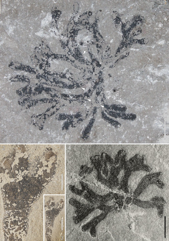
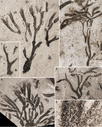
In addition to Inocladus lesquereuxi, the two aforementioned Buthotrephis species erected by White (Reference White1901), B. newlini and B. divaricata, are herein assigned to Inocladus, and a species somewhat similar to the latter, Chondrites verus Ruedemann, Reference Ruedemann1925, originally described on the basis of material from the Goat Island Formation of western New York, is also tentatively regarded as a member of this genus (Figs. 8–14). As described in detail in the following, all of these taxa share key aspects of gross thallus morphology and axial structure with Inocladus lesquereuxi.
Inocladus newlini (White, Reference White1901) from the Kokomo Limestone Member of the Wabash Formation, latest Ludlovian to Pridolian, Kokomo, Indiana: (1) complete specimen, holdfast at right, FMNH UC 875; (2) holotype, USNM P 8175 (image courtesy of F. Cochard). Specimens photographed dry. Scale bars = 20 mm.

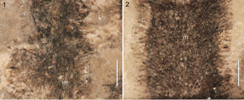
Details of axial structure for Inocladus newlini (White, Reference White1901), FMNH UC 875: (1) detail of area at lower arrow in 8.1 showing medullary (m) and cortical (c) tubes; (2) detail of area at upper arrow in 8.1 showing medullary (m) and cortical (c) tubes. Specimen photographed immersed in water with polarized light. Scale bars = 2 mm.

Inocladus divaricata (White, Reference White1901) from the Kokomo Limestone Member of the Wabash Formation, latest Ludlovian to Pridolian, Kokomo, Indiana: (1) complete specimen, neotype, arrows mark lowermost branching point of the respective primary axis, IUPC 593 (image courtesy of J. Miller-Camp); (2) enlargement of upper part of specimen shown in (3); (3) partial thallus, IUPC 11796-10A (image courtesy of J. Miller-Camp); (4) complete specimen, photo from White (Reference White1901), size corrected on the basis of White (Reference White1902). All specimens photographed dry. (1, 4) Scale bars = 20 mm; (2) scale bar = 5 mm; (3) scale bar = 10 mm.

Inocladus verus (Ruedemann, Reference Ruedemann1925) from the Goat Island Formation, Wenlockian, Gasport, New York: (1) large thallus, NYSM 19,133; (2) holotype, NYSM 6491; (3) juvenile thallus, NYSM 19,134; (4) large thallus, NYSM 19,135; (5) complete thallus, NYSM 19,136; (6) detail of area at arrow in (1) showing faint remnants of cortical tubes. All specimens photographed immersed in water with polarized light. (1–5) Scale bar in (1) = 20 mm; (6) scale bar in (1) = 2 mm.

Inocladus verus (Ruedemann, Reference Ruedemann1925) from the Goat Island Formation, Wenlockian, Gasport, New York, NYSM 19,137: (1) complete specimen; (2) detail of axis at left upper arrow in (1) showing medullary tubes; (3) detail of axis at lower arrow in (1) showing medullary tubes; (4) detail of axis at right upper arrow in (1) showing faint remnants of cortical tubes. All specimens photographed immersed in water with polarized light. (1) Scale bar = 20 mm; (2–4) scale bar in (1) = 2 mm.

Inocladus verus (Ruedemann, Reference Ruedemann1925) from the Goat Island Formation, Wenlockian, Gasport, New York, UMMP 75070: (1) complete specimen; (2) detail of area marked by arrow in (1); (3) SEM-BSE mosaic image of area at upper arrow in (2) showing cortical tubes at left; (4) SEM-BSE mosaic image of area at lower arrow in (2) showing medullary tubes. Specimen in non-SEM images photographed immersed in alcohol with polarized light. (1) Scale bar = 10 mm; (2) scale bar = 1 mm; (3, 4) scale bars = 0.2 mm.

Specimens tentatively assigned to Inocladus n. gen. from the Eramosa Formation, Wenlockian, Owen Sound Ledgerock Quarry, Wiarton, Ontario: (1) specimen tentatively assigned to Inocladus divaricata, ROMIP 65608; (2) detail of area at upper arrow in (4) showing faint remnants of cortical tubes; (3) specimen tentatively assigned to Inocladus verus, arrow marks lowermost bifurcation, ROMIP 65609; (4) partial counterpart of specimen shown in (3); (5) detail of bifurcation marked by lower arrow in (4). All specimens photographed immersed in water with polarized light. (1, 3, 4) Scale bar in (1) = 10 mm; (2, 5) scale bar in (1) = 1 mm.

It is possible that additional species currently residing within Buthotrephis should be transferred to Inocladus. These include, but are not necessarily limited to, Buthotrephis creditensis Foerste, Reference Foerste1923 from the Silurian of Ontario, B. nidarosiensis Høeg, Reference Høeg1941 from the Silurian of Norway and Podolia, B. butovi Korde, Reference Korde1993 from the Silurian of Russia, B. bifidis Ishchenko, Reference Ishchenko1985 and B. scapulus Ishchenko, Reference Ishchenko1985 from the Silurian of Podolia, a number of forms assigned to Buthotrephis from the Silurian of Australia (see Lucas, Reference Lucas1927), and B. walhalla Douglas and Jell, Reference Douglas and Jell1985 and B. trichotoma Douglas and Jell, Reference Douglas and Jell1985 from the Lower Devonian of Australia (the latter regarded by Hiller and Gess, Reference Hiller and Gess1996 as a junior synonym of Hungerfordia dichotoma Fry and Banks, Reference Fry and Banks1955). Currently, all of the aforementioned taxa are poorly known, especially with regard to internal structure, and it remains to be determined whether any possess the full suite of morphological characteristics necessary to support assignment to Inocladus. The same applies to Inocaulis akronensis Ruedemann, Reference Ruedemann1925 from the Silurian of New York, Manitobia patula Fry, Reference Fry1983 from the Ordovician of Ontario, Lenocladium rhizomatum Krassilov, Reference Krassilov and Ponomarenko2005 from the Cambrian of Siberia, and Yeaia flexuosa Douglas, Reference Douglas1983 and Y. africana Hiller and Gess, Reference Hiller and Gess1996, from the upper Silurian of Australia and the Upper Devonian of South Africa, respectively. The present work lays the methodological and taxonomic groundwork needed to proceed with such studies.
Inocladus lesquereuxi (Grote and Pitt, Reference Grote and Pitt1876) new combination
Figures 1–6
- Reference Grote and Pitt1876
Buthotrephis lesquereuxi Grote and Pitt, Reference Grote and Pitt1876, p. 88.
- Reference Pohlman1881
Buthotrephis lesquereux [sic]; Pohlman, p. 19, fig. 6.
- Reference White1901
Buthotrephis lesquereuxi; White, p. 266.
- Reference Grabau1901
Bythotrephis lesquereuxi; Grabau, p. 131, fig. 26.
- Reference Bassler1915
Buthotrephis lesquereuxi; Bassler, p. 145.
- Reference Ruedemann1916
Inocaulis lesquereuxi; Ruedemann, p. 13, pl. 4, figs. 1–4.
- Reference O'Connell1916
Buthotrephis lesquereuxi; O'Connell, p. 20.
- Reference O'Connell1916
‘Buthotrephis lesquereuxi’; O'Connell, p. 87.
- Reference Ruedemann1925
Inocaulis lesquereuxi; Ruedemann, p. 27, pl. 12, figs. 1, 2.
- Reference Monahan1931
Inocaulis lesquereuxi; Monahan, p. 383, plate 2, plate 3, fig. 1, plate 4, fig. 1.
- Reference Ruedemann1947
Inocaulis lesquereuxi; Ruedemann, p. 239, pl. 32, figs. 3–6.
- Reference Edwards1977
Inocaulis lesquereuxi; Edwards, p. 830.
- Reference Edwards, Banks, Ciurca and Laub2004
Inocaulis lesquereuxi; Edwards et al., p. 407, figs. 21–23.
- Reference Ciurca2010
Inocaulis sp.; Ciurca, p. 7, figs. 14, 15.
Holotype
BMNH 11460/E975 from the Silurian (Pridolian) Bertie Group at Buffalo, New York (figured by Ruedemann, Reference Ruedemann1916, pl. 4, fig. 2).
Diagnosis
Inocladus with sparse branching; primary axes commonly undivided or branched at only a single level, with branching not developed until 30 mm or more above base; cortical tubes with a length of 1.5–2.0 mm and a distal width of 0.16–0.22 mm.
Occurrence
Silurian Bertie Group (Pridolian) in western New York and southern Ontario.
Description
Erect, nonbiomineralized, unsegmented thallus, 100–200 mm in height, consisting of elongate flexuous axes, 2–10 or more of which extend from base; between thalli, axis width ranges from 3 to 20 mm, with most specimens falling in the range of 5–10 mm (Table 2). Axes commonly undivided or branched at only a single level; dichotomies mainly isotomous to subisotomous, not developed until 30 mm or more above thallus base; axis widths largely similar throughout a given thallus, but narrowed in basal parts and immediately above bifurcation points and occasionally expanded at tip; angle between axes at bifurcation between 20° and 30° but axes can become subparallel when traced distally; terminations obtuse to truncate (Figs. 1.1, 2.1–2.3). Axes consist of a dense array of medullary and cortical tubes without cross-walls; medulla consisting of 20 or more longitudinally arranged isodiametric tubes ranging in width from 0.05 to 0.1 mm, with most being 0.09 mm (Figs. 3.2–3.4, 4.1, 4.2), in some cases twisted around each other in a manner somewhat akin to fibers in a rope (Figs. 1.2, 3.1, 4.3, 4.4), cortical tubes closely packed, oriented at an angle of about 90° to the supporting medullary tube and arrayed along it in a single row, straight, 1.5–2 mm long, width 0.04 mm in basal part, gradually expanding to 0.16–0.22 mm near apex (most 0.2 mm), endings closed, apex obtuse to truncate (Figs. 3.1–3.3, 4.1, 4.2, 4.4, 4.5). Undamaged axis surfaces commonly show papillate texture (Fig. 1.3). Holdfast small relative to thallus, consisting of diffuse array of filaments at thallus base, each filament having a width of ~0.1 mm and a length typically less than 20 mm (Figs. 1.1, 2.4, 6.2).
Morphological data for Silurian Codium-like taxa belonging to the genera Inocladus n. gen. and Thalassocystis Taggart and Parker, Reference Taggart and Parker1976. All measurements in millimeters.

Materials
ROMIP 50692, 56896, 63964, 63965, 63966, 63971, 65539; UMMP 75068; INSM 71.1.8231.
Remarks
Grote and Pitt (Reference Grote and Pitt1876) erected Buthotrephis lesquereuxi as a new species of marine macroalga, or ‘fucoid’ in the parlance of the time, on the basis of a single specimen from the upper Silurian Bertie Group of western New York State (Buffalo area). Their brief description was limited to aspects of gross morphology, with the specimen noted to “exhibit a number of branches irregularly mingled by compression.” The branches were further described as having been cylindrical in life, sparingly dichotomous, 3–4 mm wide at the base gradually expanding to nearly 10 mm near the tip, and 13–14 cm long (Grote and Pitt, Reference Grote and Pitt1876, p. 88). A short time later, Pohlman (Reference Pohlman1881) documented a second specimen from the type locality. This report did not add any morphological details of note but did provide the first published illustration of the species (reproduced by Grabau, Reference Grabau1901), a simple outline drawing that, as noted by Ruedemann (Reference Ruedemann1916, p. 17), misleadingly shows the broken distal ends of the axes as natural terminations. White (Reference White1901, p. 266) provided the first description of the internal structure of the axes, observing a distinctive “tangle of filaments” within the axes of B. newlini and noting that this “texture” was also present in B. lesquereuxi and B. divaricata. Ruedemann (Reference Ruedemann1916, p. 13) observed similar details for a specimen then on display at the New York State Museum, noting that “the branches are composed of a twisted mass of long, fine carbonaceous (chitinous?) fibers” and further noting that, along the edges of the branches, “the terminal straight portions of the tubes or fibers are preserved in many places and are seen to project as a dense corona of processes perpendicular to the surface.” This report also provided the first illustration of the holotype. In a later report, Ruedemann (Reference Ruedemann1925, p. 27) described specimens with axes that were macerated before burial, noting that “the whole mass of fibers has been separated into ropelike bundles of twisted fibers, as many as seven of which were counted at the same level, indicating a quite complicated interior structure of the stipes.” Comparable details were observed among the material examined during the present study. However, new details were also observed. This was possible, in part, because some of the examined specimens, such as ROMIP 50692 and 63966, owing to fortuitous breakage during splitting of slabs, provided cross sections at different levels within axes (e.g., Fig. 3.3), and these, when considered collectively, provided a more complete picture of the internal structure. Polarized light images also contributed to an enhanced view since these had greater contrast and less localized glare compared with unfiltered images, thereby clarifying relationships between adjacent and overlapping tubes (Figs. 3.1–3.4, 4.3, 4.4). Further details in this regard were provided by SEM-BSE images (Fig. 4.1, 4.2). The new observations indicate that the tubes are divided into two distinct zones, a central or medullary zone, composed of longitudinally arranged medullary tubes, and an outer or cortical zone, in which closely packed cortical tubes run roughly perpendicular to the axis margin. Moreover, it is evident that the medullary tubes give rise to the cortical tubes by simple branchings spaced at more or less regular intervals, with the cortical tubes emerging at an angle of roughly 90°, showing no further branching of their own, and arrayed in a single row along the length of the medullary tube. Although these particular details for the tubes have not been reported previously, White (Reference White1901, pl. 17) provided an inset sketch with his figure of Buthotrephis newlini that conveyed a structure along these lines for this species (see the following), albeit without description in the text. It should be noted that this internal structure is not always evident in Inocladus lesquereuxi specimens. In some cases, this is because the rock split along the outer surface of the thallus, rather than along a plane passing through its interior; in others, this is attributable to poor preservation. Even when the tubes are visible, details of their arrangement are often obscured by a dense array of overlapping tubes.
To explore whether the axial structure described in the preceding could indeed generate axes comparable to those observed for this species, three-dimensional (3D) computer renderings were produced using the program Blender. For this, code written originally by Hewitt (Reference Hewitt2017) to produce a wide range of tree morphologies was utilized to provide a rough facsimile of the interpreted architecture. Specifically, parameters were set to produce an axis consisting of a bundle of longitudinally arranged tubes, with each longitudinal tube bearing a single row of roughly evenly spaced, undivided side branches (Fig. 5.1; compare with Fig. 4.2). The results provide a reasonable approximation of the appearance of actual specimens of Inocladus lesquereuxi (Fig. 5.2; compare with Fig. 3.1), including when the program is set to produce longitudinal cross sections, thereby approximating the state of some fossil specimens (Fig. 5.3; compare with Fig. 3.3). It must be noted, however, that the renderings correspond to the form of the axis in life (see the following), whereas the axes of fossil specimens have been flattened by compression. In addition, within the axes of Inocladus lesquereuxi the cortical tubes were probably always oriented outward, away from the axis center, and may not have been produced at all by medullary tubes located nearest the axis center. In the renderings, however, the cortical tubes have a somewhat random orientation, and all of the medullary tubes bear a full complement of cortical tubes.
In accordance with the detailed description provided by Ruedemann (Reference Ruedemann1916), the axes are herein viewed as having been cylindrical in life. An originally cylindrical form is indicated by a lack of twisting or folding of the axes and by a high density of cortical tubes along axis margins, which is consistent with flattening of a cylindrical axis in which the cortical tubes are arrayed around a full 360°. Ruedemann (Reference Ruedemann1916, p. 15), however, described the endings of the marginal tubes along the axis surfaces as being open in life (‘circular pores’). Examination of well-preserved material during the present investigation indicates that they were closed, a finding supported by images of specimens published by Edwards et al. (Reference Edwards, Banks, Ciurca and Laub2004, figs. 22, 23). This discrepancy in morphological interpretation is attributable to damage that occurs to specimens when slabs are split during collection since it is not uncommon for the plane of breakage to intersect the distal ends of the marginal tubes along the surface of the axis. This gives the appearance, along the axis surface, of tubes with open endings. That they were in fact closed in life is evident along the edges of the axes where the tubes run parallel to the bedding plane surface (Figs. 3.1, 3.3, 4.4, 4.5) and along axis surfaces that are undamaged (Fig. 1.3).
Elemental analysis with the SEM indicates that specimens of Inocladus lesquereuxi consist of essentially pure carbon. Carbonaceous material in two of the best-preserved specimens examined, ROMIP 50692 and 63966, appears to be concentrated in the medullar tubes because these are dark brown to black, whereas the cortical tubes are a conspicuously lighter shade of brown (Figs. 3.1–3.4, 4.3, 4.4). Even within the cortical tubes, the distribution appears uneven. In this regard, the proximal parts are darker than the distal, so that the latter typically have a ‘ghosted’ appearance apart from small isolated specks of darker material, especially near the tips. The potential biological significance of this uneven distribution of carbonaceous material is examined in a later section of the paper.
The holdfast of ROMIP 56896 (Fig. 1.1) is typical for the species. Most notably, it is small in relation to overall thallus size and consists of a loose array of thin filaments, each with a width of 0.1 mm. Monahan (Reference Monahan1931) described and illustrated an oval structure at the base of two specimens, BSNS 13291/E1620 and 13289/E1618, from the Bertie Group in the Bennett quarries, north of Buffalo, New York. He regarded these structures as the remains of thin-walled “inflated sacs” developed from the basal part of the thallus and used for flotation (Monahan, Reference Monahan1931, p. 385). During the present study, a similar structure was observed in association with a specimen from the Bertie Group exposed in the Ridgemount Quarry of southern Ontario (Fig. 6.1). A distinctive, scale-like pattern is evident in one part of this oval structure (Fig. 6.2). On the basis of overall form, together with the distinctive pattern, this object is herein regarded as a phyllocarid carapace, the pattern being an overprint of that present on the underlying abdomen. The objects associated with 13291/E1620 and 13289/E1618 are poorly defined and lack distinctive structure, but it is inferred that they, too, represent arthropod material, albeit showing a greater degree of decay. Notably, in all of these cases, a bundle of filaments comparable to that of a typical holdfast can be observed at the point of attachment to the oval object (Fig. 6.2). The interpretation of these oval structures as arthropod material is consistent with the observation of O'Connell (Reference O'Connell1916, p. 20) that “many of the specimens of Eurypterus [at the Buffalo locality] are found lying embedded in Buthotrephis lesquereuxi, and in one case there is a large mass of Buthotrephis at the side and on top of a Eurypterus.” Thus, it appears that, in at least some instances and in a fashion not unlike how modern Codium attaches to bivalve shells, this species affixed itself to the exoskeletons of arthropods. Whether it did so before or after the death of the arthropod remains unknown. Such an association has not been observed among material of the other species assigned herein to Inocladus, but this could simply reflect a scarcity of arthropod material and other skeletal debris in the respective specimen-bearing intervals compared with the Bertie Group occurrences.
Inocladus newlini (White, Reference White1901) new combination
Figures 8, 9
- Reference White1901
Buthotrephis newlini White, p. 266, pls. 17, 18.
- Reference Cumings and Shrock1928
Buthotrephis newlini; Cumings and Shrock, p. 121.
- Reference Cumings1929
Buthotrephis newlini; Cumings, p. 206.
- Reference Monahan1931
Buthotrephis newlini; Monahan, p. 384.
- Reference Johnson and Konishi1959
Buthotrephis newlini; Johnson and Konishi, p. 106, pl. 21.
- Reference Edwards1977
Buthotrephis newlini; Edwards, p. 830.
- Reference Fry1983
Buthotrephis newlini; Fry, p. 325.
Holotype
USNM P 8175 from the Kokomo Limestone Member of the Wabash Formation (latest Ludlovian to Pridolian) at Kokomo, Indiana (White, Reference White1901, pl. 17) (Fig. 8.2).
Diagnosis
Inocladus with frequent branching and long intervals between successive dichotomies, each primary axis commonly developing at least two and often three levels of dichotomous branching, the first level not initiated until 30 mm or more above thallus base; cortical tubes with a distal width of 0.13–0.17 mm and a length of 1.0–1.5 mm.
Occurrence
Kokomo Limestone Member of the Wabash Formation (latest Ludlovian to Pridolian) at Kokomo, Indiana.
Description
Thallus of FMNH UC 875 erect, nonbiomineralized, unsegmented, 180 mm in height, consisting of numerous elongate flexuous axes 5–6 mm wide (Fig. 8.1). Each primary axis with up to two levels of dichotomous branching; dichotomies mainly isotomous to subisotomous, the first level not developed until 30 mm or more above thallus base, with a comparable interval between first and second level; axis widths largely uniform throughout thallus, but ultimate branches slightly expanded distally; angle between axes at bifurcation between 20° and 30°; axial terminations obtuse to truncate. Axes consist of a dense array of medullary and cortical tubes (Fig. 9.1, 9.2); medulla consisting of numerous longitudinally arranged isodiametric tubes ranging in width from 0.05 to 0.1 mm, with most being 0.07 mm, cross-walls not evident; cortical tubes closely packed, oriented at an angle of about 90° to the supporting medullary tube and arrayed along it in a single row, straight, 1.0–1.5 mm long, with a distal width of 0.13–0.17 mm (most 0.15 mm), endings closed, apex rounded to truncate. Axis surfaces commonly show a papillate texture (Fig. 8.1). Thallus of USNM P 8175 similar, but primary axes more strongly divergent at base (Fig. 8.2).
Remarks
White (Reference White1901, p. 266) noted that this species differs from Inocladus lesquereuxi primarily in details of thallus branching, the branches of this species being “much more frequently forking.” More specifically, as circumscribed herein by the provided diagnosis, the primary axes on a given thallus typically display at least two and often three levels of dichotomous branching. By comparison, in Inocladus lesquereuxi the axes often remain undivided, and the rare branchings are typically limited to a single level along a particular axis. White (Reference White1901) also noted that the axes are somewhat narrower than those of Inocladus lesquereuxi. In the present investigation, it was determined that the axes of the largest Inocladus lesquereuxi are indeed wider than those of the largest Inocladus newlini (up to 20 mm versus 6 mm, respectively). Smaller examples of the former, however, have axis widths that overlap with the range shown by the latter (Table 2). As noted by White (Reference White1901), internal details are essentially the same between these taxa. This includes the morphology of the cortical tubes, as shown well by FMNH UC 875, a previously undescribed Field Museum specimen cataloged as Buthotrephis newlini and examined during the present study (Figs. 8.1, 9.1, 9.2). The well-preserved cortical tubes of this specimen have a width and shape closely comparable to those of Inocladus lesquereuxi but differ in having a slightly shorter length (1–1.5 mm versus 1.5–2 mm; Table 2).
White (Reference White1901) assigned two specimens to this species, both of which were described and figured. One of these, the holotype (USNM P 8175; Fig. 8.2), is currently housed at the Smithsonian. The internal structure for this specimen, as shown in a sketch that accompanied the original figure (White, Reference White1901, pl. 17), agrees with the description provided herein for FMNH UC 875. The other specimen (White, Reference White1901, pl. 18) is notable in having axes only about half as wide as those of USNM P 8175. White (Reference White1901, p. 266) regarded this specimen as a “more delicate or perhaps younger example” of the species. Material of a further species considered herein as belonging to Inocladus, Inocladus verus, from the Goat Island Formation of New York, supports White's assessment because it also shows both robust and gracile forms, as well as thallus morphologies intermediate between these endpoints. At the time that White described this specimen, it resided in the private collection of C.E. Newlin of Irvington, Indiana, and its current whereabouts could not be determined.
Inocladus divaricata (White, Reference White1901) new combination
Figures 10, 14.1
- Reference White1901
Buthotrephis divaricata White, p. 265, pl. 16.
- Reference White1902
Buthotrephis speciosa; White, p. 86.
- Reference Cumings and Shrock1928
Buthotrephis divaricata; Cumings and Shrock, p. 121.
- Reference Cumings1929
Buthotrephis divaricata; Cumings, p. 206.
- Reference Monahan1931
Buthotrephis divaricata; Monahan, p. 384.
- Reference Edwards1977
Buthotrephis divaricata; Edwards, p. 830.
- Reference Fry1983
Buthotrephis divaricata; Fry, p. 325.
- Reference Douglas and Jell1985
Buthotrephis divaricata; Douglas and Jell, p. 161.
Neotype
IUPC 593 from the Kokomo Limestone Member of the Wabash Formation (latest Ludlovian to Pridolian) at Kokomo, Indiana (Fig. 10.1).
Diagnosis
Inocladus with frequent branching and short intervals between successive dichotomies, each primary axis commonly developing two levels of dichotomous branching, the distance between dichotomies being about three times the width of the axis or less, the first level of branching typically initiated within 20 mm of thallus base.
Occurrence
Kokomo Limestone Member of the Wabash Formation (latest Ludlovian to Pridolian) at Kokomo, Indiana; Eramosa Formation (Wenlockian) in the Bruce Peninsula, Ontario.
Description
Erect nonbiomineralized unsegmented thallus 60–100 mm in height, consisting of elongate axes 5–8 mm wide arising from a broad base (Fig. 10.1, 10.4). Each primary axis typically develops two levels of dichotomous branching; dichotomies isotomous, spacing between dichotomies about three times axis width, the first level of branching typically initiated within 20 mm of thallus base; axis widths largely uniform but often expanded at tip; angle between axes at bifurcation typically greater than 30° (divaricate); terminations obtuse to truncate.
Materials
IUPC 593, 11796-10A; ROMIP 65608.
Remarks
White's (Reference White1901, p. 265–266) descriptions of this species and Inocladus newlini are largely in accord, but differ in two key areas, both having to do with branching morphology. First, branching angle for the former was said to be “divaricate, usually at a wide angle” whereas a “moderate or wide angle” was specified in this regard for Inocladus newlini. Second, the first bifurcation for the former occurs “near the base” with the spacing in this regard and between higher-level bifurcations being “about three times the width of the lamina [ = axis],” whereas for Inocladus newlini bifurcations were said to be developed “generally distantly.” Herein, only the second difference is regarded as having taxonomic significance since burial conditions can have a marked impact on branching angle for specimens preserved as compressions and White's description of Inocladus. divaricata was based on a single specimen. White (Reference White1901, p. 266) also noted that the internal structure (“texture”) of this species agreed with that of both Inocladus lesqueruexi and Inocladus newlini but differed slightly in that the tubes were “finer.”
During the present study, an effort was made to locate the specimen upon which the species was erected by White (Reference White1901), which would constitute the holotype. White (Reference White1901, p. 265) noted that the specimen was in private hands, in the “collection of Mr. [C.E.] Newlin in Irvington, Indiana.” Examination of online databases and consultation with curators indicate that this specimen is not at the Smithsonian, American Museum of Natural History, Purdue University–Fort Wayne, Indiana State Museum, Field Museum, or Indiana University, and its current whereabouts is unknown. A complete specimen from the type locality assignable to this species was, however, located in the Indiana University Paleontology Collections (J. Miller-Camp, personal communication, 2019). This specimen, which was collected by R.R. Shrock in 1961 and carries the label ‘Buthotrephis divaricata’ written on the slab, is herein proposed as the neotype (Fig. 10.1). The preservation of IUPC 593 is poor, but the overall size and branching pattern are comparable to that of the specimen figured by White (Reference White1901). In particular, the initial branching point for the primary axes occurs near the base (indicated by white arrows in Fig. 10.1), and branching intervals are spaced at a distance of approximately three times the axis width. A further specimen from the same collection, IUPC 11796-10A, is also formally assigned herein to Inocladus divaricata (Fig. 10.3).
A specimen from the Eramosa Formation near Wiarton, Ontario, ROMIP 65608, is tentatively assigned herein to Inocladus divaricata (Fig. 14.1). The branching morphology of this thallus is consistent with that shown by Inocladus divaricata from the type locality, in that it exhibits characteristically short intervals between dichotomies, but internal details of the axes are poorly preserved, with large areas appearing as a uniform black film. Hints of what appear to be cortical tubes are, however, evident in areas. Regarded as Inocladus divaricata, this specimen is important in being the first representative of the species from outside the type locality and extends the stratigraphic range slightly.
White (Reference White1902), in a follow-up note to the original description, observed that the name Buthotrephis divaricata was already occupied by Buthotrephis divaricata Kidston, Reference Kidston1886 and proposed the name Buthotrephis speciosa as a replacement. The latter name does not appear to have been used in any subsequent publication. Therefore, herein the original specific epithet is retained, and assignment to the new genus Inocladus eliminates any nomenclatural conflict.
Inocladus verus (Ruedemann, Reference Ruedemann1925) new combination
Figures 11–13, 14.2–14.5
- Reference Ruedemann1925
Chondrites verus Ruedemann, p. 15, pl. 4, fig. 5.
Holotype
NYSM 6491, from the Goat Island Formation of the Lockport Group at Gasport, New York (Ruedemann, Reference Ruedemann1925, pl. 4, fig. 5) (Fig. 11.2).
Diagnosis
Inocladus with frequent branching and long intervals between dichotomies, each primary axis commonly developing at least two and often three levels of dichotomous branching; axes above ultimate branch point often wider than those immediately below and commonly conspicuously expanded distally; cortical tubes narrow, with a width ranging from 0.07 to 0.11 mm.
Occurrence
Goat Island Formation of the Lockport Group (Wenlockian) at Gasport, New York; Eramosa Formation of the Lockport Group (Wenlockian) at Wiarton, Ontario.
Description
Erect nonbiomineralized unsegmented thallus, 40–120 mm in height, consisting of two to four primary axes, each 2–5 mm wide, arising from a bulbous base (Figs. 11.1, 11.2, 11.4, 11.5, 12.1, 13.1, 14.3; Table 2). Each primary axis with at least two levels of dichotomous branching; dichotomies mainly isotomous to subisotomous, intervals between branch points 20–30 mm; axis widths largely uniform throughout thallus, but ultimate branches typically expanded relative to subtending axes and often with distinctly lobate terminations; angle between axes at bifurcation between 20° and 30°, but axes can become subparallel when traced distally. Axes consist of a dense array of medullary and cortical tubes; medulla consisting of numerous longitudinally arranged isodiametric tubes, estimated to number 5–15, ranging in width from 0.15 to 0.19 mm (Figs. 12.2, 12.3, 13.4), cross-walls not evident; cortical tubes closely packed, narrow, with width of 0.07 to 0.11 mm (most 0.09 mm), these imparting a coarsely hirsute texture to axis surfaces and margins (Figs. 11.6, 12.4, 13.2, 13.3). Holdfast small relative to thallus, bulbous, with a fringe of fine filaments (Figs. 11.1–11.3, 12.1, 13.1).
Materials
NYSM 19,133–19,137; UMMP 75070; ROMIP 65609.
Remarks
Ruedemann (Reference Ruedemann1925) assigned a single specimen to this species, NYSM 6491, from the Goat Island Formation at Gasport, New York (Fig. 11.2). The accompanying description did not provide details about the internal structure of the axes. Since that initial report, no other specimens have been formally assigned to the species, and with the exception of LoDuca (Reference LoDuca1995, p. 374) noting a “siphonous construction” for the axes, no further descriptions have been provided. The branching habit of 6491 is similar to that of Inocladus newlini, particularly the ‘delicate’ specimen assigned to this species by White (Reference White1901), in that the axes have a similar width and develop two or more levels of dichotomous branching with relatively large intervals between successive dichotomies. In addition, the distalmost parts of the axes of Inocladus newlini occasionally show a lobate form similar to that of 6491. Internal details for 6491 are not clearly evident, but faint remnants of tubes can be detected in places.
During the present study, a number of previously undescribed specimens from the type locality comparable to NYSM 6491 were examined, all of which are herein formally assigned to Inocladus verus. Most of these were purchased from amateur collectors E. Reinhard and G. Reimann by the New York State Museum in 1928 and 1934, respectively. The remainder were collected by the senior author during study of this locality in the late 1980s. Some of these specimens, most notably NYSM 19,133, 19,137, and UMMP 75070, provide more information about internal structure than the holotype, but some key details, including relationships between the medullary and cortical tubes, remain unresolved. Notably, these specimens do show that, compared with Inocladus newlini, the cortical tubes are narrower and the medullary tubes are wider, in each case by a factor of about two (Figs. 11.6, 12.2–12.4, 13.2–13.4; Table 2). On the basis of these differences, Inocladus verus is maintained herein as a separate species. Because some key details of internal structure are currently unknown, it is possible that better preserved material, should it be discovered, will reveal that this species belongs to a genus apart from Inocladus, albeit closely related to it.
As with specimens of Inocladus newlini, specimens of Inocladus verus show variation in gross thallus morphology, ranging from gracile forms (e.g., Fig. 11.5) to those with a more robust morphology (e.g., Figs. 11.1, 12.1). Thalli with a morphology intermediate between these endpoints are also known (Fig. 11.2, 11.4). Notably, some of the Gasport specimens are small and show only a single level of branching (e.g., Fig. 11.3). These are regarded herein as early growth stages of Inocladus verus.
A specimen from the Eramosa Formation near Wiarton in the Bruce Peninsula of Ontario, ROMIP 65609, is tentatively assigned to Inocladus verus (Fig. 14.2–14.5). Considered as such, it constitutes the first material for the species from an area outside of the type locality. The branching habit is similar to that of the Inocladus verus holotype in that several levels of dichotomous branching are present and the terminal branches are lobate and expanded relative to those below. Internal details of the axes, however, are not clearly evident. What appear to be poorly preserved cortical tubes are present in places. These impart a coarsely hirsute texture to axis surfaces and margins (Fig. 14.3) similar in character to specimens of Inocladus verus from the type locality (compare with Fig. 12.4).
Genus Thalassocystis Taggart and Parker, Reference Taggart and Parker1976
Type species
Thalassocystis striata Taggart and Parker, Reference Taggart and Parker1976, by monotypy.
Thalassocystis striata Taggart and Parker, Reference Taggart and Parker1976
Figures 15.1–15.5, 15.7–15.10, 16
- Reference Taggart and Parker1976
Thalassocystis striata Taggart and Parker, p. 1390, figs. 1–4.
- Reference Johnson and Campbell1980
Inocaulis manistiquensis; Johnson and Campbell, p. 1046.
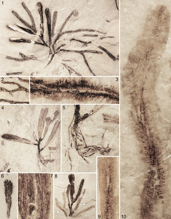
(1–10) Thalassocystis striata Taggart and Parker, Reference Taggart and Parker1976 (1–5, 7–10) and Medusaegraptus sp. (6) from the Schoolcraft Formation, Llandoverian, Carmeuse Lime & Stone quarry (formerly Inland Quarry), Gulliver area, Michigan: (1) large complete specimen, UMMP 75071; (2) counterpart of area at arrow in (1); (3) enlargement of area at arrow in (2) showing medullary and cortical tubes within axis; (4) complete specimen with holdfast at base, UMMP 75072; (5) enlargement of lower part of specimen in (4) showing holdfast; (6) Medusaegraptus sp., complete thallus, UMMP 75073; (7) detail of wide axis showing medullary tubes within, DMF 5183; (8) nearly complete specimen, DMF 4781; (9) enlargement of axis at right arrow in (8) showing medullary and cortical tubes; (10) enlargement of axis at left arrow in (8) showing medullary and cortical tubes. All specimens photographed immersed in water with polarized light. (1, 2, 4, 8) Scale bar in (1) = 10 mm; (6) scale bar in (1) = 5 mm; (5) scale bar in (1) = 3.3 mm; (7) scale bar in (1) = 2.5 mm; (9) scale bar in (1) = 2 mm; (3, 10) scale bar in (1) = 1 mm.

(1–12) Thalassocystis striata Taggart and Parker, Reference Taggart and Parker1976 from the Schoolcraft Formation, Llandoverian, Lake Michigan shoreline type locality (1–9, 11, 12) and White Marble Lime Company quarry (10), vicinity of Manistique, Michigan: (1) nearly complete specimen, UMMP 75075; (2) partial specimen, UMMP 75076; (3) enlargement of area at arrow in (2) showing papillate surface and compression rim around margin; (4) detail of wide axis showing medullary tubes within, UMMP 75077; (5) enlargement of lower part of thallus in (1) showing medullary tubes; (6) partial specimen, UMMP 75078; (7) enlargement of area at arrow in (6) showing papillate surface; (8) partial specimen, UMMP 75079; (9) enlargement of area at arrow in (8) showing papillate surface; (10) nearly complete specimen, previously illustrated by Ehlers (Reference Ehlers1973, pl. 22, fig. 6), UMMP 11137; (11) enlargement of area at arrow in (12) showing medullary tubes in lower part of thallus; (12) partial specimen, UMMP 75080. All specimens photographed immersed in water with polarized light. (1, 2, 6, 8, 10, 12) Scale bar in (1) = 10 mm; (3–5, 7, 9, 11) scale bar in (1) = 2 mm.

Holotype
FMNH PP 43982 from the Schoolcraft Formation (Llandoverian) in an exposure along the Lake Michigan shoreline two miles east of Manistique, Michigan (Taggart and Parker, Reference Taggart and Parker1976, fig. 1).
Diagnosis
Erect nonbiomineralized unsegmented thallus comprising dichotomously branched axes with a clavate to obovate form; branching highly irregular (anisotomous). Axes consist of a dense array of medullary and cortical tubes; medulla consisting of numerous narrow longitudinally arranged tubes in an intertwined configuration; cortical tubes closely spaced, arising from the supporting medullary tube at an angle of ~90° and arrayed along it in a single row, straight, cylindrical, 0.09–0.12 mm wide, with closed endings, apex blunt. Holdfast small, consisting of a loose bundle of thin filaments.
Occurrence
Schoolcraft Formation of the Manistique Group (Llandoverian) in the White Marble Lime Company quarry in the city of Manistique (Locality 105 in Ehlers, Reference Ehlers1973); in exposures along the Lake Michigan shoreline two miles east of Manistique (Locality 104 in Ehlers, Reference Ehlers1973; see also Taggart and Parker, Reference Taggart and Parker1976); in a temporary exposure approximately 1 km due east of Manistique (locality 1958/S-3 in the UMMP collection); in the southernmost part of east pit of the Carmeuse Lime & Stone quarry at 46°03'33.9''N, 85°49′33.2″W (this is an expansion of the quarry at Locality 73 in Ehlers, Reference Ehlers1973).
Description
Erect nonbiomineralized unsegmented thallus, 35–70 mm in height, comprising dichotomously branched axes with a clavate to obovate form; branching highly irregular (anisotomous). Individual axes with a maximum width of 2–11 mm and reaching 50 mm long; proximal parts with a width of 1–3 mm; terminations obtuse (Figs. 15.1, 15.4, 15.8, 16.1, 16.2, 16.10, 16.12; Table 2). Angle between axes at bifurcation typically between 30° and 40°, but axes often become subparallel when traced distally. Axes consist of a dense array of medullary and cortical tubes; medulla consisting of 5–15 longitudinally arranged isodiametric tubes, each with a width of 0.13–0.18 mm, cross-walls not evident (Figs. 15.3, 15.7, 15.9, 15.10, 16.4, 16.5, 16.11); cortical tubes closely packed, oriented at an angle of about 90° to the supporting medullary tube and arrayed along it in a single row, with a spacing between adjacent tubes of less than one tube width, straight, cylindrical, 0.5–1.0 mm long and 0.10–0.13 mm wide, with blunt endings (Fig. 15.3, 15.9, 15.10), coalesced distally to form a continuous outer surface with a finely papillate texture (Fig. 16.3, 16.4, 16.7, 16.9). Holdfast small relative to thallus, typically 4–6 mm wide and 10–20 mm long, consisting of a loose array of thin filaments, each having a width of ~0.3 mm (Fig. 15.5).
Materials
DMF 4781, 5183, 5184, 5186, 5187; UMMP 11137, 74056, 74058, 74060, 74063, 75071, 75072, 75075–75080.
Remarks
The emended diagnosis presented in the preceding differs markedly from the original diagnosis provided by Taggart and Parker (Reference Taggart and Parker1976, p. 1390). In particular, they noted that the branches are “terminated by an enlarged bladder,” this formed by a “sheath of tissue,” and further noted that “the surface of the bladders show numerous fine parallel longitudinal striations.” During the present investigation, well-preserved specimens showing new details were collected from a new locality for the species at the Carmeuse Lime & Stone quarry near Gulliver, Michigan (Fig. 15.1–15.5, 15.7–15.10). The best preserved of these specimens show that the axes have a structure very much like those of Inocladus lesqueureuxi, with the surface striations described by Taggert and Parker (Reference Taggart and Parker1976) instead corresponding to internal, longitudinally arranged medullary tubes, each of which bears short cortical tubes arrayed in a single row (Fig. 15.3, 15.9, 15.10). As with specimens of Inocladus lesqueureuxi, the cortical tubes often contain much less carbonaceous material than the medullary tubes. In many specimens, only the medullary tubes are clearly evident (Figs. 15.7, 16.4). This is interpreted to reflect a lower fossilization potential for the cortical tubes. In some cases the presence of cortical tubes is manifested by a distinctly papillate surface on the axes (Fig. 16.3, 16.4, 16.7, 16.9). This previously undocumented detail, which is present only on the best preserved material, is comparable to that evident on axis surfaces of Inocladus lesquereuxi (e.g., Fig. 1.3) as well as pressed herbarium specimens of Codium, with each subtle circular protuberance corresponding to the distal end of a cortical tube.
Morphometric details pertaining to the medullary and cortical tubes correspond closely to those of Inocladus verus (Table 2). Specifically, the cortical tubes of both taxa are similar in terms of width and shape, these being narrow compared with those of the other Codium-like taxa described herein, and the medullary tubes of both taxa are similar in terms of width, with these being wider than those of the other Codium-like taxa described herein. Despite these similarities, T. striata is maintained in a genus apart from Inocladus. The key determining factor in this regard is the marked difference in branching habit between T. striata and that diagnostic of Inocladus. Specifically, the former is characterized by highly irregular dichotomous branching, whereas bifurcations for the latter are isotomous or very nearly so. Whether such a difference is sufficient justification to obviate the alternative, congeneric viewpoint is open to question, since Codium includes species with isotomous as well as highly irregular branching, with both types of branching in some cases occurring within a single species (e.g., C. isthmocladum Vickers, Reference Vickers1905). However, because Thalassocystis striata and all species assigned to Inocladus differ from Codium in lacking external reproductive structures along the cortical tubes and in details of medullary tube arrangement, Codium may not serve as a reliable guide in this context. With this in view, the two-genus approach is regarded as the most pragmatic.
An additional novel observation is that some specimens consist entirely of conspicuously wide axes, with axis widths reaching 9–10 mm (e.g., Fig. 16.1), whereas others show only comparatively narrow axes, with maximum axis widths at or below 5 mm (e.g., Fig. 15.1). One might infer that these wide and narrow morphs, which are present in all occurrences, represent different species, but two factors argue against such an interpretation. First, forms showing axis widths midway between these endpoints are known (e.g., Fig. 15.4). Second, some specimens show both wide and narrow axes on the same thallus (e.g., Fig. 16.8; see also Ehlers, Reference Ehlers1973, plate 16, fig. 2). Consequently, without further information to the contrary, all of the material at hand is regarded as representing a single species of Thalassocystis.
The terminal branches of some specimens do appear rather bladder-like, in agreement with the description provided by Taggart and Parker (Reference Taggart and Parker1976). The branches in these cases have a strongly 3D, roughly ovoid form. Such a morphology is particularly well developed among specimens from the type locality (Fig. 16.2) but is also known for some specimens from the new locality. These inflated terminal branches resemble those of some living Codium species, where trapped oxygen produced during photosynthesis results in bladder-like terminations, these providing positive buoyancy thought to assist in the dispersal of detached branches during episodes of vegetative reproduction (Gagnon et al., Reference Gagnon, McKindsey and Johnson2011). The fact that some axes retain a degree of three-dimensionality indicates that the initial burial layer was thin and lithified very rapidly after deposition. Many specimens, however, are completely flattened. These often show a distinct rim or margin (e.g., Fig. 16.3) comparable to that produced along the edges of pressed herbarium specimens of Codium.
Taggart and Parker (Reference Taggart and Parker1976) apparently were unaware that material belonging to this species had been previously illustrated by Ehlers (Reference Ehlers1973, pl. 16, fig. 1, 2, pl. 22, figs. 6–8). In that earlier report, Ehlers (Reference Ehlers1973) astutely recognized that the material represented a new species, listing it under the manuscript name ‘Inocaulis manistiquensis,’ but he did not describe it. The referral to Inocaulis is significant because it suggests that Ehlers observed internal details for the specimens along the lines of those noted by Ruedemann (Reference Ruedemann1916) in his treatment of Inocaulis lesqueruexi (= Inocladus lesqueruexi, herein), a point of similarity also observed in the present investigation and a key point of departure from the formal description provided by Taggart and Parker (Reference Taggart and Parker1976). Stratigraphic section descriptions, faunal inventories, and figure captions by Ehlers (Reference Ehlers1973) list this species as a graptolite. This, too, is in keeping with the description of Inocaulis lesqueruexi provided by Ruedemann (Reference Ruedemann1916). During the present study, one of the four specimens figured by Ehlers (Reference Ehlers1973) was located in the collections of the University of Michigan after a search through uncataloged material (Fig. 16.10); the other three specimens could not be located and may be lost (D. Miller, personal communication, 2019). Within the cataloged University of Michigan collections, additional material collected by Ehlers, but not previously figured, was also examined. This includes material from the Lake Michigan shoreline type locality (UMMP 74056, 74058, 74063) and four specimens, all cataloged as UMMP 74060, that were recovered from a previously unreported occurrence of the species approximately 1 km due east of Manistique. The latter were collected when the Schoolcraft Formation was briefly exposed during the construction of a sewage pumping station. All of this material is herein formally referred to Thalassocystis striata.
Previously, material assignable to T. striata had been reported only from the Manistique area, with Ehlers (Reference Ehlers1973) noting material from the White Marble Lime Company quarry within the city limits and Taggart and Parker (Reference Taggart and Parker1976) describing material from exposures along the Lake Michigan shoreline, some 3 km to the east of the city center. The assignment herein of material from the new locality within the Carmeuse Lime & Stone quarry near Gulliver expands the geographic range of the species eastward along the Silurian outcrop belt in northern Michigan by some 30 km.
At the Carmeuse Lime & Stone quarry, material was collected from a thin interval within the Schoolcraft Formation that, at the time of collection, formed the cap unit at the extreme southern end of the east pit. As with intervals that produced T. striata in the Manistique area, the specimen-bearing interval at the new locality consists of thin-bedded to laminated, finely crystalline (<20 μm) dolostone with conchoidal fracture, few cavities, and an absence of chert. Numerous large and mostly complete specimens of T. striata were recovered, including individuals with holdfasts. Typically, these are associated with concentrations of other fossils, mainly leperditid ostracods but also crinoid columnals, styginid and encrinurid trilobites (mainly cephala and pygidia), pentamerid and atrypid brachiopods, scolecodonts, and filamentous algae. Less commonly, fragmentary T. striata material was encountered. This material tended to occur without associated shelly biota. Overall, T. striata specimens and other fossil material show a distinctly patchy distribution, with large areas devoid of fossils of any kind. The large, complete specimens of T. striata could be explained by two scenarios. First, they may represent in situ remains. The associated shelly material could have been transported and concentrated by earlier storms, thereby providing a firm substrate on which T. striata germlings could initiate growth, a habit well known for living Codium (Thomsen and McGlathery, Reference Thomsen and McGlathery2007; Dawes and Mathieson, Reference Dawes and Mathieson2008). Alternatively, both the large thalli and the shelly material could have been transported by storm-generated currents, becoming concentrated in shallow depressions on the seafloor. In this regard, an experimental study of the effects of storms on modern Codium indicated a greater risk of dislodgment with increasing thallus size (D'Amours and Scheibling, Reference D'Amours and Scheibling2007). Both scenarios could explain the association of the largest and most complete T. striata specimens with concentrations of shelly material. Even if the complete T. striata specimens experienced postmortem transport, however, it must have been limited, given the rather delicate nature of the material. The patches of highly fragmented T. striata devoid of shelly material indicate that at least some specimens experienced transport. Mater (Reference Mater1991) reported an uneven, patchy distribution of shelly material in the lower Manistique Group in Mason and Oceana Counties in west-central lower Michigan, which she attributed to postmortem transport by currents.
Cores from near the collection site at the Carmeuse Lime & Stone quarry indicate that the depth to the contact with the underlying Hendricks Formation in this area ranges from 2.5 to 2.8 m (J. Groves, personal communication, 2019). This would place the Thalassocystis striata-bearing interval within the ‘Lower Laminated’ unit of the Schoolcraft Formation, as defined by Johnson and Campbell (Reference Johnson and Campbell1980), which is situated between the ‘Lower Pentamerus’ and ‘Upper Pentamerus’ units of Ehlers (Reference Ehlers1973). The aforementioned material illustrated by Ehlers (Reference Ehlers1973) from the White Marble Lime Company quarry in Manistique, however, was recovered from strata situated above the Upper Pentamerus unit, from the ‘Upper Laminated’ unit of Johnson and Campbell (Reference Johnson and Campbell1980). Johnson and Campbell (Reference Johnson and Campbell1980) regarded the laminated intervals as marking the regressive maxima of small-scale transgressive–regressive cycles recorded within the Schoolcraft, with the wavy laminations and associated carbonaceous partings considered indicative of microbial mat development. Both of the laminated intervals within the Schoolcraft Formation bear the ‘fucoid-ostracode community’ of Johnson and Campbell (Reference Johnson and Campbell1980), which they regarded as indicating a very shallow marine setting with poor circulation, low oxygen levels, and high salinity, somewhat akin to present-day conditions at Castle Roads, Bermuda, where microbial mats grow in quiet waters 1–1.5 m deep. The presence of T. striata within both the Lower Laminated and Upper Laminated intervals indicates that conditions conducive to the growth and preservation of this taxon recurred during Schoolcraft time with oscillations in sea level.
In LoDuca et al. (Reference LoDuca, Bykova, Wu, Xiao and Zhao2017), Thalassocystis striata was regarded as the only early Paleozoic representative of FFG-4, which is comparable to the ‘thick blades and branches’ functional-form group of Littler and Arnold (Reference Littler and Arnold1982). This assignment was based on the description provided in the Taggart and Parker (Reference Taggart and Parker1976) report. The revised description provided herein, developed from examination of better-preserved material, indicates that this species should instead be included within the ‘coarsely branched’ functional-form group of Littler and Arnold (Reference Littler and Arnold1982) (= FFG-3 in LoDuca et al., Reference LoDuca, Bykova, Wu, Xiao and Zhao2017), in agreement with their assignment of living Codium and with the assignment of the other Codium-like taxa described herein in the study by LoDuca et al. (Reference LoDuca, Bykova, Wu, Xiao and Zhao2017).
Paleoecology
To construct a detailed picture of the paleoecology of Silurian Codium-like taxa, data pertaining to depositional environment, stratigraphic context, and associated biota were gleaned from previously published reports that provide these details for the various specimen-bearing intervals from the Schoolcraft Formation (Ehlers, Reference Ehlers1973; Taggart and Parker, Reference Taggart and Parker1976; Johnson and Campbell, Reference Johnson and Campbell1980), Goat Island Formation (LoDuca, Reference LoDuca1990, Reference LoDuca1997; LoDuca and Brett, Reference LoDuca, Brett, Brett and Baird1997), Eramosa Formation (von Bitter et al., Reference von Bitter, Purnell, Tetreault and Stott2007; LoDuca and Tetreault, Reference LoDuca and Tetreault2017), Bertie Group (O'Connell, Reference O'Connell1916; Ruedemann, Reference Ruedemann1925; Vrazo et al., Reference Vrazo, Brett and Ciurca2016), and Kokomo Limestone Member (O'Connell, Reference O'Connell1916; Cumings and Shrock, Reference Cumings and Shrock1928; Cumings, Reference Cumings1929). This information was supplemented by qualitative study of specimens from all occurrences and additional field study, key exceptions for the latter being the specimen-bearing intervals from the Kokomo Limestone Member at Kokomo, Indiana, and the Schoolcraft Formation at the White Marble Company quarry in Manistique, Michigan, which are no longer exposed.
Three key patterns emerge from the assembled paleoecological data. The first pertains to depositional environment. In this regard, all of the specimen-bearing units were deposited within a distinctive nearshore marine setting characterized by an expansive carbonate mud substrate developed in an area of restricted circulation. Restricted circulation in each case was promoted by associated reefs; in many instances, however, these were relict structures draped over by a relatively thin veneer of sediment during the time the Codium-like taxa were alive (e.g., sediments that make up the lower part of the Schoolcraft Formation accumulated atop an undulating surface formed by bioherms within the upper part of the Hendricks Formation). Second, the associated biotas are similar and consist of a very low abundance and diversity of shelly taxa and an assortment of nonbiomineralized taxa. With regard to the latter, all but the Schoolcraft and Kokomo have yielded complete body fossils of worms (the former does include scolecodonts), and all but the Schoolcraft and Goat Island (i.e., all but the two oldest units) include eurypterids. The associated biotas are also similar in their inclusion of dasycladalean algae, a key indicator of shallow water depths. In this regard, cyclocrinitids have been reported from the Lower Laminated interval of the Schoolcraft (Johnson and Campbell, Reference Johnson and Campbell1980), noncalcified dasyclads have been described from all of the other occurrences except the Kokomo (LoDuca, Reference LoDuca1990, Reference LoDuca1997; LoDuca and Tetreault, Reference LoDuca and Tetreault2017), and a medusaegraptid was recovered from the T. striata-bearing interval of the Schoolcraft at the Carmeuse Lime & Stone quarry during the present investigation (Fig. 15.6). Conversely, graptolites are generally lacking. Ehlers (Reference Ehlers1973) listed two new graptolite species from the Schoolcraft Formation at the White Marble Company quarry in Manistique under the manuscript names Hippurograptus ruedemanni and Mastigograptus manistiquensis, but these were not illustrated or described, and no specimens labeled as such could be located in the UMMP collections. One of these might correspond to the aforementioned medusaegraptid specimen. A dictyonemid dendroid graptolite was recovered from the Carmeuse Lime & Stone quarry occurrence during the present investigation, but extensive collecting produced only a single specimen. Dendroid graptolites are locally abundant in the Goat Island Formation at the same locality that produced all of the Inocladus verus material from this unit, but they occur mainly in graptolite-rich patches developed atop localized seafloor highs (produced by bioherms in the underlying Gasport Formation) that lack Inocladus verus. Instead, specimens of Inocladus verus are associated with patches of macroalgae dominated by Medusaegraptus mirabilis Ruedemann, Reference Ruedemann1925 that are spatially limited to areas corresponding to poorly circulated depressions between the localized highs (LoDuca, Reference LoDuca1990; LoDuca and Brett, Reference LoDuca, Brett, Brett and Baird1997). Finally, specimens of Inocladus and Thalassocystis tend to be complete, or nearly so, thereby indicating that specimens experienced little or no transport before burial. A key exception in this regard is material from the type locality of T. striata. Here, specimens are consistently highly fragmentary and are occasionally concentrated in what appear to be windrows.
Stable carbon isotope analysis
To further explore the paleobiology of Silurian Codium-like taxa, stable carbon isotope composition was determined for a specimen of Inocladus lesquereuxi from the Bertie Group exposed in the Ridgemount Quarry in Ontario (UMMP 75068; Fig. 6.1, 6.2). Results for this specimen, as well as those for a specimen of Inocaulis plumulosa analyzed from the Rochester Shale at Middleport, New York, are presented in Table 3.
Fossil and associated whole-rock organic matter δ13C values for the samples analyzed in this study. See text for details.

For the Inocladus lesquereuxi specimen, replicates derived from the same batch of scrapings yielded nearly identical δ13C values, ranging from –28.1 to –28.3. These δ13C values show minimal variation from those obtained from the surrounding whole-rock organic carbon, which range from –28.3 to –28.4. They are also similar to values reported for both whole-rock organic carbon samples (–28.3 to –28.7; Hladikova et al., Reference Hladikova, Hladil and Kribek1997) and organic microfossils (–29.1 to –30.0; Lecuyer and Paris, Reference Lecuyer and Paris1997) from roughly coeval strata in the Pridolian stratotype area. The large oval object at the base of the specimen (Fig. 6.2) yielded a markedly different δ13C value, at –25.8 (Table 3).
As discussed in the preceding, the oval object at the base of UMMP 75068 is herein regarded on morphological grounds as the carapace of a phyllocarid, but Monahan (Reference Monahan1931, p. 385) interpreted similar objects at the base of two other Inocladus lesquereuxi specimens as the remains of “thin-walled, inflated sacs” produced for flotation. The large difference in δ13C composition determined during the present study is in line with the oval object having a biological affinity separate and apart from the Inocladus lesquereuxi specimen. By extension, then, isotopic data argues against the Sargassum-like, surface-drifting habit envisioned for this species by Monahan (Reference Monahan1931).
The Inocaulis plumulosa specimen yielded δ13C values lighter by about 1‰ than those obtained from the Inocladus lesquereuxi specimen, with values ranging between replicates from –26.8 to –27.1 (Table 3). This difference, by itself, is not necessarily informative from a biological standpoint because it could simply reflect differences in the dissolved inorganic carbon of the associated seawater, especially given that the specimens are from different units separated by a considerable interval of time (LoDuca and Pratt, Reference LoDuca and Pratt2002). It is notable in this regard, however, that the mean Inocaulis plumulosa value is comparable to those reported for three graptolite species from the same bed and locality (for the latter, see LoDuca and Pratt, Reference LoDuca and Pratt2002, table 2). The implications of this finding will be examined in detail in the following.
Phylogenetic affinity of Silurian Codium-like taxa
One of the key morphological features of the Silurian Codium-like taxa examined herein is that the endings of the marginal tubes were closed in life. Such a morphology indicates that the tubes could not have functioned as theca-like structures to house the polyps of cnidarians or the zooids of graptolites or bryozoans, nor could they have functioned in a manner akin to the ostia or oscula of sponges. It is possible that polyps were developed along the surfaces of the marginal tubes, as this is known to occur along the hydrocladia of some hydroids, but no evidence of polyps was observed, including in SEM-BSE images (e.g., Fig. 4.1, 4.2, 4.5). In addition, the closely packed nature of the marginal tubes would seem to be antithetic to such an interpretation.
Stable carbon isotope compositions of Inocladus lesquereuxi and Inocladus verus also point away from an animal affinity for the Codium-like taxa examined in this study. In this regard, both Inocladus lesquereuxi and Inocladus verus show stable carbon isotopic signatures distinctly lighter than those of directly associated metazoan material, as reported herein for the former (Table 3) and by LoDuca and Pratt (Reference LoDuca and Pratt2002, table 2) for the latter. The specimen of Inocaulis plumulosa analyzed during the present study, however, showed a δ13C composition essentially identical to that determined for three directly associated graptolite taxa (LoDuca and Pratt, Reference LoDuca and Pratt2002, table 2). Collectively, although not definitive, these findings support an animal affinity for Inocaulis but run counter to such an affinity for the Codium-like taxa assigned herein to Inocladus.
In contrast to the foregoing, all aspects of Inocladus and Thalassocystis can be accommodated by bryopsidalean green algae. Most crucially, the morphology and arrangement of the tubes that comprise the axes of these taxa, including the lack of evident cross walls and the closed nature of the endings, agrees with general anatomical details characteristic of living members of this clade. The specifics of thallus branching habit also have counterparts among living bryopsidalean algae. The elongate, seldom-branched axes of Inocladus lesquereuxi have an approximate counterpart in Codium giraffa Silva, Reference Silva1979, the compact thallus of Inocladus divaricata with its short intervals between dichotomies is reminiscent of Codium contractum Kjellman, Reference Kjellman1897, thalli of Inocladus newlini and Inocladus verus are not unlike those known for Codium decorticatum (Woodward) Howe, Reference Howe1911, and the irregular branching pattern and clavate terminal segments characteristic of Thalassocystis striata are not far removed from the form of Codium fernandezianum Setchell, Reference Setchell and Skottsberg1937 and C. profundum Silva and Chacana, Reference Silva and Chacana2010 (see Oliveira-Carvalho et al., Reference Oliveira-Carvalho, Pereira and Pedroche2010, fig. 27).
Molecular clock estimates indicate that bryopsidalean algae originated during the early Paleozoic (Verbruggen et al., Reference Verbruggen, Ashworth, LoDuca, Vlaeminck, Cocquyt, Sauvage, Zechman, Littler, Littler, Leliaert and De Clerk2009; Del Cortona et al., Reference Del Cortona, Jackson, Bucchini, Van Bel, D'hondt, Škaloud, Delwiche, Knoll, Raven, Verbruggen, Vandepoele, De Clerck and Leliaert2020), and fossils of both calcified (e.g., Dimorphosiphon) and noncalcified Bryopsidales have been described from Ordovician strata (LoDuca, Reference LoDuca2019). Thus, there is nothing about the age of Silurian Codium-like taxa that is antithetic to an affinity within the order Bryopsidales. As noted, the absence of external reproductive structures along the sides of the cortical tubes of Silurian Codium-like taxa precludes assignment of these forms to Codium. It should not, however, be viewed as precluding an affinity with bryopsidalean algae in general, since some living members of this clade lack external structures dedicated to reproduction, including Bryopsis and Caulerpa. Notably in this regard, dasycladalean algae with specialized reproductive outgrowths do not appear until a considerable time after the origination of the group (Elliott, Reference Elliott1972; Berger and Kaever, Reference Berger and Kaever1992). Thus, it is conceivable that such a pattern applies to the evolutionary history of bryopsidalean algae as well.
As described in the preceding, a distinctive aspect of some well-preserved specimens of Inocladus lesquereuxi is enhanced pigmentation of the medullary tubes compared with the cortical tubes. Likewise, the cortical tubes of these specimens are often darker near the base than in distal areas. Such a distribution of pigmentation, too, is consistent with these remains belonging to an early form of siphonous green alga. Specifically, large-scale chloroplast migrations have been documented in both dasycladalean algae (Koop et al., Reference Koop, Schmid, Hennert and Milthaler1978) and bryopsidalean algae (Dawes and Barilotti, Reference Dawes and Barilotti1959; Drew and Abel, Reference Drew and Abel1990, Reference Drew and Abel1992). The migrations are daily in nature and involve basipetal migration of chloroplasts in darkness. In Halimeda, the chloroplasts migrate into proximal ends of the cortical tubes and, primarily, the medullary tubes (Drew and Abel, Reference Drew and Abel1990, Reference Drew and Abel1992). If Inocladus taxa employed a similar system of chloroplast migration, then it is conceivable that, if in vivo burial occurred (1) at night or (2) during the day beneath a sediment layer thick enough to block sunlight but not so thick as to cause immediate flattening and death, chloroplasts would be largely or entirely confined to the medullary tubes and basal parts of cortical tubes, thereby enriching those areas in organic material. In similar fashion, enrichment in “carbonaceous residue” noted by White (Reference White1901, p. 266) near the apices of the Inocladus divaricata holotype is consistent with this specimen having been buried and immediately flattened during daylight hours.
Finally, the paleoecology of Silurian Codium-like taxa conforms well with an algal affinity. Most important in this regard, all occurrences except that within the Kokomo Limestone Member are known to be associated with dasycladalean algae, indicating a very shallow-water environment well within the upper part of the photic zone. In addition, the quiet, restricted tropical marine settings associated with Silurian Codium-like taxa are among the places in the modern biosphere where bryopsidalean algae occur in their largest numbers (Taylor, Reference Taylor1960; Littler and Littler, Reference Littler and Littler2000; Dawes and Mathieson, Reference Dawes and Mathieson2008). The attachment of Silurian Codium-like taxa to hard biogenic material, as documented herein for Inocladus lesquereuxi and inferred for Thalassocystis striata, also finds a counterpart in living bryopsidalean algae. Specifically, Codium prefers to attach to hard substrates. Often times, this is shell material, which can then be dislodged and transported when Codium is entrained by storm currents, hence its common name of ‘oyster thief’ (Mathieson et al., Reference Mathieson, Dawes, Harris and Hehre2003; Thomsen and McGlathery, Reference Thomsen and McGlathery2007).
In arguing against an algal affinity for Inocladus lesquereuxi, Ruedemann (Reference Ruedemann1916, p. 15) expressed doubt that the siphons of Codium could be preserved, noting “it is quite doubtful that the unicellular filaments composing that alga would be able to furnish the strong threads or tubes seen in Inocaulis lesquereuxi …” Much has been learned about the algal fossil record since that time, and dozens of undoubted occurrences of noncalcified macroalgae have been described from strata of Neoproterozoic and early Paleozoic age within the past forty years that are preserved in a fashion similar to that observed for the Codium-like taxa examined herein (summarized in LoDuca et al., Reference LoDuca, Bykova, Wu, Xiao and Zhao2017; Bykova et al., Reference Bykova, LoDuca, Ye, Marusin, Grazhdankin and Xiao2020). These include delicate and complex thalli comparable to those of living Bryopsidales (LoDuca, Reference LoDuca2019). Such examples clearly illustrate that high-resolution morphological preservation of nonbiomineralized siphonous algae, although rare, is possible under a select set of conditions.
Given all of the available evidence, it is concluded that Silurian Codium-like taxa are best regarded as early bryopsidalean algae with complex thalli. The various evolutionary implications of such an interpretation are examined in detail in the next section.
Evolutionary significance of Silurian Codium-like taxa
If Silurian Codium-like taxa are correctly interpreted as early bryopsidalean algae, two quite different evolutionary scenarios are possible. The first is that Silurian Codium-like taxa belong to a lineage leading to modern Codium, with reproductive structures external to the siphons originating within this lineage sometime after the Silurian. Doubt is cast on this scenario by molecular data, which indicate that Codium diverged from an ancestor with a relatively simple thallus, perhaps similar to Bryopsis, during the late Paleozoic (Verbruggen et al., Reference Verbruggen, Ashworth, LoDuca, Vlaeminck, Cocquyt, Sauvage, Zechman, Littler, Littler, Leliaert and De Clerk2009). In this context, however, it should be noted that although Verbruggen et al. (Reference Verbruggen, Ashworth, LoDuca, Vlaeminck, Cocquyt, Sauvage, Zechman, Littler, Littler, Leliaert and De Clerk2009) argued against a direct ancestral relationship between early Paleozoic calcareous algae assigned to Bryopsidales (e.g., Palaeoporella) and all living members of this clade on the basis of temporal patterns of calcification shown by living bryopsidalean lineages, this would not necessarily apply in the present case, since Silurian Codium-like taxa did not calcify. The other scenario entails that Silurian Codium-like taxa represent an early experiment in complex thallus organization by bryopsidalean algae that went extinct. Such a scenario agrees with the molecular evidence cited in the preceding and is supported by additional molecular evidence indicating that complex thalli consisting of a medulla and a cortex evolved from simple siphonous thalli three times independently within the Bryopsidales (Verbruggen et al., Reference Verbruggen, Ashworth, LoDuca, Vlaeminck, Cocquyt, Sauvage, Zechman, Littler, Littler, Leliaert and De Clerk2009). They evolved once in the Bryopsidineae (Codium), a second time in the Dichotomosiphonaceae (Avrainvillea), and a third time in the core Halimedineae (Halimedaceae, Udoteaceae, and Pseudocodiaceae), the last including the Codium lookalike, Pseudocodium. Thus, it is conceivable that it may have happened a fourth time, in a lineage that included the Codium-like taxa described herein and for which there are no living descendants.
Apart from the preceding scenarios, results of a recent molecular study of green algae by Del Cortona et al. (Reference Del Cortona, Jackson, Bucchini, Van Bel, D'hondt, Škaloud, Delwiche, Knoll, Raven, Verbruggen, Vandepoele, De Clerck and Leliaert2020) point to an additional possibility. Silurian Codium-like taxa may represent a completely independent evolution of siphonous thalli among green algae. Such a scenario is supported by the finding that Bryopsidales and Dasycladales are not sister taxa (Del Cortona et al., Reference Del Cortona, Jackson, Bucchini, Van Bel, D'hondt, Škaloud, Delwiche, Knoll, Raven, Verbruggen, Vandepoele, De Clerck and Leliaert2020). Instead, the data show that siphonous thalli evolved independently twice during the phylogeny of the Chlorophyta. This, then, introduces the possibility that such thalli evolved independently a third time, with this additional clade, represented by the Silurian Codium-like taxa, perishing before the present.
Of all of the preceding scenarios, the most parsimonious is that Silurian Codium-like taxa are early complex members of the Bryopsidales that represent an evolutionary dead end. In view of this, these taxa are assigned to Bryopsidales but not to an extant lineage within the order. On the basis of all of the available information, however, this must be regarded as a well-supported inference rather than a certainty.
Regardless of their precise placement within the Chlorophyta, interpretation of Silurian Codium-like taxa as complex green algae is consistent with fossil data and molecular evidence that points to a major early Paleozoic radiation among macroscopic marine members of this group, beginning during the latest Cambrian or Ordovician (LoDuca et al., Reference LoDuca, Bykova, Wu, Xiao and Zhao2017; Del Cortona et al., Reference Del Cortona, Jackson, Bucchini, Van Bel, D'hondt, Škaloud, Delwiche, Knoll, Raven, Verbruggen, Vandepoele, De Clerck and Leliaert2020). Origination of novel macroalgal morphologies during this interval, as described by LoDuca et al. (Reference LoDuca, Bykova, Wu, Xiao and Zhao2017), appears to have been driven by an evolutionary arms race between macroalgae and grazing animals, this initiating a positive feedback loop of accelerated diversification for both groups and thereby forming an important, if minor, driver of the Great Ordovician Biodiversification Event through the top-down mechanism embodied in the cropping hypothesis of Stanley (Reference Stanley1973). That herbivory, in general, was an important driver of macroalgal diversification at this time is indicated, in part, by a proliferation of thallus morphologies that conveyed increased grazing tolerance at the expense of productivity. For siphonous green algae, this included the rise of a wide range of forms that developed a cortex, such as the aforementioned calcified Ordovician bryopsidalean alga Dimorphosiphon and the noncalcified Silurian dasycladalean alga Palaeocymopolia LoDuca, Melchin, and Verbruggen, Reference LoDuca, Melchin and Verbruggen2011. Thus, in the context of this evolutionary scenario, Silurian Codium-like taxa find ready accommodation as but one more variation on a well-established major theme.
Acknowledgments
Assistance with museum specimens and collections was provided by J. Wingerath and F. Cochard (Smithsonian); J. Miller-Camp (Indiana University); P. Fisherkeller and J. Sheff (Indiana State Museum); L. Amati (New York State Museum); J.-B. Caron, M. Akrami, and D. Rudkin (Royal Ontario Museum); D. Miller, A. Rountrey, and J. Bauer (University of Michigan); K. Page (Buffalo Museum of Science); P. Mayer and A. Klymiuk (Field Museum). O. Neill assisted with SEM work. D. Troyer, J. Groves, and K. Seeley aided studies at the Carmeuse Lime & Stone quarry. M. Lask, P. Chinnici, F. Barber, and W. and K. Davey generously donated specimens vital to the study. Work on this project by S.T.L. was supported in part by NSF grant EAR 1250756. S.S. received support from the Symposium Undergraduate Research Fellow (SURF) program at EMU through a kind gift of the D. Jacobs family. This manuscript was improved by thoughtful reviews and comments provided by B. Granier, an anonymous reviewer, associate editor P. Noble, and editor E. Currano.