I. INTRODUCTION
Smart polymer gels are macromolecular networks that can exhibit significant and reversible conformational rearrangements upon variable changes in the local environment. Hydrogels are examples of such materials that can swell or deswell in aqueous solutions upon application of an external stimulus such as temperature or pH.Reference Tanaka, Fillmore, Sun, Nishio, Swislow and Shah1 Owing to their large water content and soft texture, hydrogels bear a close resemblance to natural tissue and possess good biocompatibility.Reference Ratner and Hoffman2 These properties make hydrogels ideal candidates for use in targeted drug delivery, tissue engineering, and biosensing applications.Reference Peppas, Bures, Leobandung and Ichikawa3–Reference Ulijn, Bibi, Jayawarna, Thornton, Todd, Mart, Smith and Gough5
Hydrogels containing ionic pendant groups that are immersed in aqueous solutions of suitable pH and ionic strength can be ionized to induce electrostatic repulsion within the polymer network. This results in pH dependent swelling behavior.Reference Gupta, Vermani and Garg6 Cationic hydrogels swell in acidic pH and have been used for controlled delivery of cancer therapeutics in tumor cellsReference Zhang, Mardyani, Chan and Kumacheva7,Reference Lee, Kim, Youn, Oh and Bae8 and release of antibiotics in the stomach owing to the low pH of the target environment.Reference Patel and Amiji9 Chitosan is a cationic polysaccharide and is produced from thermochemical alkaline deacetylation of chitin, a natural and abundant polymer found in the exoskeletons of many invertebrates.Reference Tolaimate, Desbrières, Rhazi, Alagui, Vincendon and Vottero10 Chitosan is known to be biocompatible, nontoxic, biodegradable, antimicrobial, mucoadhesive, and a strong candidate for drug release.Reference Munjeri, Collett and Fell11–Reference Kean and Thanou16 However, owing to the fragility and uncontrollable porosity of chitosan matrices, they have limited use.Reference Risbud, Hardikar, Bhat and Bhonde17 To enhance stability, chitosan is usually crosslinked with agents such as glutaraldehyde,Reference Monteiro and Airoldi18 formaldehyde,Reference Singh, Narvi, Dutta and Pandey19 or epoxy compounds,Reference Wan Ngah, Endud and Mayanar20 although all are cytotoxic and are inappropriate for biomedical applications. Genipin is an alternative natural crosslinking agent with low cytotoxicityReference Liang, Chang, Liang, Lee and Sung21 (10,000 times less cytotoxic than glutaraldehydeReference Sung, Huang, Huang and Tsai22) and has a slower degradation rate.Reference Mi, Tan, Liang, Huang and Sung23 Genipin is obtained from its parent compound, geniposide, via enzymatic hydrolysis with β-glucosidaseReference Butler, Ng and Pudney24 and reacts with primary amines to form a fluorescent structureReference Hwang, Larsen, Krasieva and Lyubovitsky25 with a blue pigmentation.Reference Chen, Wu, Mi, Lin, Yu and Sung26 Such fluorescent attributes may be capitalized upon to aid structural analysis. Poly(vinyl pyrrolidone) (PVP) is a haemocompatible,Reference Higuchi, Shirano, Harashima, Yoon, Hara, Hattori and Imamura27 water soluble polymer known to form a high degree of hydrogen bonding between aqueous and nonaqueous solvent systemsReference Türker, Güner, Yigit and Güven28 and has been shown to control the porosity of chitosan structures.Reference Zeng, Fang and Xu29
Temperature and pH sensitive behaviors of chitosan–PVP hydrogels crosslinked with genipin have been previously studied via a gravimetric swelling characterization method in which samples were dried at 55 °C to form thin films and swollen in various buffer solutions where the swollen mass was compared to that of the original.Reference Khurma, Rohindra and Nand30 Swelling is found to increase with increasing temperature owing to dissociation of intra and inter molecular hydrogen bonds in chitosan, allowing more solvent to permeate into the hydrogel network. Increased swelling is also observed at low pH due to protonation of the amine groups of chitosan and the nitrogen heteroatom of PVP, leading to electrostatic repulsion between polymer chains and eventual network expansion. Similar findings have also been observed for genipin-crosslinked chitosan–poly(ethylene oxide) hydrogels where the same gravimetric regime was also used to evaluate smart behavior.Reference Liu, Chen and Kim31,Reference Khurma and Nand32 Previous reports on the mechanical properties of genipin-crosslinked chitosan hydrogels show promise toward the development of a biomedical application.Reference José Moura, Margarida Figueiredo and Helena Gil33 However, long-term stability remains a challenge. This has been addressed by preparing freeze-thawed hydrogels whereby due to formation of crystalline regions, facilitated in some cases by phase separation,Reference Holloway, Lowman and Palmese34 ice crystals will most likely concentrate the polymers in the crystal boundaries, leading to formation of insoluble hydrogen-bonded complexes of polymers.Reference Nugent and Higginbotham35 This method has been shown to enhance the overall mechanical properties of the network, improving the suitability of hydrogels for a wide range of applications.Reference Hassan and Peppas36,Reference Calvert37
To produce a stable and responsive hydrogel that can be used in many applications, it is essential to consider the effect sample constituents and preparatory regimes have on the porous architecture of the network. Scanning electron microscopy (SEM) is commonly used to assess the morphology of these structures where images are collected by accelerating and focusing electrons (0.5–40 keV) into a narrow beam that interacts with a sample in a controlled pattern (raster). Inelastically scattered particles are then detected and displayed as an image via the use of a cathode ray tube.Reference McMullan38 However, either critical point drying (CPD) or freeze drying (FD) preparatory techniques must be used prior to sample analysis, hence the native network structure is altered.Reference Kishi, Kihara and Miura39 Environmental scanning electron microscopy (ESEM) has been explored as an alternative as sample pretreatment is not required (images can be collected when samples are “wet”/uncoated owing to the gaseous environment in the specimen chamber).Reference Gattás-Asfura, Weisman, Andreopoulos, Micic, Muller, Sirpal, Pham and Leblanc40,Reference Lu, Ling, Zhao, Xu, Deng, Zheng, Huang and Chen41 Confocal laser scanning microscopy (CLSM) utilizes optical sectioning to acquire images from selected sample depths, allowing three-dimensional reconstructions of the specimen architecture. Use of reflectance and/or fluorescence can be used during CLSM to analyze samples.Reference Paddock42 CLSM has been successfully applied for morphological analysis whereby hydrogel samples can remain in an aqueous environment during image acquisition.Reference Fergg, Keil and Quader43–Reference Bodugoz-Senturk, Macias, Kung and Muratoglu45 However in such cases, conjugation of a fluorescent probe is required, which may affect network morphology.
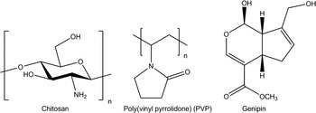
In this work, SEM, ESEM, and CLSM are used to determine the structural characteristics of smart, genipin-crosslinked chitosan–poly(vinyl pyrrolidone) hydrogels (Fig. 1). These techniques are compared for suitability for morphological characterization. This is an important study as porosity measurements are easy to misinterpret as results can be highly dependent on both the sample preparation and the technique used. In contrast with previous investigations, CLSM is performed purely in reflectance mode so as not to change the network in any way, and as this hydrogel autofluoresces, images are also acquired in fluorescence mode without the need to add a fluorescent probe. The smart characteristics of the hydrogel are studied at low pH (2) where the variation in macrostructure is evaluated gravimetrically and optically. These techniques are subsequently compared. The changes in gel microstructure during swelling are also examined via CLSM.
Chemical structures of the constituents of a chitosan–PVP hydrogel crosslinked with genipin.

II. EXPERIMENTAL
A. Hydrogel synthesis
Chitosan was dissolved at room temperature in 1% aqueous acetic acid solution with continuous mechanical stirring for 24 h to obtain a 1.5% [w/v (weight by volume)] pale yellow, viscous solution. PVP was dissolved in distilled water at 85 °C with continuous mechanical stirring for 1 h to yield a 5% (w/v), transparent, homogeneous solution. Genipin solution [0.5% (w/v)] was produced by dissolving genipin in distilled water for 15 min using a sonication bath at room temperature. One milliliter of chitosan solution was combined with 1 mL PVP solution in a polyethylene vial, and 100 µL genipin solution was slowly added over a period of 30 min with continuous mechanical stirring at room temperature. Samples were allowed to undergo gelation at room temperature for 24 h and further polymerization at 37 °C for 24 h to form a viscous gel. All gels were frozen by placing samples in a freezer at −10 °C for 30 min and were slowly thawed and stored at 2 °C before use. The gels were removed from the containers by inserting a cork borer through the vial after the base was cut-off. A cylindrical gel of fixed diameter (15 mm) could then be cut into discs of 3 mm thickness using a Stanley knife.
B. Morphology, critical point drying sample preparation
Hydrogel samples were fully swollen in distilled water and subsequently dehydrated by being subjected to a series of graded ethanol solutions, culminating with drying using a critical point dryer (Baltec, Schalksmühle, Germany). Liquid carbon dioxide was added to the ethanol-filled pressure chamber containing the samples cooled to 10 °C. Upon mixing, the ethanol was replaced with liquid carbon dioxide and the chamber was heated up to 41 °C past the critical point of carbon dioxide. Excessive pressure was vented and the samples were immersed in carbon dioxide gas, without being subjected to harmful surface tension forces.
1. FD sample preparation
Hydrogel samples were fully swollen in distilled water, rapidly frozen in liquid nitrogen and then dried in a freeze dryer (Edwards, West Sussex, UK) with the ice condenser temperature at −55 °C, until all the solvent sublimed.
2. Scanning electron microscopy
Following CPD and FD, each sample was mounted on a carbon disk supported by an aluminum stub and coated with a 15 nm layer of gold (Polaron SEM. Coating Unit). Images of the polymer gel structure were taken on a SEM (Cambridge Stereoscan 240).
3. Environmental scanning electron microscopy
Hydrogel morphology was characterized using an ESEM (FEI XL30 ESEM-FEG, Philips, Eindhovan, Netherlands) operated at 15 kV in “wet mode.” Samples fully swollen in distilled water were mounted on carbon disks supported by an aluminum stub (Agar Scientific, Stansted, UK) and were placed on the ESEM cooling stage adjusted to 3–5 °C and maintained above a saturated water pressure of 0.76–0.84 kPa. The images were subsequently observed at a vapor pressure of 0.33–0.56 kPa. A gaseous secondary electron detector was used to collect the images.
4. Confocal laser scanning microscopy
The pore structure was examined using a Leica TCS SP2 UV (Leica Microsystems, Heidelberg, Germany) confocal microscope. Samples swollen and immersed in distilled water were examined using a 20x dipping lens in reflectance and fluorescence modes. Throughout the experiments, the entire sample was fully submerged in the transparent solution. During sample scanning, cross-sectional images of the hydrogel morphology along the z-plane were taken to construct the native three-dimensional network structure. The sample penetration depth was 100 μm in all experiments. For fluorescence measurements, samples were excited using 488 nm light in the presence of a 500–550 nm filter. In all cases, all samples were transparent to the wave lengths of the excitation and fluorescent light selected.
C. Smart characteristics
Hydrogel samples were contracted in methanol and subsequently swollen in a pH 2 glycine buffer solution to ascertain the smart behavior.
1. Gravimetric measurement
Swelling of samples was measured gravimetrically by immersing preweighed hydrogels in a pH 2 glycine buffer at constant temperature (20 °C). At predetermined time intervals, swollen hydrogels were removed and excess solution was blotted from the sample surfaces. To facilitate transfer of the samples from solution to weighing scales, hydrogels were enclosed in a PTFE cage. The change in mass was then recorded, and the gravimetric swelling ratio (ϕg) was calculated according to Eq. (1), where W s is the weight of the swollen hydrogel and W 0 is the weight of the hydrogel immediately before the sample was allowed to swell in the pH 2 buffer.
 $$\phi _{\rm{g}} = (W_{\rm{s}} - W_0 )/W_0 \quad .$$
2. Optical measurement
Smart behavior was determined by measuring the change in surface area of hydrogel samples using a Nikon AZ100 microscope (Surrey, UK) using a 0.5x 0.05 numerical aperture AZ Plan Apo lens equipped with a green excitation fluorescence filter with an excitation wavelength range between 510 and 560 nm and a dichromatic mirror with a 575 nm cut-on value. Samples were immersed in a transparent pH 2 glycine buffer and maintained at a constant temperature (20 °C). At predetermined time intervals, the change in surface area of the samples was measured. The optical swelling ratio ( $\phi _0$) was determined according to Eq. (2), where the initial and final surface areas of the gel are (a0) and (a) respectively.
 $$\phi _{\rm{0}} = (a - a_0 )/a\quad .$$
Variation in fluorescence intensity was determined using LCS Software Version 2.61.
3. Confocal laser scanning microscopy
An identical setup was used as previously detailed to determine the smart behavior of the same samples. Hydrogels were immersed in a transparent pH 2 glycine aqueous buffer solution and allowed to swell for 30 min. Images were acquired every 5 min. Pore analysis of samples acquired in reflectance mode was performed using the public domain ImageJ program (http://rsbweb.nih.gov/ij/) developed at the US National Institutes of Health (NIH). Porosity was determined using the measure and label plug-in developed by Wayne Rasband at NIH. Individual pore areas were summated and divided by the total area of each image. Sample topography was determined following the three-dimensional structure construction from stacked sliced images.
III. RESULTS AND DISCUSSION
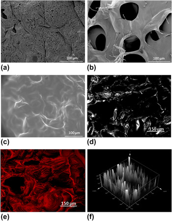
Genipin-crosslinked chitosan–PVP hydrogels were prepared, and the porous structure was evaluated (Fig. 2). SEM imaging following CPD sample treatment generated a porous architecture [Fig. 2(a)], although it is clear that extensive network collapse occurred following ethanol dehydration as evidenced by the small pores (<10 μm in diameter) and dense surface skin. Contraction could also be induced via exchange of ethanol for liquid carbon dioxide.Reference Trieu and Qutubuddin46 In contrast, FD is found to preserve the native structure to a greater extent [Fig. 2(b)] whereby pores are larger (50–100 μm in diameter) and the surface skin appears to be smoother. Images collected via ESEM were difficult to elucidate structural detail from [Fig. 2(c)]. Although it appears that there is a porous structure present with pore sizes corresponding to freeze-dried samples, the interaction with the electron beam on the surface of the hydrated network seems to cause damage to samples and local evaporation, resulting in an inconclusive representation of gel morphology.Reference Kishi, Kihara and Miura39 Furthermore, sample analysis occurs in the vacuum chamber of the microscope and not in an aqueous/solvent environment where the hydrogel will find application. CLSM images generated in situ reveal a continuous, three-dimensional pore architecture that is fully representative of the hydrogel morphology when it is applied in the aqueous target environment [Fig. 2(d)]. As images have been collected by utilizing the reflectance mode of the microscope, addition of a fluorescent probe is not required, which could potentially alter the network structure. Such a network change may take place in nonfluorescent materials where a fluorescent probe such as fluorescein isothiocyanate must be added. This process can occur via blending or covalent linking, which may restrict functional groups necessary for crosslinking or swelling.Reference Lamprecht, Schäfer and Lehr47 Furthermore probes may not bind permanently if simply blended, which could lead to migration and unreliable results. However, as this particular hydrogel autofluoresces upon successful crosslinking, fluorescence can be used to increase the amount of structural detail during image acquisition [Fig. 2(e)]. Utilization of CLSM can also provide further topographical information about such porous networks [Fig. 2(f)].
Genipin-crosslinked chitosan–PVP hydrogel structure analyzed by (a) SEM via CPD, (b) SEM via FD (c) ESEM and (d) CLSM in reflectance mode, (e) CLSM in fluorescence mode, and (f) CLSM topography evaluation. Square edges in (f) are 750 μm in length and total depth is 255 μm.

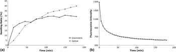
Smart behavior of the cationic hydrogel was confirmed (Fig. 3), both gravimetrically and optically, by immersing the network in a pH 2 buffer solution [Fig. 3(a)]. Owing to protonation of the nitrogen heteroatoms of the gel at low pH, a sufficient amount of electrostatic repulsion is generated within the network to induce swelling. From [Fig. 3(a)], there is a significant discrepancy between the characterization techniques used. Initial expansion of the network is greater during gravimetric measurement, in contrast to using an optical technique, where a gradual increase in the sample area that continues over time is observed. This may be because the mass of the hydrogel is representative of the whole sample, whereas optical measurement only considers expansion in the surface area of the top of the material.
Swelling of genipin-crosslinked chitosan–PVP in pH 2 buffer evaluated via (a) gravimetric and optical techniques and (b) by assessing macro-fluorescence intensity variation with time.

Owing to the autofluorescence of these networks, this attribute has been further utilised to monitor swelling [Fig 3(b)]. As the autofluorescence originates from the crosslinks in the network, upon swelling, the gel volume increases, leading to a decrease in the crosslink density, which is represented by a decrease in overall fluorescence intensity.
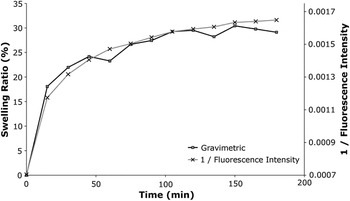
A strong correlation was noted between swelling recorded gravimetrically and the inverse of fluorescence intensity (Fig. 4). This is expected because as in gravimetric measurement, the degree of fluorescence is also representative of the whole sample, perhaps indicating that measuring a mass change is a preferable technique to observing variation in the surface area.
Correlation between swelling behavior evaluated gravimetrically and variation in the inverse of fluorescence intensity during network expansion.

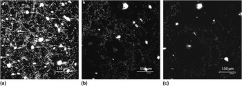
It is possible to infer structural information about hydrogel networks by observing variation in the bulk structure of samples during swelling. However by determining the change in the microporous structure of these smart materials during expansion, a more in-depth analysis can be achieved to inform as to how the internal structure varies as a function of time. This has been achieved by observing in situ genipin-crosslinked chitosan–PVP hydrogels swelling in acidic media via CLSM in purely reflectance mode (Fig. 5).
CLSM images of a genipin-crosslinked chitosan–PVP hydrogel swelling in pH 2 buffer at (a) 0 min, (b) 15 min and (c) 30 min acquired in reflectance mode.

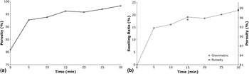
It is clear from Fig. 5 that as time progresses and swelling occurs, on the micro-scale, the size of the pores increase quite dramatically. As this experiment was conducted in reflectance mode, CLSM is applicable for monitoring the swelling of hydrogels that do not fluoresce. The change in porosity during this period is represented in Fig. 6(a), and clearly, the behavior is aligned with previous gravimetric measurements [Fig. 6(b)].
Variation in (a) porosity of genipin-crosslinked chitosan–PVP hydrogels as a function of time during swelling in pH 2 and (b) correlation of the porosity with gravimetric measurements.

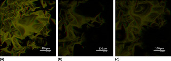
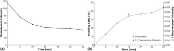
The variation in the porous architecture of genipin-crosslinked chitosan–PVP hydrogels during swelling has also been evaluated using CLSM in fluorescence mode (Fig. 7). Interestingly, variation in porosity is not as clear as when reflectance was used, and hence, a porosity study could not be conducted. Consequently, it may be concluded that reflectance is a superior technique to fluorescence for analyzing how the porous architecture varies during swelling. However, as previously shown to be the case on the macroscale (Figs. 3 and 4), fluorescence intensity decreases with hydrogel swelling [Fig. 8(a)]. Furthermore, upon inversion of fluorescence intensity data, there is a strong correlation with gravimetric measurements [Fig. 8(b)].
CLSM images of a genipin-crosslinked chitosan–PVP hydrogel swelling in pH 2 buffer at (a) 0 min, (b) 15 min and (c) 30 min acquired in fluorescence mode.

Variation in (a) micro-fluorescence intensity of genipin-crosslinked chitosan–PVP hydrogels during swelling in a pH 2 buffer and (b) correlation with gravimetric measurements.

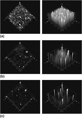
Further to three-dimensional pore architecture detail, CLSM has been used to investigate how hydrogel topography varies with swelling (Fig. 9). As expected, upon swelling, the gel surface becomes smoother owing to a higher degree of hydration. Variation in pore size can also be visualized by this method.
Topography variation of genipin-crosslinked chitosan–PVP hydrogels collected in reflectance mode during swelling in a pH 2 buffer after (a) 0 min, (b) 15 min and (c) 30 min. Square edges are 750 μm in length, and total depth is 255 μm.

IV. CONCLUSIONS
A stable and responsive genipin-crosslinked chitosan–PVP hydrogel has been synthesized, and the optimum method for morphological characterization has been identified. While SEM imaging following FD preserves the native hydrogel structure better than samples subjected to CPD, both techniques alter network morphology. CLSM is shown to be a valuable characterization technique owing to the in situ nature of image acquisition and the extra three-dimensional and topographical information that can be ascertained. CLSM is also beneficial for investigating pore structure variation of smart hydrogels during swelling/contraction. All findings were in agreement with gravimetric measurements. When operated in reflectance mode, there is no requirement for an additional fluorescent probe, which may alter network morphology. Upon using fluorescence to monitor pore structure during swelling, a lower degree of structural detail was observed compared to using reflectance.
ACKNOWLEDGMENTS
The authors wish to acknowledge the UK Engineering and Physical Sciences Research Council (EPSRC) Grant No. EP/H003908/1 for funding this research. G.A.H would also like to acknowledge the School of Chemical Engineering and Advanced Materials for funding his PhD studies. We are grateful to A. Lane for the use of FD equipment, T. Davey and B. Walker of the Electron Microscopy Research Services at Newcastle University for their assistance with SEM imaging, P Carrick of the Advanced Material and Chemical Analysis facility for ESEM imaging, and T. Booth and A. Laude of the Bio-Imaging Unit at Newcastle University for their help with confocal microscopy studies.