Introduction
Oocyte differentiation is a very long process starting during fetal life and culminating in preovulatory follicle as the oocyte reaches its full competence to resume meiosis and successfully accomplish both nuclear and cytoplasmic aspects of maturation (reviewed by Gosden et al., Reference Gosden, Krapez and Briggs1997). Meiotically competent oocytes aspirated from ovarian follicles and cultured in a suitable culture medium spontaneously reinitiate meiosis, which initiates with the germinal vesicle breakdown (GVBD) and proceeds to metaphase of the second meiotic division (MII). Accompanying nuclear maturation, many cytoplasmic changes occur, such as cortical granule (CG) migration and mitochondria repositioning. Both nuclear and cytoplasmic maturation are considered essential for normal embryo development after fertilization (Yang et al., Reference Yang, Kubota, Suzuki, Taneja, Bols and Presicce1998).
However, the ability of the in vitro matured and fertilized bovine oocytes to develop to the morula/blastocyst stage is less than that obtained in in vivo produced embryos (Rizos et al., Reference Rizos, Ward, Duffy, Boland and Lonergan2002). The decrease in the developmental competence of such oocytes may be due to the insufficient cytoplasmic maturity, which is gradually acquired in vivo during follicular growth. During this period, which has been termed oocyte ‘capacitation’, the oocyte undergoes structural changes that could be related to oocyte competence for development (Hyttel et al., Reference Hyttel, Fair, Callesen and Greve1997).
Thus, it can be hypothesized that oocytes prematuration in the presence of meiotic inhibitors, a procedure that could more precisely mimic in vivo oocyte capacitation (Ponderato et al., Reference Ponderato, Lagutina, Crotti, Turini and Lazzari2001), could give them the opportunity to acquire greater developmental competence (Avery et al., Reference Avery, Hay-Schmidt, Hyttel and Greve1998). By using this procedure, the oocyte could undergo the repositioning of organelles and storing of newly synthesized proteins and ribonucleoproteins, which in turns could increase its developmental competence (Rodriguez et al., Reference Rodriguez and Farin2004). Some pharmacological inhibitors utilized for the maintenance of meiotic arrest that have been studied include isobutyl-methylxanthine (IBMX) and roscovitine.
Purine derivatives more specifically directed toward the inhibition of cell cycle regulating cyclin-dependent kinases (CDKs) have been used for preventing cell division and meiotic resumption (Marchal et al., Reference Marchal, Tomanek, Terqui and Mermillod2001). Roscovitine, a purine known to specifically inhibit M-phase promoting factor (MPF) kinase activity in numerous cell systems, has been successfully used in maintaining cattle oocytes at the germinal vesicle stage without affecting their developmental potential (Mermillod et al., Reference Mermillod, Tomanek, Marchal and Meijer2000). This drug is a selective cdc2 inhibitor (kinase subunit of MPF complex), which has been reported to arrest cells in late G1 and G2/M cell cycle transition and inhibit resumption of meiosis (Mermillod et al., Reference Mermillod, Tomanek, Marchal and Meijer2000). It acts as a competitive inhibitor for ATP and when complexed with cdk2, it binds to the ATP-binding pocket of cdk2.
Cyclic AMP (cAMP) maintains meiotic arrest in GVBD-competent oocytes and helps growing oocytes to acquire competence (Chesnel et al., Reference Chesnel, Wigglesworth and Eppig1994). IBMX, a phosphodiesterase inhibitor by increasing cAMP (Tsafriri et al., Reference Tsafriri, Chun, Zhang, Hsueh and Conti1996), can maintain bovine cumulus-oocyte complexes (COC) in meiotic arrest for a short period (Lonergan et al., Reference Lonergan, Khatir, Carolan and Mermillod1997). However, as the action of cAMP and its analogues may be modified by the presence of serum, growth factors and hormones added to the culture media (Lohman & Walter, Reference Lohmann, Walter and Greengard1984), synthetic and defined supplement sources of culture media may be used as substitutes. The serum replacer Knockout SR® (SR; BRL) is a protein source used for stem cell culture and whose defined formula is protected by fabricant, but that do not have serum in its composition (Goldsborough et al., Reference Goldsborough, Tilkins, Price, Lobo-Alfonso, Morrison, Stevens, Meneses, Pedersen, Koller and Latour1998).
The objectives of this study were first, to analyze the kinetics of bovine oocyte nuclear maturation following release from meiosis inhibition by IBMX and roscovitine in media supplemented with Knockout SR® and hormones; and second, to assess the developmental capacity of such oocytes.
Material and methods
Reagents, media and culture conditions
Chemicals were purchased from Sigma Chemical Co. (St. Louis, MO, USA), unless otherwise stated.
Inhibitors were prepared as a stock solution of 5 mM in dimethylsulphoxide (DMSO), aliquoted and stored at –20°C until use. For meiotic block culture, inhibitors were diluted to the appropriate concentration in in vitro maturation (IVM) medium (see below): 25 μM roscovitine (Calbiochem, EMD Biosciences Inc.) and 0.5 mM IBMX.
The IVM medium consisted of TCM-199 (Gibco BRL) supplemented with 0.2 mM sodium pyruvate, 25 mM sodium bicarbonate, 75 μg/ml kanamycin (Gibco BRL), 0.5 μg/ml FSH (Pluset®, Hertape Calier), 100 IU/ml hCG (Profasi®, Serono), 1.0 μg/ml estradiol and 10% (v/v) Knockout SR® (SR).
In vitro fertilization (IVF) medium consisted of Tyrode's albumin lactate pyruvate (TALP) supplemented with 0.2 mM sodium pyruvate, 6 mg/ml fatty acid-free BSA, 25 mM sodium bicarbonate, 13 mM sodium lactate, 75 μg/ml kanamycin, 4 μl/ml PHE solution (2 mM penicillamine, 1 mM hypotaurine and 250 μM epinephrine) and 10 μg/ml heparin.
In vitro culture (IVC) medium was synthetic oviductal fluid (SOF) supplemented with 0.2 mM l-glutamine, 0.34 mM sodium citrate, 2.8 mM myo-inositol, 2% MEM essential amino acid solution, 1% MEM non-essential amino acid solution, 0.2 mM sodium pyruvate, 75 μg/ml kanamycin, 5 mg/ml fraction V fatty acid free BSA and 2.5% FCS.
Cultures (meiosis block, IVM, IVF and IVC) were carried out at 38.5°C with maximum humidity and under atmosphere of 5% CO2 in air.
Oocyte recovery
Abattoir-derived ovaries were transported to the laboratory in saline solution at 30–35°C. The follicles (2–8 mm) were aspirated using an 18-gauge needle attached to a 20 ml syringe. Oocytes with at least four layers of cumulus cells were selected for the experiments.
Meiosis block (prematuration culture)
Oocytes where the meiotic block was applied were cultured in vitro in IVM medium supplemented with 10% SR, hormones and 25 μM roscovitine or 0.5 mM IBMX. The culture was performed in 100 μl droplets (approximately 20 oocytes per droplet) under mineral oil (Dow Corning Co.) during 8 h or 16 h, according to the experimental approach.
In vitro maturation
For IVM, oocytes were washed and cultured in IVM medium supplemented with 10% SR and hormones. The IVM culture was performed in 100 μl droplets (approximately 20 oocytes per droplet) under mineral oil for 24 h (control group) or for 8, 16 and 24 h (blocked groups).
Assessment of nuclear maturation
Oocytes were stripped from their cumulus cells by vortexing in phosphate-buffered saline (PBS) with 0.1% hyaluronidase for 3 min. Denuded oocytes were then stained with 10 μg/ml Hoechst 33342 for 10 min, placed between slide and coverslip and visualized under epifluorescence microscopy (330–385 nm; at ×200 magnification). The oocytes were classified as germinal vesicle stage (GV, immature oocytes), metaphase I (MI), anaphase I (AI), telophase I (TI) or metaphase II (MII, matured oocytes). Oocytes remaining at the GV stage after meiotic block culture were considered to be completely blocked and oocytes in MII after IVM were considered to be fully reversed of the meiotic inhibition. Oocytes reaching MI, AI and TI were considered as intermediate stages (INT).
In vitro fertilization
Motile spermatozoa were obtained by centrifugation of frozen-thawed spermatozoa on a Percoll (Pharmacia) discontinuous density gradient (2 ml of 45% Percoll over 2 ml of 90% Percoll) for 30 min at 900 g at room temperature. The supernatant was discarded and the spermatozoa were resuspended in IVF medium after being counted on a hemocytometer to obtain a final concentration of 2 × 10⁶ cells/ml. Oocytes and sperm were co-incubated for 18 h under the same temperature and atmosphere conditions used for IVM.
In vitro development culture and embryo evaluation
Following fertilization, the presumptive zygotes were transferred to IVC medium. Zygotes were incubated under mineral oil up to 48 h for assessment of cleavage rates under stereoscopic microscopy (at ×40 magnification), when 2- and 4-cell embryos were counted. Blastocyst and hatched blastocyst development rates were observed, respectively, at 168 (day 7) and 192 (day 8) hours post insemination (hpi).
Experimental design
Experiment I: Effects of IBMX or roscovitine diluted in IVM medium supplemented with SR and hormones on effectiveness of meiotic block and meiosis progression after meiotic block of bovine oocytes
Oocytes were blocked for 8 h (first part of this experiment; n = 412 in 11 replicates) and for 16 h (second part of this experiment; n = 236 in seven replicates) with 0.5 mM IBMX (group IBMX) or 25 μM roscovitine (group ROSC) diluted in IVM medium (TCM-199) supplemented with 10% SR and hormones (0.5 μg/ml FSH, 100 IU/ml hCG and 1.0 μg/ml estradiol). As controls, a group of oocytes was only in vitro matured during 8 h (n = 203) and 16 h (n = 130), respectively, without prior meiotic block. At the end of cultures, all oocytes were assessed for maturation rates. A group of oocytes was fixed after aspiration (n = 89; Control 0 h).
After meiotic block culture for 8 h (first part; n = 2543 in 11 replicates) and for 16 h (second part; n = 2054 in 11 replicates), oocytes were IVM for 24 h. As controls, a group of oocytes was only in vitro matured during 24 h without prior meiotic block. During IVM, all oocytes were assessed for maturation rates at 8 h intervals (0, 8, 16 and 24 h IVM).
Experiment II: Effects of meiotic block on embryo development in vitro
Oocytes (n = 1640 in 5 replicates) were cultured during 8 h with IBMX or roscovitine as previously mentioned in Experiment I and then were submitted to IVM for 16, 20 or 24 h. As controls, a group of oocytes was only in vitro matured during 24 h without prior meiotic block. At the end of IVM culture, all oocytes were fertilized in vitro and cultured up to 8 days (D8). Cleavage, blastocyst development and hatching rates were recorded.
Statistical analysis
In this study, data were reported as mean ± standard error (SEM). Data in percentages were arcsine transformed and analysed by ANOVA. When a statistical significant effect was found, multiple comparisons of means were determined using Tukey's test (SAS Program V.8). A p-value of less than 0.05 was considered to be statistically significant.
Results
Experiment I
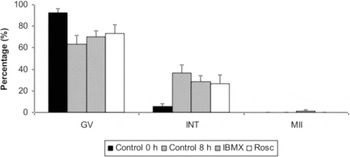
In the first part of this experiment, most oocytes treated with IBMX and roscovitine with SR and hormones during 8 h culture remained in GV stage (70.3 ± 5.6% and 73.1 ± 8.1% for IBMX and ROSC groups, respectively; p > 0.05) (Fig. 1). The majority of control oocytes cultured for 8 h with SR and hormones but without meiotic inhibitors were also in GV (63.5 ± 7.7%). Similarly, most control immature oocytes (Control 0 h) evaluated soon after follicle aspiration were in GV stage (92.5 ± 3.9%). However, some oocytes progressed through meiosis and reached the intermediate stages of meiotic maturation (5.3–36.5%; p > 0.05) and MII stage (0.0–1.4%; p > 0.05; Fig. 1).
Meiotic status in bovine oocytes cultured for 8 h with IBMX or roscovitine in TCM-199 with Knockout SR® and hormones. Control 0 h: oocytes evaluated soon after follicle aspiration; Control 8 h: oocytes cultured in maturation medium (MM–TCM-199 with 10% Knockout, 0.5 μg/ml FSH, 100 IU/ml hCG and 1.0 μg/ml estradiol) for 8 h without meiotic inhibitors; IBMX: oocytes cultured in MM for 8 h with 0.5 mM IBMX; ROSC: oocytes cultured in MM for 8 h with 25 μM roscovitine. GV: germinal vesicle; INT: intermediate stages of nuclear maturation, including metaphase I, anaphase I and telophase I; MII: metaphase II. a, bDifferent letters indicate differences among treatments (p < 0.05).

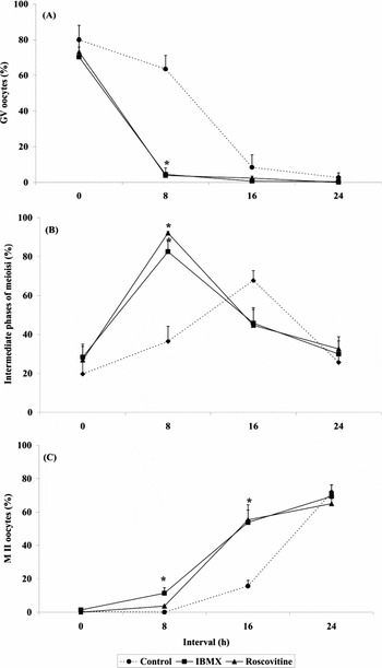
After meiotic block for 8 h, oocytes were IVM for 24 h in inhibitor-free media and oocytes were assessed for maturation rates at 8 h intervals. GV rates after 8 h IVM were greater (p < 0.05) for control oocytes (63.5 ± 7.7%) than for the treated ones (3.9–4.5%), which were similar (p > 0.05) between themselves (Fig. 2). At this moment, most oocytes of IBMX (82.5 ± 5.5%) and ROSC (92.1 ± 0.8%) were in intermediate stages of meiosis (p > 0.05), but the controls had lesser rates (36.5 ± 7.7%; p < 0.05). A small proportion of oocytes in IBMX group (11.4 ± 3.2%) were in MII stage at 8 h IVM, but Control and ROSC had lesser rates (0.0% and 3.7 ± 0.7%, respectively; p < 0.05). At 16 h, the rates of oocytes in the intermediate stages of meiosis were similar (p > 0.05) between treatments (44.7–67.7%) (Fig. 2). On the other hand, a higher proportion (p < 0.05) of oocytes of IBMX (53.8 ± 7.4%) and ROSC (55.3 ± 9.0%) groups had reached MII stage when compared with Control (15.6 ± 3.5%). MII rates at 24 h IVM (Fig. 2) were similar (p > 0.05) between treatments (65.0–71.7%).
Kinetics of oocyte maturation after 8 h, 16 h and 24 h IVM in bovine oocytes previously submitted to meiotic block for 8 h in TCM-199 with Knockout (10%), hormones (0.5 μg/ml FSH, 100 IU/ml hCG and 1.0 μg/ml estradiol) and meiotic inhibitors (0.5 mM IBMX or 25 μM roscovitine). (a) Percentage of germinal vesicle (GV) oocyte. (b) Percentage of oocytes in the intermediate stages of nuclear maturation, including metaphase I, anaphase I and telophase I. (c) Percentage of metaphase II (MII) oocytes. *Asterisk indicates difference from control within each time point (p < 0.05).

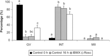
When the meiotic block was conducted for 16 h (second part of this experiment), no oocytes remained in GV stage at similar rates to those recently aspirated (p < 0.05) (Fig. 3). GV rates in oocytes treated with ROSC (32.4 ± 6.9%) were higher (p < 0.05) than in Control (8.6 ± 6.2%), but similar (p > 0.05) to IBMX (9.7 ± 5.0%). In this experiment, the majority of oocytes progressed through meiosis, reaching the intermediate stages of meiotic maturation (67.6–86.9%). Similar rates (p > 0.05) of oocytes reaching MII stage (0.0–7.1%) were observed between treatments (Fig. 3).
Meiotic status in bovine oocytes cultured for 16 h with IBMX or roscovitine in TCM-199 with Knockout and hormones. Control 0 h: oocytes evaluated soon after follicle aspiration; Control 16 h: oocytes cultured in maturation medium (MM– TCM199 with 10% Knockout, 0.5 μg/ml FSH, 100 IU/ml hCG and 1.0 μg/ml estradiol) for 16 h without meiotic inhibitors; IBMX: oocytes cultured in MM for 16 h with 0.5 mM IBMX; ROSC: oocytes cultured in MM for 16 h with 25 μM roscovitine. GV: germinal vesicle; INT: intermediate stages of nuclear maturation, including metaphase I, anaphase I and telophase I; MII: metaphase II. a, bDifferent letters indicate differences among treatments (p < 0.05).

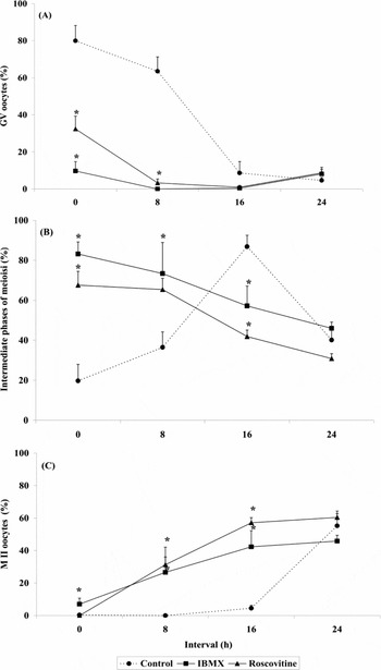
Subsequently to the meiotic block for 16 h, oocytes were IVM for 24 h in inhibitor-free media. At the beginning of culture (0 h), GV rates were greater (p < 0.05) for Control (79.9 ± 8.3%) than for IBMX (9.7 ± 5.0%) and ROSC (32.4 ± 6.7%), which were similar (p > 0.05) between themselves (Fig. 4). At this moment, the majority of IBMX (83.2 ± 5.9%) and ROSC (67.6 ± 6.9%) oocytes were in intermediate stages of meiosis (p > 0.05), which were greater (p < 0.05) than Control (19.7 ± 8.3%). After 8 h IVM, most oocytes of IBMX (73.4 ± 15.5%) and ROSC (65.4 ± 5.6%) were in intermediate stages of meiosis (p > 0.05), but the controls had lesser rates (36.5 ± 7.7%; p < 0.05; Fig. 4). At this moment, some oocytes of IBMX (26.6 ± 15.5%) and ROSC (31.4 ± 4.7%) had reached MII (p > 0.05), while no control oocytes had reached this stage (0.0%; p < 0.05). At 16 h, the majority control oocytes were in intermediate stages of meiosis (86.9 ± 5.6%), but IBMX (57.2 ± 9.9%) and ROSC (41.9 ± 3.2%) had lesser rates (p < 0.05) (Fig. 4). However, a higher proportion (p < 0.05) of oocytes of IBMX (42.3 ± 9.9%) and ROSC (57.2 ± 3.1%) groups had reached MII stage when compared with Control (4.5 ± 1.8%). MII rates at 24 h IVM (Fig. 4) were similar (p > 0.05) between treatments (45.9–60.4%).
Kinetics of oocyte maturation after 8 h, 16 h and 24 h IVM in bovine oocytes previously submitted to meiotic block for 16 h in TCM-199 with Knockout (10%), hormones (0.5 μg/ml FSH, 100 IU/ml hCG and 1.0 μg/ml estradiol) and meiotic inhibitors (0.5 mM IBMX or 25 μM roscovitine). (a) Percentage of germinal vesicle (GV) oocytes. (b) Percentage of oocytes in the intermediate stages of nuclear maturation, including metaphase I, anaphase I and telophase I. (c) Percentage of metaphase II (MII) oocytes. *Asterisk indicates difference from control within each time point (p < 0.05).

Experiment II
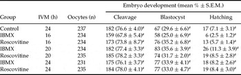
To evaluate developmental competence of oocytes treated with IBMX and roscovitine in medium with SR and hormones during 8 h, these oocytes were in vitro matured for 16, 20 or 24 h, fertilized and cultured. Data for cleavage and embryonic development to blastocyst and hatched blastocyst are presented in Table 1. Cleavage rates (67.8–78.2%), embryonic development in D7 (25.0–35.6%) and hatching rates in D8 (2.5–11.3%) were similar for all groups (p > 0.05).
Embryo development of bovine oocytes previously submitted to meiotic block for 8 h in TCM-199 with Knockout SR® (10%), hormones (0.5 μg/ml FSH, 100 IU/ml hCG and 1.0 μg/ml estradiol) and meiotic inhibitors (0.5 mM IBMX or 25 μM roscovitine).

aDifferent letters within columns indicate significant differences (p < 0.05).
Discussion
Oocyte nuclear maturation together with the cytoplasmic maturation prepares oocytes for being fertilized and to acquire embryonic developmental competence (Thibault et al., Reference Thibault, Szöllösi and Gérard1987; Sirard et al., Reference Sirard, Florman, Leibfried-Rutledge, Barnes, Sims and First1989). Currently, the effectiveness of in vitro culture systems used for embryo production (30–40% blastocyst rate) is lower than that obtained in vivo (approximately 70% blastocyst rate). Among several factors associated with culture systems that can explain such poor results, we can cite the short period that oocytes have to acquire developmental competence: 24 h of IVM culture. For comparison, in vivo, meiotically competent oocytes are maintained at the GV stage by the follicular environment until the preovulatory gonadotropin surge, which allow them to differentiate while included in a follicle as small as 2 mm (the size at which oocytes are punctured for IVM purposes: 2–8 mm) until ovulation (Marchal et al., Reference Marchal, Tomanek, Terqui and Mermillod2001).
One way to offer the oocytes additional time for cytoplasmic maturation and developmental competence acquisition is to prematurate them in the presence of meiotic inhibitors, which could better induce the oocyte capacitation (Ponderato et al., Reference Ponderato, Lagutina, Crotti, Turini and Lazzari2001). However, several studies have recently demonstrated that prematuration of bovine oocytes in simple medium did not improve oocyte competence, but also did not cause major structural alterations in oocytes and embryos (Barretto et al., Reference Barretto, Caiado Castro, Garcia and Mingoti2007; Adona et al., Reference Adona, Pires, Quetglas, Schwarz and Leal2008). Then, it can be supposed that the addition of stimulatory components to the prematuration culture medium, such as hormones, could create a more suitable environment for promoting the developmental potential of the oocytes (Ponderato et al., Reference Ponderato, Crotti, Turini, Duchi, Galli and Lazzari2002). For that reason, prematuration of bovine oocytes with meiotic inhibitors (IBMX and roscovitine) in medium supplemented with SR and hormones were used to assess their effects on meiotic block, meiosis progression after meiotic reversal and developmental competence.
GV rates after 8 h meiotic block (prematuration) were similar between groups, which did not differ from both the Control 8 h (8 h of IVM) and those evaluated soon after follicle aspiration (Control 0 h). This finding was expected at this moment, since meiosis resumption in vitro is observed just after 6–8 h of IVM in inhibitor-free media (Sirard et al., Reference Sirard, Florman, Leibfried-Rutledge, Barnes, Sims and First1989).
Nuclear maturation kinetics after meiotic block for 8 h was then followed. Meiotic resumption was accelerated similarly in both IBMX and ROSC treatments. At 8 h of IVM, most treated oocytes reached the intermediate phases of meiosis, while most control oocytes were still at GV stage. At 16 h of IVM, MII rates in treated oocytes were superior to control, indicating that the acceleration is mostly due to the premature meiotic resumption. These results may support the hypothesis that the activity of MPF and mitogen activated protein kinases (MAPK) are inhibited by roscovitine, but not their activation pathway (Marchal et al., Reference Marchal, Tomanek, Terqui and Mermillod2001). According to these authors, roscovitine prevents MPF kinase activity by competing with ATP for binding cdk1 but does not prevent the formation of active MPF itself. Then, after roscovitine removal, active MPF is already present and could induce meiotic resumption more rapidly than in untreated oocytes deprived of active MPF. Besides CDK inhibitors, protein synthesis (Mayes & Sirard, Reference Mayes and Sirard2001) and phosphodiesterase inhibitors (Grupen et al., Reference Grupen, Fung and Armstrong2006), like IMBX, also accelerated meiotic resumption. Even short periods of meiotic block, as that used in the present study, cause such effects on nuclear maturation kinetics (Mayes & Sirard, Reference Mayes and Sirard2001).
To assess the effects of meiotic inhibitors in media supplemented with SR and hormones during more prolonged periods, oocytes were prematured for 16 h. Nevertheless, IBMX could not maintain bovine oocytes at GV stage during 16 h in such culture system. Although GV rates in ROSC were higher than in Control 16 h, the efficiency in blocking meiosis was reduced when compared with Control 0 h and to results of previous reported studies (Ponderato et al., Reference Ponderato, Lagutina, Crotti, Turini and Lazzari2001). The majority of oocytes (treated and control 16 h) progressed meiosis until intermediate phases, as expected in a IVM culture free of meiotic inhibitors (10–18 h of IVM, according to Sirard et al., Reference Sirard, Florman, Leibfried-Rutledge, Barnes, Sims and First1989).
Although adenylate cyclase activators and phosphodiesterase inhibitors, such as IBMX, had demonstrated to be capable of maintaining mouse oocytes at the GV stage (Eppig et al., Reference Eppig, O'Brien and Wiggleworth1996) and also assist growing oocytes to acquire GVBD competence (Chesnel et al., Reference Chesnel, Wigglesworth and Eppig1994), pharmacological elevation of intracellular cAMP in isolated COCs or denuded oocytes transiently delays spontaneous maturation in bovine oocytes (Schultz, Reference Schultz and Wasserman1991), maintaining the meiotic arrest for only approximately 3 h from GV (Lonergan et al., Reference Lonergan, Khatir, Carolan and Mermillod1997). This transient effect is due to the interference of some substances present in the maturation media such as serum, hormones and growth factors (Harper & Brackett, Reference Harper and Brackett1993), which act upon the adenylate cyclase pathway. For that reason, prematuration media were supplemented with Knockout SR® in the present study, in order to avoid such undesirable effect. SR was developed for isolation and maintenance of embryonic stem cells (Goldsborough et al., Reference Goldsborough, Tilkins, Price, Lobo-Alfonso, Morrison, Stevens, Meneses, Pedersen, Koller and Latour1998; Moore et al, Reference Moore, Rodriguez-Sallaberry, Kramer, Johnson, Wroclawska, Goicoa and Niasari-Naslaji2007). The chemical formula of SR is protected by fabricant, but it is marketed as a defined serum-free supplement and is known to contain amino acids, glucose, insulin, transferring, antioxidants, inorganic salts, trace elements and a proprietary mix of proteins and vitamins. It does not contain growth factors, cytokines, hormones, immunoglobulins or the macromolecules PVA or PVP.
Unfortunately, replacement of serum by SR could not maintain meiosis blocked for periods as long as 16 h, but it remains to be investigated if this undesirable effect was due to some interaction of meiotic inhibitors with SR and/or the hormones present in prematuration media.
Effective meiotic blockage has already been obtained using drugs that affect MPF activity, like roscovitine (Ponderato et al., Reference Ponderato, Lagutina, Crotti, Turini and Lazzari2001). On the contrary, our data demonstrated that roscovitine was unable to block meiosis for periods as long as 16 h when prematuration media was supplemented with SR and hormones. Based in the present results, we can suppose that SR and/or the stimulatory components (hormones) added to the prematuration media have negatively affected the effect of both meiotic inhibitors used in this study (IBMX and roscovitine).
Nuclear maturation kinetics after meiotic block for 16 h was evaluated and it was observed acceleration in meiotic progression during IVM. As previously discussed, these results may be a consequence of the premature meiotic resumption, mainly in this specific experiment where most treated oocytes were at intermediate stages of meiosis in the beginning of IVM culture.
In summary, we observed that oocyte prematuration with meiotic inhibitors supplemented with SR and hormones was not efficient in maintaining meiotic block for periods longer than 8 h, however, this culture system did not compromise subsequent progression to MII. Moreover, previous results from our laboratory have demonstrated that prematuration of bovine oocytes in similar culture conditions did not impair oocyte cytoplasmic maturation, assessed by the analysis of CG repositioning (Barretto et al., Reference Barretto, Caiado Castro, Garcia and Mingoti2007). Then, acquisition of developmental competence was assessed. Oocytes were prematured for 8 h and then submitted to IVM (for 16, 20 and 24 h; to test the best moment for fertilization, due to the meiosis acceleration after prematuration culture), IVF and IVC. Independently of treatment and IVM duration, the results obtained for cleavage (67.8–78.2%) and development to blastocyst (25.0–35.6%) and hatched blastocyst (2.5–8.5%) were similar to the average results described in the literature and even superior to results of Moore et al. (Reference Moore, Rodriguez-Sallaberry, Kramer, Johnson, Wroclawska, Goicoa and Niasari-Naslaji2007) who have obtained 16.5% of blastocyst rate after IVM with SR. Then, present results indicate that both replacement of serum by SR during IVM of bovine oocytes and prematuration with meiotic inhibitors with SR and hormones had no adverse effect on embryo development.
It is well known that embryo development depends on the quality of nuclear and cytoplasmic maturation. Some studies (Ponderato et al., Reference Ponderato, Crotti, Turini, Duchi, Galli and Lazzari2002; Shimada et al., Reference Shimada, Nishibori, Isobe, Kawano and Terada2003) suggested that mammalian oocytes can respond to the manipulation of factors applied during meiotic arrest that can increase oocyte developmental competence. Shimada et al. (Reference Shimada, Nishibori, Isobe, Kawano and Terada2003) demonstrated that induction of LH receptors by exposure of porcine COC to FSH during induced meiotic arrest with IBMX resulted in an increase in developmental competence.
Regarding roscovitine, it is supposed that its inhibitory effect on MPF activity may also interfere with cytoplasmic maturation, since MPF also modulates the properties of proteins included in the regulation of cytoskeletal organization (Parrish et al., Reference Parrish, Kim and Bae1992). In fact, a recent work (Adona et al., Reference Adona, Pires, Quetglas, Schwarz and Leal2008) has demonstrated that butyrolactone, a cdk inhibitor that selectively inhibits MPF like roscovitine does, reversibly blocked CG migration and partially blocked mitochondria translocation, demonstrating that MPF and/or MAPK can be implicated in organelle movement control. However, other studies (Vigneron et al., Reference Vigneron, Perreau, Dupont, Uzbekova, Prigent and Mermillod2004) suggested that cytoplasmic maturation is not completely blocked during meiotic block and that other factors besides MPF could participate in this control. Then, based in the knowledge that CG migration and mitochondrial aggregation during porcine oocyte maturation showed a dose-dependant relationship with gonadotropin concentrations (Sha et al., Reference Sha, Xu, Li, Liu, Feng and Sun2009), we tried to improve cytoplasmic maturation and, consequently, developmental competence during meiotic block by adding hormones to the prematuration culture. However, in the present work, gonadotropins did not improve developmental competence during meiotic block of bovine oocytes and even have negatively affected the meiotic block itself. Then, the results suggest that stimulatory substances, like gonadotropins and estradiol were not suitable supplements for meiotic block culture.
To conclude, this research has demonstrated that addition of Knockout SR® and hormones to meiotic block culture with IBMX and roscovitine negatively affected meiotic arrest, but did not impair oocyte nuclear maturation and acquisition of developmental competence.
Acknowledgements
This work was supported by the Fundação de Amparo a Pesquisa do Estado de São Paulo (FAPESP), Brazil, Grant #01/06137-0. V.S.D. Caiado Castro was recipient of studentship from CAPES, Brazil and L.S.S. Barretto was recipient of studentship from FAPESP, Brazil.