Introduction
Posthodiplostomum minimum (MacCallum, Reference MacCallum1921) Dubois, Reference Dubois1936, is a diplostomid trematode with a life cycle involving aquatic snails (Physidae) as first intermediate hosts, freshwater fish as second intermediate hosts, and birds, particularly members of Ardeidae, as definitive hosts (Ferguson Reference Ferguson1937). The metacercaria, commonly known as ‘white grub’, encysts in the visceral organs of freshwater fish and can reduce growth, damage organs, and cause mortality (Hoffman Reference Hoffman1958; Smitherman Reference Smitherman1968; Mitchell et al. Reference Mitchell, Smith and Hoffman1982). Putative metacercariae of P. minimum have been reported in almost 200 fish species representing 30 families, mainly in North and Middle America (Hoffman Reference Hoffman1958; Dubois Reference Dubois1970; Dechtiar Reference Dechtiar1972; Nepszy Reference Nepszy1988; McDonald and Margolis Reference McDonald and Margolis1995; Gibson Reference Gibson1996; Aguirre-Macedo et al. Reference Aguirre-Macedo, Scholz, González-Solís, Vidal Martínez, Posel, Arjona-Torres, Siu-Estrada and Dumailo2001; Salgado-Maldonado Reference Salgado-Maldonado2006; Pérez-Ponce de León et al. Reference Pérez-Ponce de León, García-Prieto and Mendoza-Garfias2007) and occasionally in the Caribbean (Bunkley-Williams and Williams Reference Bunkley-Williams and Williams1994), South Asia (Chandra Reference Chandra2006) and Africa (Hamouda and Bazh Reference Hamouda and Bazh2019; Vandi and Daniel Reference Vandi and Daniel2023).
The taxonomic history of P. minimum is one of ongoing revision. The first records were misidentifications of a European species that causes black spots in the skin of Palearctic leuciscids (minnows), namely Diplostomum cuticola (von Nordmann, 1832) Diesing, 1850, a species later moved to Posthodiplostomum by Dubois (Reference Dubois1936). In North America, ‘D. cuticola’ was reported from leuciscids (Cooper Reference Cooper1915), centrarchids (Leidy Reference Leidy1856; Linton Reference Linton1898; Stafford Reference Stafford1904), and other fish families (Ward Reference Ward, Ward and Whipple1918). Without mentioning these studies, Agersborg (Reference Agersborg1926) described Diplostomum vancleavei Agersborg, Reference Agersborg1926, based on metacercariae from pugnose shiner (Miniellus anogenus; Leuciscidae) in Illinois and considered worms from centrarchid (sunfish) hosts to be similar or identical. Hughes (Reference Hughes1928) recognised the species described by Agersborg (Reference Agersborg1926) to be distinct from D. cuticola, considered the many earlier North American reports of D. cuticola to be synonyms of D. vancleavei, and transferred this species to the larval genus Neascus.
In a key contribution to the taxonomic history of P. minimum, Ferguson (Reference Ferguson1936, Reference Ferguson1937) fed bluntnose minnow (Pimephales notatus; Leuciscidae) and pumpkinseed (Lepomis gibbosus; Centrarchidae) infected with N. vancleavei to domestic chicks (Gallus gallus domesticus). Based on the morphology of experimentally obtained adults, as well as other specimens from naturally infected birds, Ferguson (Reference Ferguson1937) concluded that metacercariae of N. vancleavei were conspecific with adult worms that MacCallum (Reference MacCallum1921) described from a great blue heron (Ardea herodias; Ardeidae) from the Zoological Park in New York. MacCallum (Reference MacCallum1921) had named this species Diplostomum minimum. Dubois (Reference Dubois1935) transferred D. minimum to the genus Neodiplostomum Railliet, 1919, then (Dubois Reference Dubois1936) to Posthodiplostomum.
Owing to a wide range of reported hosts, metacercariae of P. minimum initially appeared to be compatible with diverse fishes (Ferguson Reference Ferguson1937). However, Klak (Reference Klak1940) observed leuciscids heavily infected with P. minimum in a pond where centrarchids were uninfected, which was inconsistent with the concept of P. minimum as generalist. This prompted Ferguson (Reference Ferguson1943) to infect black-crowned night herons (Nycticorax nycticorax) with metacercariae of P. minimum from either leuciscid or centrarchid hosts. Ferguson (Reference Ferguson1943) used the resulting miracidia to infect laboratory-reared snails, obtain cercariae, and attempted to infect leuciscid and centrarchid fish. The experimental leuciscid fish became infected only with cercariae originating from naturally infected leuciscid hosts, while centrarchid fish could only be infected with those originating from centrarchid hosts; cross-infections among fish families were unsuccessful. This host specificity was corroborated by similar findings by Hunter and Hunter (Reference Hunter and Hunter1940), Miller (Reference Miller1954), Avault and Smitherman (Reference Avault and Smitherman1965), Hoffman (Reference Hoffman1958), and Palmieri (Reference Palmieri1976). The experiments of Hoffman (Reference Hoffman1958) included transplantation of metacercariae among fish families, and led him to conclude that two strains exist within P. minimum; he proposed the names P. minimum minimum for the minnow-infecting strain (leuciscids) and P. minimum centrarchi for the sunfish-infecting strain (centrarchids). Many subsequent studies did not make this distinction, and P. minimum continued to be reported from a wide variety of fish hosts in North and Middle America, including centrarchids, leuciscids, and numerous other families (Gibson Reference Gibson1996; Hoffman Reference Hoffman1999; Salgado-Maldonado Reference Salgado-Maldonado2006).
Sequences of DNA are now available from numerous species of Posthodiplostomum, many of which are unidentified (Locke et al. Reference Locke, McLaughlin and Marcogliese2010; Boone et al. Reference Boone, Laursen, Colombo, Meiners, Romani and Keeney2018; Achatz et al. Reference Achatz, Chermak, Martens, Pulis, Fecchio, Bell, Greiman, Cromwell, Brant, Kent and Tkach2021; Díaz-Pernett et al. Reference Díaz-Pernett, Brant and Locke2022; Pérez-Ponce de León et al. Reference Pérez-Ponce de León, Sereno-Uribe, Pinacho-Pinacho and García-Varela2022). Stoyanov et al. (Reference Stoyanov, Georgieva, Pankov, Kudlai, Kostadinova and Georgiev2017) considered the genetically distinguished Posthodiplostomum sp. 3 equivalent to Hoffman’s sunfish line, P. minimum centrarchi, based largely on the specificity of its metacercariae for Lepomis spp. (sunfish) in molecular surveys. Stoyanov et al. (Reference Stoyanov, Georgieva, Pankov, Kudlai, Kostadinova and Georgiev2017) elevated Hoffman’s subspecies to P. centrarchi, a taxonomic change that has been accepted (Locke et al. Reference Locke, Van Dam, Caffara, Pinto, López-Hernández and Blanar2018; Čech et al. Reference Čech, Sándor, Molnár, Paulus, Papp, Preiszner, Vitál, Varga and Székely2020; Achatz et al. Reference Achatz, Chermak, Martens, Pulis, Fecchio, Bell, Greiman, Cromwell, Brant, Kent and Tkach2021; Ondračková et al. Reference Ondračková, Tkachenko, Bartáková, Bryjová, Janáč, Zięba and Kvach2023). This change implies a similar fate for Hoffman’s minnow line, i.e., from P. minimum minimum to P. minimum. Among genetically distinguished, unidentified species, the best candidates for correspondence with Hoffman’s minnow line are Posthodiplostomum sp. 4 (sequenced from Pi. notatus, Locke et al., Reference Locke, McLaughlin and Marcogliese2010) and Posthodiplostomum Lin II (from Pimephales promelas, Pérez-Ponce de León et al., Reference Pérez-Ponce de León, Sereno-Uribe, Pinacho-Pinacho and García-Varela2022). Achatz et al. (Reference Achatz, Chermak, Martens, Pulis, Fecchio, Bell, Greiman, Cromwell, Brant, Kent and Tkach2021) considered Posthodiplostomum sp. 4 to be P. minimum based on the morphology of adult specimens, including samples from A. herodias, the type host species. Achatz et al. (Reference Achatz, Chermak, Martens, Pulis, Fecchio, Bell, Greiman, Cromwell, Brant, Kent and Tkach2021) relied on both the original description by MacCallum (Reference MacCallum1921) and a more detailed redescription of P. minimum based on new material by Dubois and Rausch (Reference Dubois and Rausch1948). Unfortunately, the original description by MacCallum (Reference MacCallum1921) is superficial and whether the material described by Dubois and Rausch (Reference Dubois and Rausch1948) represents the same species is unclear, particularly considering high species diversity of Posthodiplostomum revealed in recent molecular studies. The other species that may correspond to P. minimum is Posthodiplostomum Lin II, which Pérez-Ponce de León et al. (Reference Pérez-Ponce de León, Sereno-Uribe, Pinacho-Pinacho and García-Varela2022) considered possibly conspecific with P. minimum due to its presence in the leucisid Pi. promelas. Unfortunately, different regions of the cytochrome c oxidase I (CO1) mtDNA gene and different portions of the nuclear rDNA array (ITS versus 28S) have been sequenced in Posthodiplostomum sp. 4 and Posthodiplostomum Lin II, which prevents genetic comparisons between these species.
In the present study, our main goal was to clarify the identity of P. minimum, the minnow-infecting line of Hoffman (Reference Hoffman1958), Posthodiplostomum sp. 4 from Pi. notatus in Quebec, Canada (Locke et al., Reference Locke, McLaughlin and Marcogliese2010), and Posthodiplostomum Lin II (from Pi. promelas in Durango, Mexico (Pérez-Ponce de León et al., Reference Pérez-Ponce de León, Sereno-Uribe, Pinacho-Pinacho and García-Varela2022)). We sampled in Ontario and Quebec (Canada) and New York and Mississippi (USA), i.e., a region overlapping localities in northeastern North America where important early studies were conducted by MacCallum (Reference MacCallum1921), Agersborg (Reference Agersborg1926), and Ferguson (Reference Ferguson1937). Our aim was to determine the relative abundance of Posthodiplostomum sp. 4 or Lin II in this region. A prior survey of metacercariae of Posthodiplostomum from leuciscids in this region yielded 13 specimens of Posthodiplostomum sp. 4 sequenced from nine Pi. notatus in Quebec (Locke et al. Reference Locke, McLaughlin and Marcogliese2010), but this does not compellingly exclude the presence of Posthodiplostomum Lin II, known from sequences of two worms from an unstated number of Pi. promelas in Durango, Mexico (Pérez-Ponce de León et al. Reference Pérez-Ponce de León, Sereno-Uribe, Pinacho-Pinacho and García-Varela2022). To determine whether metacercariae sequenced from leuciscid hosts correspond to P. minimum of MacCallum (Reference MacCallum1921), we compared the type specimens to adults from experimentally and naturally infected avian hosts. Our second goal was to provide a basis of genetic comparison between Posthodiplostomum sp. 4 and Lin II by sequencing the complete mitochondrial genome and rDNA operon from a representative specimen from a leuciscid host. The mitochondrial genome and rDNA operon are available from only one species in the genus Posthodiplostomum, and these new sequences could also clarify the unexpected or unresolved placement of this genus within the Diplostomoidea Poirier, 1886 in phylogenetic studies (Blasco-Costa and Locke Reference Blasco-Costa and Locke2017; Locke et al. Reference Locke, Van Dam, Caffara, Pinto, López-Hernández and Blanar2018; Achatz et al. Reference Achatz, Chermak, Martens, Pulis, Fecchio, Bell, Greiman, Cromwell, Brant, Kent and Tkach2021; Achatz and Tkach Reference Achatz and Tkach2025).
Materials and Methods
Sample Collection and Experimental Infections
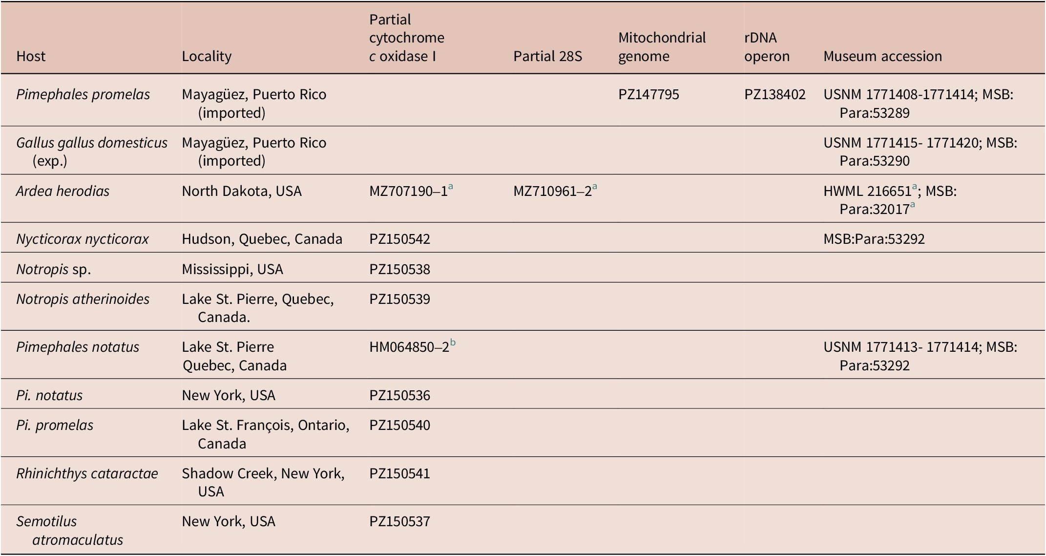
Metacercariae of Posthodiplostomum sp. were obtained from wild-caught fish from Ontario and Quebec, Canada, and New York and Mississippi, USA (Table 1), broadly overlapping the type locality of P. minimum (New York; MacCallum, Reference MacCallum1921), and the localities where Agersborg (Reference Agersborg1926) and Hoffman (Reference Hoffman1958) obtained material from leuciscid hosts (Urbana, Illinois and Turtle River, North Dakota, USA, respectively). Among the specimens studied are paragenophores for Posthodiplostomum sp. 4 sequenced by Locke et al. (Reference Locke, McLaughlin and Marcogliese2010; sequences HM064850–2). Additional metacercariae were taken from fathead minnows (Pi. promelas) purchased from a pet shop in Mayagüez, Puerto Rico, which staff stated were imported from two suppliers: one based in Florida and the other in Georgia, USA. All Pi. promelas at the pet store were rosy red minnows, an aquaculturally sourced variety often sold as feeder fish for larger carnivorous aquarium fishes (Stone et al. Reference Stone, Roy and Park2019).
Origins of specimens of Posthodiplostomum vancleavei n. comb. and associated DNA sequence and morphological voucher accessions from the Smithsonian Instituion, National Museum of Natural History (USNM), the Howard W. Manter Laboratory (HWML) and the Museum of Southwestern Biology, Division of Parasites (MSB:Para)

a Sequences from Achatz et al. (Reference Achatz, Chermak, Martens, Pulis, Fecchio, Bell, Greiman, Cromwell, Brant, Kent and Tkach2021); vouchers reexamined in present study.
b Sequences from Locke et al. (Reference Locke, McLaughlin and Marcogliese2010).
Adult worms were obtained from the salvaged carcass of a naturally infected black-crowned night heron, which had been stored frozen for several months at Le Nichoir, an avian rehabilitation centre in Hudson, Quebec, Canada. One specimen from this N. nycticorax was used for molecular work and paragenophores were retained as morphological vouchers. Other adult worms were obtained from domestic chicks that within 24–72 hours of hatching were fed metacercariae from Pi. promelas from the pet shop in Puerto Rico. Adult worms were taken from sacrificed-chick intestines 1–2 days post-infection. In addition, we re-examined adult specimens of “P. minimum” sequenced (MZ710961–2, MZ707190–1) by Achatz et al. (Reference Achatz, Chermak, Martens, Pulis, Fecchio, Bell, Greiman, Cromwell, Brant, Kent and Tkach2021) from great blue heron and black-crowned night heron from North Dakota.
Morphological Analysis
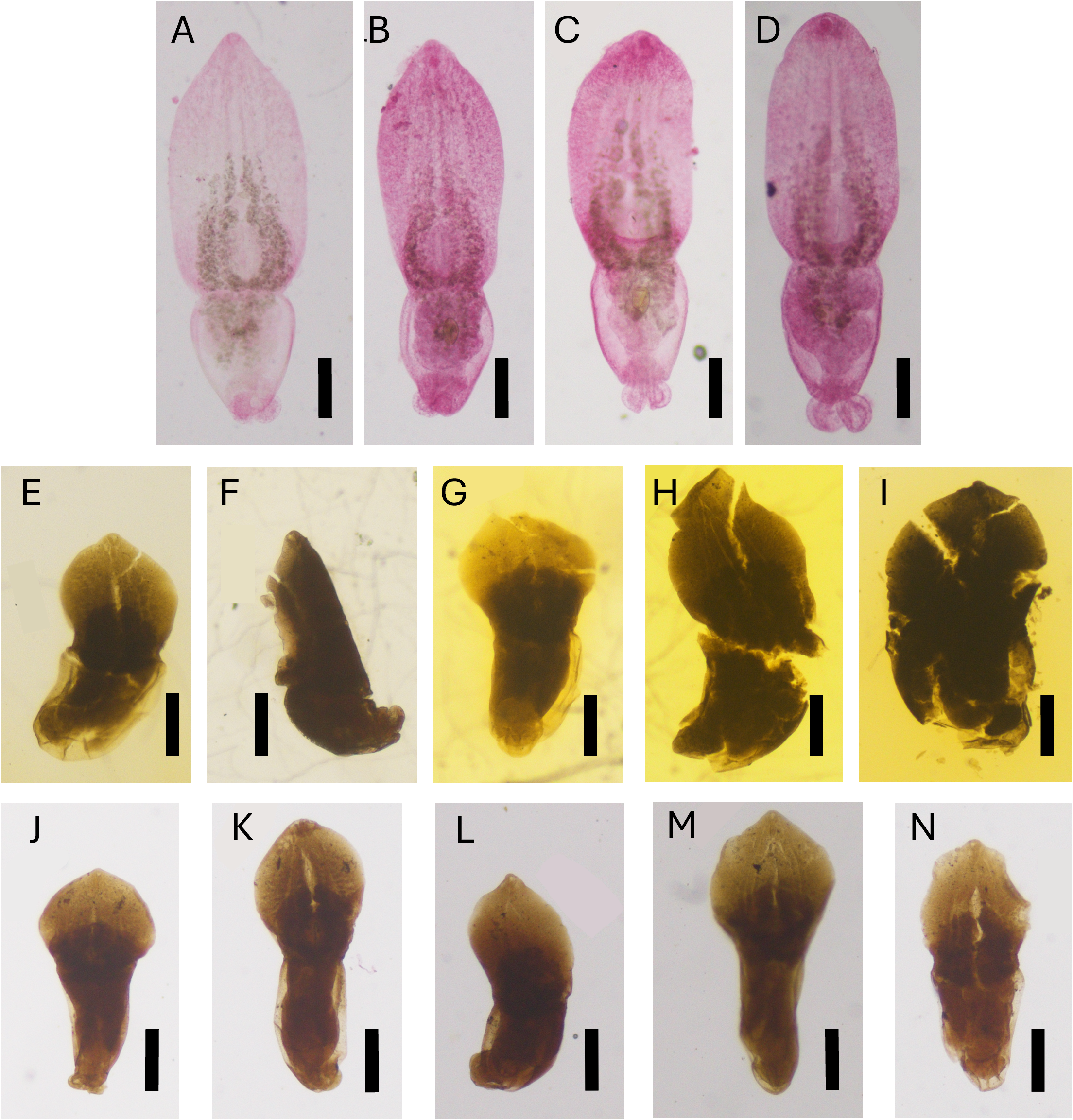
Adult worms from experimental and natural infections were compared to the type specimens of P. minimum deposited by MacCallum (Reference MacCallum1921), which were loaned by the Smithsonian National Museum of Natural History. Specimens deposited by MacCallum (Reference MacCallum1921) comprised seven fixed mounts (including one sectioned specimen) and 36 worms preserved in 70% ethanol. How MacCallum (Reference MacCallum1921) originally preserved, mounted, or handled the specimens is unknown. Five of the worms in 70% ethanol from the type material were newly mounted in Canada balsam, after clearing in lactophenol for several days, serial rehydration in an ethanol to water series, acetocarmine stain, dehydration in ethanol, and clearing in clove oil. Because many features could not be clearly distinguished in newly or originally mounted specimens of MacCallum (Reference MacCallum1921), drawings and descriptions of MacCallum’s (Reference MacCallum1921) original material by earlier authors were also considered (fig. 4 and measurements in Dubois, Reference Dubois1935; figs. 192, 204, 205, 207, 208, 209 in Ferguson, Reference Ferguson1937).
Both experimentally obtained adults and mechanically excysted metacercariae from Pi. promelas from the pet shop in Mayagüez were heat-fixed in 10% formalin and subjected to the same clearing and staining process described above before mounting on slides. Adults from thawed, naturally infected N. nycticorax were preserved in 95% ethanol before staining, clearing, and mounting on slides. Specimens were measured in micrometres (μm) using a Nikon Eclipse TS100 and NIS-Elements Microscope Imaging Software, version 4.51 (Nikon Metrology Inc., Texas, USA). Line drawings were made with assistance of a drawing tube.
Molecular and Phylogenetic Analysis
One metacercaria from Pi. promelas from the pet store in Puerto Rico was excysted, preserved in 95% ethanol, and subjected to DNA purification using a NucleoSpin Tissue XS kit (Macherey-Nagel, Allentown, Pennsylvania) following the manufacturer’s instructions. The DNA from this worm was fragmented and analysed at Azenta (South Plainfield, New Jersey, USA) in a fraction of a lane on an Illumina HiSeq 4000 machine yielding 150-bp paired-end reads. The mitochondrial genome was assembled using, as an initial scaffold, a consensus derived from an alignment of sequences of partial CO1 from Posthodiplostomum sp. 4 (HM064844) and the mitochondrial genome of P. centrarchi (MH536512). Similarly, the rDNA operon was assembled iteratively, starting with a consensus scaffold made from P. centrarchi (MH521251) and Posthodiplostomum sp. UG1 (LC511183). Iterative assemblies for both markers employed the Geneious assembler and BBduk (Bushnell Reference Bushnell2014), implemented in Geneious Prime (Biomatters Ltd., New Zealand). Sequences were annotated using MITOS (Bernt et al., Reference Bernt, Donath, Jühling, Externbrink, Florentz, Fritzsch, Pütz, Middendorf and Stadler2013, for the mitochondrial genome) and based on alignments with published sequences from related digeneans (e.g., Locke et al., Reference Locke, Van Dam, Caffara, Pinto, López-Hernández and Blanar2018, both mitochondrial genome and rDNA operon).
In the metacercariae from Notropis sp., emerald shiner (Notropis atherinoides), longnose dace (Rhinichthys cataractae), Pi. notatus, Pi. promelas, creek chub (Semotilus atromaculatus), and one adult from N. nycticorax (Table 1), the barcode region of cytochrome c oxidase subunit I (CO1) was amplified using polymerase chain reaction (PCR) with the primers and protocols of Moszczynska et al. (Reference Moszczynska, Locke, McLaughlin, Marcogliese and Crease2009) and sequenced at the Canadian Centre for DNA Barcoding (Guelph, Ontario, Canada). All mitochondrial DNA sequences identified as Posthodiplostomum spp. were obtained from GenBank and aligned with data from this study using MUSCLE (Edgar Reference Edgar2004) implemented in Geneious Prime. Short and non-overlapping sequences were removed from the alignment with barcode region CO1, and pairwise uncorrected p-distances were calculated in MEGA11 (Tamura et al. Reference Tamura, Stecher and Kumar2021). The best-fitting nucleotide substitution model for phylogenetic analysis of the partial CO1, mitochondrial genome, and rDNA operon alignments, as determined using the Bayesian Information Criterion in MEGA11, was the general time-reversible model with estimates of gamma-distributed among-site variation and invariant sites (GTR + G + I). Phylogenetic trees were constructed in Geneious Prime using RAxML (Maximum Likelihood, ML, Stamatakis, Reference Stamatakis2014, for 1,000 bootstrapped pseudoreplicates) and MrBayes (Bayesian Inference, BI, Huelsenbeck & Ronquist, Reference Huelsenbeck and Ronquist2001, for 2,000,000 generations sampled every 2,000 steps, with the initial 20% discarded as burn-in).
Results
Morphological Analysis
As described below, CO1 sequences indicated all material sequenced in this study (metacercariae from leuciscids in Ontario, Quebec, New York, Mississippi, and those used to infect chicks, adults from N. nycticorax) corresponded to Posthodiplostomum sp. 4. However, the adults of this species differed morphologically from P. minimum of MacCallum (Reference MacCallum1921) (see remarks below). We therefore restore the name Agersborg (Reference Agersborg1926) created for the species with metacercariae in North American leuciscids, D. vancleavei, and transfer it to Posthodiplostomum.
Posthodiplostomum vancleavei (Agersborg, Reference Agersborg1926) n. comb.
Synonyms: Diplostomum vancleavei Agersborg, Reference Agersborg1926, Neascus vancleavei (Agersborg, Reference Agersborg1926) Hughes, Reference Hughes1928, Posthodiplostomum minimum minimum Hoffman, Reference Hoffman1958; Posthodiplostomum minimum sensu Achatz et al. (Reference Achatz, Chermak, Martens, Pulis, Fecchio, Bell, Greiman, Cromwell, Brant, Kent and Tkach2021); Posthodiplostomum sp. 4 sensu Locke et al. (Reference Locke, McLaughlin and Marcogliese2010)
Adult
Hosts: Gallus gallus domesticus (experimental), Nycticorax nycticorax, Ardea herodias
Site in host: Intestine.
Localities: Hudson/Montreal Area, and Grande Île, Quebec, Canada (45° 27′ 30.8″ N, 74° 08′ 26.4″ W; 45° 16′ 45″ N, 74° 06′ 50″ W); Mississippi, USA; Nelson County, North Dakota, USA.
Voucher specimens: USNM 1771415-1771415; HWML 216651, 216653; MSB:Para:32017, 53290, 53292.
GenBank: 28S: MZ710961, MZ710962 (Achatz et al. Reference Achatz, Chermak, Martens, Pulis, Fecchio, Bell, Greiman, Cromwell, Brant, Kent and Tkach2021); CO1: PZ150542 (present study); HM064844, HM064845, HM0648466 (Locke et al. Reference Locke, McLaughlin and Marcogliese2010); MZ707190, MZ707191 (Achatz et al. Reference Achatz, Chermak, Martens, Pulis, Fecchio, Bell, Greiman, Cromwell, Brant, Kent and Tkach2021)
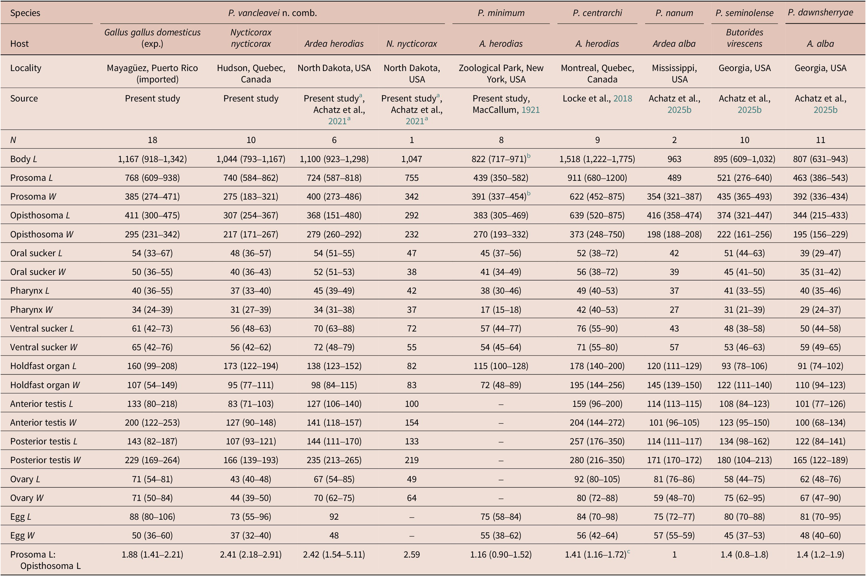
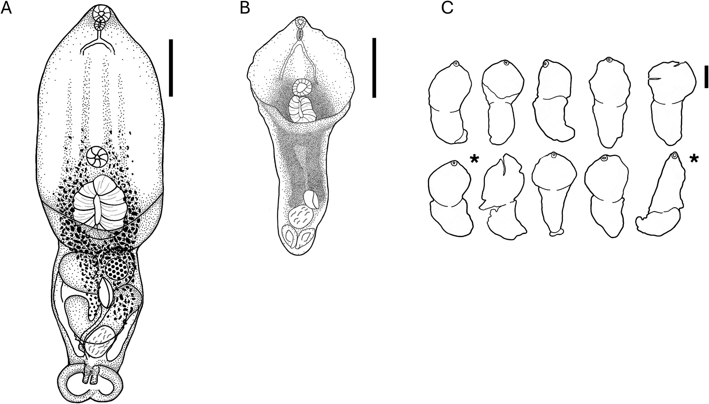
Description based on 18 adult worms obtained from chicks experimentally infected with metacercariae from Pi. promelas, 11 worms from naturally infected N. nycticorax and 6 worms from naturally infected A. herodias (Table 2, Figs. 1, 2). Body distinctly bipartite, consisting of distinct prosoma and opisthosoma. Prosoma foliate, flat or weakly concave near prosoma-opisthosoma junction, longer than opisthosoma; opisthosoma oval or subcylindrical. Tegument of prosoma armed with fine spines; opisthosoma unarmed. Oral sucker subterminal, round, muscular. Ventral sucker round, muscular, larger than oral sucker (ventral:oral sucker width ratio 1.2). Holdfast organ posterior to ventral sucker, round, with median slit as aperture. Proteolytic gland distinct, consisting of 2 defined lobes. Prepharynx not observed. Pharynx oval, muscular. Oesophagus similar in length to or slightly longer than pharynx. Cecal bifurcation in anterior-most quarter of prosoma length. Ceca slender, extending near posterior margin of posterior testis.
Morphometrics of adult specimens of Posthodiplostomum from present and other studies: mean (range in parentheses) in μm

a Specimens reexamined in the present study, collected and sequenced by Achatz et al. (Reference Achatz, Chermak, Martens, Pulis, Fecchio, Bell, Greiman, Cromwell, Brant, Kent and Tkach2021), in which they are identified as P. minimum.
b MacCallum (Reference MacCallum1921) states worms are ‘about 1000 μm long and 600 μm wide’, which exceeds our measurements of the same specimens.
c Calculated from Locke et al. (Reference Locke, Van Dam, Caffara, Pinto, López-Hernández and Blanar2018), who called this ‘Forebody/Hindbody’, but reported the inverse (hindbody/forebody).
A) Drawing of adult Posthodiplostomum vancleavei n. comb. from chicks fed metacercariae from Pimephales promelas from a pet store in Mayagüez, Puerto Rico, in this study, and of (B, C) Posthodiplostomum minimum from Ardea herodias in the New York Zoological Park (MacCallum, Reference MacCallum1921). Asterisks indicate specimens drawn by Dubois (Reference Dubois1935) and Ferguson (Reference Ferguson1937). Scale bars = 200 μm.

A–D) Adult Posthodiplostomum vancleavei n. comb. obtained from chicks fed metacercariae from Pimephales promelas from a pet store in Mayagüez, Puerto Rico, in this study. E–N) Type specimens of Posthodiplostomum minimum from Ardea herodias from the New York Zoological Park (MacCallum, Reference MacCallum1921): E–I) original mounts; J–N) specimens mounted in the present study. Scale bars = 200 μm.

Testes tandem, bilobed; anterior testis positioned near prosoma-opisthosoma junction; posterior testis with distinct cleft. Seminal vesicle primarily post-testicular, ventral to posterior testis, compact, continues as short ejaculatory duct. Ejaculatory duct joins metraterm dorsally to form hermaphroditic duct near proximal part of genital cone. Hermaphroditic duct opens at tip of genital cone into genital atrium. Terminal reproductive complex consisting of genital cone surrounded by prepuce, often protruding from genital pore. Genital pore subterminal, dorsal.
Ovary round, positioned near prosoma-opisthosoma junction, ventral to anterior testis. Ootype and Mehlis’ gland intertesticular. Laurer’s canal not observed. Vitellarium extending from near level of or anterior to level of ventral sucker to level of posterior testis, forming two ventral fields in opisthosoma. Vitelline reservoir intertesticular. Uterus ventral to testes and seminal vesicle. Eggs large, few.
Excretory pore terminal.
Metacercaria
Type host: Miniellus anogenus
Other hosts: Pimephales notatus, Pimephales promelas, Pseudaspius hakonensis, Rhinichthys cataractae, Semotilus atromaculatus
Site in host: Encysted in body cavity
Type locality: Crystal Lake, Urbana, Illinois, USA
Other localities: Lakes St. Francois, St. Louis, and St. Pierre, Quebec, Canada; Otsego Lake, New York, USA; Nagano, Japan
Voucher specimens: USNM 1771408-1771414; MSB:Para:53289, 53291
GenBank accessions: mitochondrial genome: PZ147795; rDNA operon: PZ138402 CO1: PZ150536, PZ150537, PZ150538, PZ150539, PZ150540, PZ150541 (present study); FJ477218 (Moszczynska et al. Reference Moszczynska, Locke, McLaughlin, Marcogliese and Crease2009); HM064847, HM064848, HM064849, HM064850, HM064851, HM064852, HM064853, HM064854, HM064855, HM064856 (Locke et al. Reference Locke, McLaughlin and Marcogliese2010); LC511186 (Komatsu et al. Reference Komatsu, Itoh and Ogawa2020); ITS rDNA: LC511183 (Komatsu et al. Reference Komatsu, Itoh and Ogawa2020).
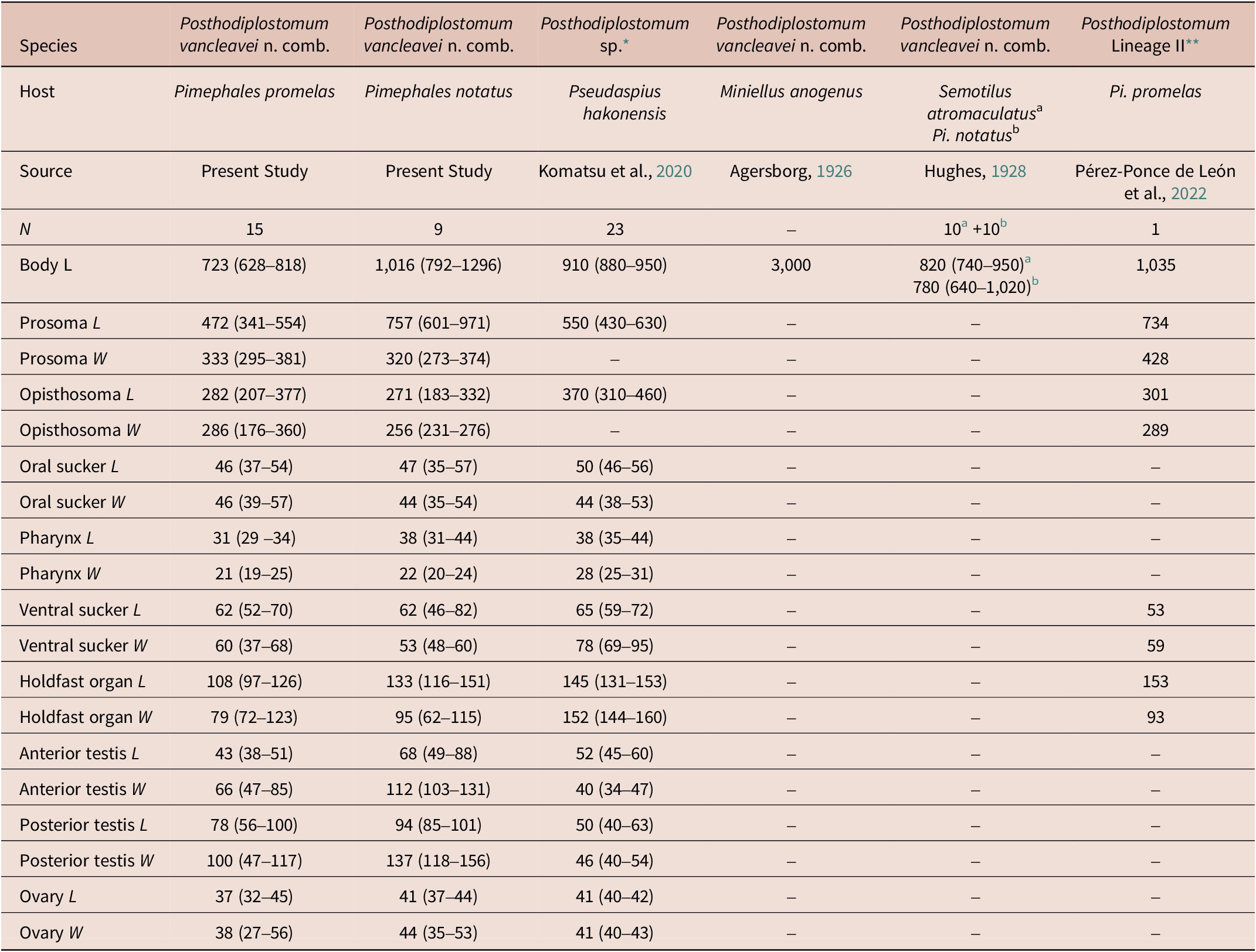
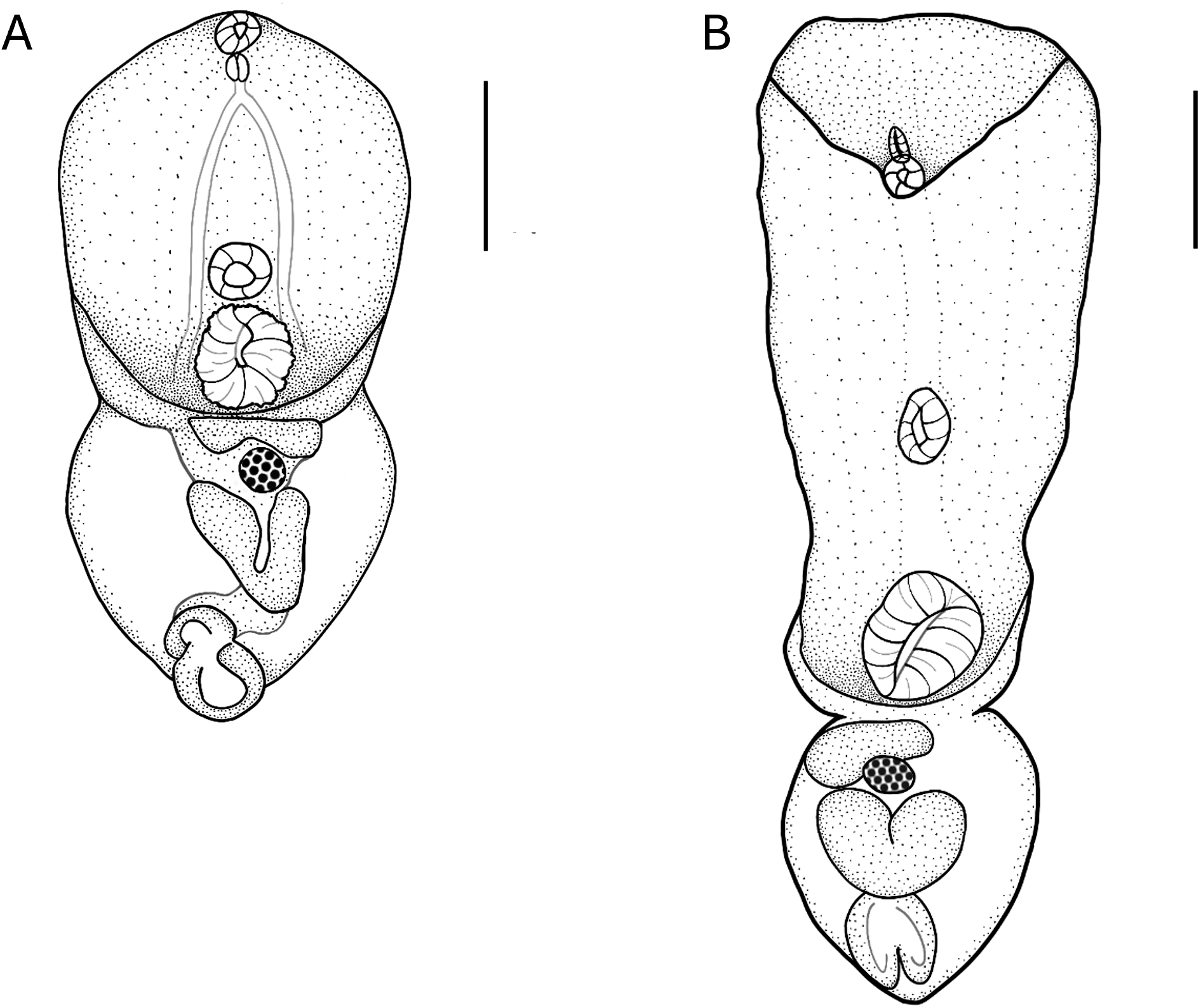
Description based on 24 metacercariae from Pi. promelas and Pi. notatus (Table 3, Fig. 3). Body distinctly bipartite. Prosoma round to oval-elongate, separated from spherical opisthosoma by distinct constriction. Oral sucker round, muscular, at anterior extremity. Prepharynx not observed; pharynx oval, elongate, immediately posterior to oral sucker. Ventral sucker round, muscular, larger than oral sucker. Holdfast organ large, muscular, oval, in posterior half of prosoma. Testes and ovary underdeveloped. Genital atrium round at posterior end of opisthosoma. In transparent, spacious, thin-walled cysts in body cavity.
Morphometrics of metacercariae of Posthodiplostomum from Leuciscidae in the present and other studies: mean (range in parentheses) in μm

* Unclear whether measurements reported in Komatsu et al. (2019) are of Posthodiplostomum vancleavei n. comb. or Posthodiplostomum sp. 16.
** Measurements estimated from fig. 4B of Pérez-Ponce de León et al. (Reference Pérez-Ponce de León, Sereno-Uribe, Pinacho-Pinacho and García-Varela2022).
a 10 worms from Semotilus atromaculatus with Body L 820 (740–950)
b 10 worms from Pi notatus with Body L 780 (640–1,020)
Metacercaria of Posthodiplostomum vancleavei n. comb. from A) Pimephales promelas from a pet store in Mayagüez, Puerto Rico, and B) from Pimephales notatus from Lake St. Pierre, St. Lawrence River, Quebec, Canada. Scale bars = 200 μm.

Remarks
Posthodiplostomum vancleavei n. comb. is here distinguished from P. minimum and other species based on the morphology of adults. Palmieri (Reference Palmieri1977a,Reference Palmierib,Reference Palmieric), who worked with P. centrarchi, found variation in many characters of adult worms (organ size and position, body dimensions) cultivated in different definitive host taxa, which suggests that species of Posthodiplostomum cannot reliably be distinguished based on adult morphology. However, the morphological variations observed by Palmieri (Reference Palmieri1977a,Reference Palmierib,Reference Palmieric) were associated with the class of experimental host (e.g., Aves, Mammalia, Amphibia), and the adult worms considered in this study all originate from birds, and include A. herodias, the type host of P. minimum. Moreover, the adults of P. vancleavei n. comb. showed no important variation across naturally and experimentally infected hosts (Table 2).
Adults of P. vancleavei n. comb. are longer than the specimens of P. minimum of MacCallum (Reference MacCallum1921) (Table 2, Figs. 1, 2), primarily due to a longer prosoma in P. vancleavei n. comb., while the opisthosoma is similar in length in both species. As a result, the prosoma-to-opisthosoma length ratio is higher in P. vancleavei n. comb. (average of all specimens in all hosts = 2.2) than in P. minimum (≤1.5). In addition to being longer, the prosoma of adult P. vancleavei n. comb. is generally foliate in shape, i.e., longer than wide, although prosoma length may only exceed its width by a small amount in contracted specimens. In comparison, the prosoma of P. minimum has uniformly subspherical to round shape (see description below). Prosoma shape is a diagnostic feature of several Posthodiplostomum spp. (Achatz et al. Reference Achatz, Von Holten, Pritchard and Keel2025b).
Morphological differences between adults of P. vancleavei n. comb. and P. centrarchi are more subtle, but include generally smaller total lengths, narrower prosoma, and a prosoma twice as long as the opisthosoma in P. vancleavei n. comb., versus 1.4 times as long in P. centrarchi (Table 2). The mitochondrial genomes and rDNA operons of these two species differ by 19.34% and 1.22%, respectively.
Compared to other species of Posthodiplostomum from the New World, a wider or more robust body distinguishes adults of P. vancleavei n. comb. from the elongated and slender forms of Posthodiplostomum prosostomum Dubois et Rausch, Reference Dubois and Rausch1948, Posthodiplostomum boydae Dubois, 1969, Posthodiplostomum opisthosicya Dubois, 1969, and Posthodiplostomum macrocotyle Dubois, Reference Dubois1937 (data from Dubois Reference Dubois1970, Achatz et al. Reference Achatz, Von Holten, Pritchard and Keel2025b). The width of the prosoma in adults of P. vancleavei n. comb. is only approximately 25% greater than that of the opisthosoma, whereas the relative width of the prosoma is greater in the following species: Posthodiplostomum aztlanensis González-García et al., Reference González-García, López-Jiménez, Ortega-Olivares, Sereno-Uribe, de León G and García-Varela2024, Posthodiplostomum dawnsherryae Achatz, Von Volten, Pritchard et Keel, 2025, Posthodiplostomum grande (Diesing, 1850), Posthodiplostomum nanum Dubois, Reference Dubois1937, Posthodiplostomum pacificum Achatz, Chermak, Kent et Tkach 2021, Posthodiplostomum microsicya Dubois, Reference Dubois1936, Posthodiplostomum seminolense Achatz, Von Holten, Pritchard et Keel, 2025 (data from Dubois Reference Dubois1970; González-García et al. Reference González-García, López-Jiménez, Ortega-Olivares, Sereno-Uribe, de León G and García-Varela2024; Achatz et al. Reference Achatz, Von Holten, Pritchard and Keel2025b). In P. vancleavei n. comb., the prosoma of the adult is separated from the opisthosoma by a marked constriction, which is less pronounced or absent in Posthodiplostomum anterovarium (Dronen, Reference Dronen1985), Posthodiplostomum eurypygae Achatz, Chermak, Bell, Fecchio et Tkach 2021, Posthodiplostomum ptychocheilus (Faust, 1917), Posthodiplostomum podicipitis (Yamaguti, 1939), and Posthodiplostomum recurvirostrae Achatz, Chermak et Tkach 2021 (data from Dubois, Reference Dubois1970; Dronen Reference Dronen1985; Achatz et al. Reference Achatz, Chermak, Martens, Pulis, Fecchio, Bell, Greiman, Cromwell, Brant, Kent and Tkach2021, Reference Achatz, Von Holten, Pritchard and Keel2025b). The prosoma of adults of P. vancleavei n. comb. is approximately twice the length of the opisthosoma, which distinguishes it from P. boydae, Posthodiplostomum erickgreenei Achatz, Chermak, Cromwell et Tkach Reference Achatz, Chermak, Martens, Pulis, Fecchio, Bell, Greiman, Cromwell, Brant, Kent and Tkach2021, P. grande, and Posthodiplostomum orchilongum (Noble, 1936) (data from Dubois Reference Dubois1970; Achatz et al. Reference Achatz, Chermak, Martens, Pulis, Fecchio, Bell, Greiman, Cromwell, Brant, Kent and Tkach2021, 2025b). Adults of P. vancleavei n. comb. are also smaller than those of P. prosostomum, P. grande, P. microsicya, and Posthodiplostomum giganteum Dubois, Reference Dubois1988 (data from Dubois Reference Dubois1970, Reference Dubois1988). Finally, in P. vancleavei n. comb., the posterior extent of the vitellaria does exceed the middle of the posterior testis, whereas the vitellaria extend further in P. boydae, P. dawnsherryae, P. nanum, P. opisthosicya, and Po. macrocotyle (data from Dubois Reference Dubois1970; Achatz et al. Reference Achatz, Chermak, Martens, Pulis, Fecchio, Bell, Greiman, Cromwell, Brant, Kent and Tkach2021, Reference Achatz, Von Holten, Pritchard and Keel2025b).
Agersborg (Reference Agersborg1926) reported metacercariae of P. vancleavei n. comb. to be circa 3 × 1 mm. We suspect these anomalously large values may be of cysts flattened under pressure. Cysts from Pi. promelas under pressure were only approximately 0.5 mm × 1 mm and excysted, fully extended live worms from this host reached a maximum total length of about 2 mm. However, for reasons described in the Discussion, we believe metacercariae and cysts in Pi. promelas obtained in the study were probably recently acquired and not fully developed, as also indicated by the larger forms in naturally infected hosts (Fig. 3).
Partial CO1 and 28S sequences of P. vancleavei n. comb. differ from several of these species (P. anterovarium, P. dawnsherryae, P. erickgreenei, P. eurypygae, P. macrocotyle, P. nanum, Posthodiplostomum pricei (Krull, 1934), P. seminolense, P. orchilongum, P. pacificum, P. ptychocheilus) and congeners from other regions (Posthodiplostomum brevicaudatum (von Nordmann, 1832), P. cuticola, Posthodiplostomum scardinii (Shulman, 1952)), by at least 11.8% and 1.5%, respectively.
Posthodiplostomum minimum (MacCallum, Reference MacCallum1921)
Adult
Type host: Ardea herodias L. (Pelecaniformes: Ardeidae)
Site in host: Intestine
Type locality: Bronx Zoo (referred to as the Zoological Park) in New York City, USA
Type material: USNM 1335821
Description based on eight adult worms (Table 2, Figs. 1, 2). Body bipartite, consisting of weakly distinct prosoma and opisthosoma. Prosoma subspherical or otherwise rounded, weakly concave near prosoma-opisthosoma junction, similar in length or shorter than opisthosoma; opisthosoma oval or subcylindrical. Oral sucker subterminal, round, muscular. Ventral sucker round, muscular, larger than oral sucker (ventral:oral sucker ratio 1.25). Holdfast organ posterior to ventral sucker, round, with median slit as aperture. Proteolytic gland not observed. Prepharynx not observed. Pharynx oval, muscular. Oesophagus shorter than pharynx. Caecal bifurcation in anterior-most quarter of prosoma length. Ceca slender, posterior extent not observed.
Testes lobed. Seminal vesicle, in posterior opisthosoma. Terminal reproductive complex consisting of genital cone surrounded by prepuce. Genital pore subterminal, dorsal.
Ovary, ootype, Mehlis’ gland, and Laurer’s canal not observed. Vitellarium extending from near level of or anterior to level of ventral sucker to posterior margin of posterior testis. Vitelline reservoir not observed. Eggs large, few.
Excretory pore not observed.
Remarks
The original description and illustration of P. minimum by MacCallum (Reference MacCallum1921) were superficial and differed from the type specimens. For example, MacCallum (Reference MacCallum1921) depicts the oesophagus to be more than twice the length of the pharynx, but it is much shorter than the pharynx in the original specimens. MacCallum (Reference MacCallum1921) mentioned spines on the prosoma, but these have not been observed (possibly lost due to fixation). In MacCallum’s (Reference MacCallum1921) illustration, the vitellaria extend well anterior to the ventral sucker and in the opisthosoma reach the genital pore, but they do not extend as far in either body region of the deposited specimens. The testes illustrated by MacCallum (Reference MacCallum1921) are round and smaller than those in the type specimens, as seen in illustrations of the type material by Dubois (Reference Dubois1935) and Ferguson (Reference Ferguson1937). The holdfast organ illustrated by MacCallum (Reference MacCallum1921) is smaller than the ventral sucker, whereas it is twice the size of this structure in the specimens he deposited.
The specimens of P. minimum deposited by MacCallum (Reference MacCallum1921) are in poor condition and many structures could not be observed, even in specimens newly mounted in the present study. However, the subspherical or otherwise rounded prosoma, with a width and length about 1.2 times that of the opisthosoma, is a feature shared by only four Nearctic congeners: P. aztlanensis, P. dawnsherryae, P. nanum, and P. seminolense. The prosoma of P. centrarchi can occasionally be somewhat rounded and of similar relative size, but adults of P. minimum are much smaller than those of P. centrarchi in terms of total length, including both prosoma and opisthosoma lengths, and the ventral sucker is also much smaller in P. minimum than in P. centrarchi. Collection of fresh, well-fixed specimens of P. minimum is necessary to provide an adequate description and differential diagnosis of this species.
Molecular and phylogenetic analysis
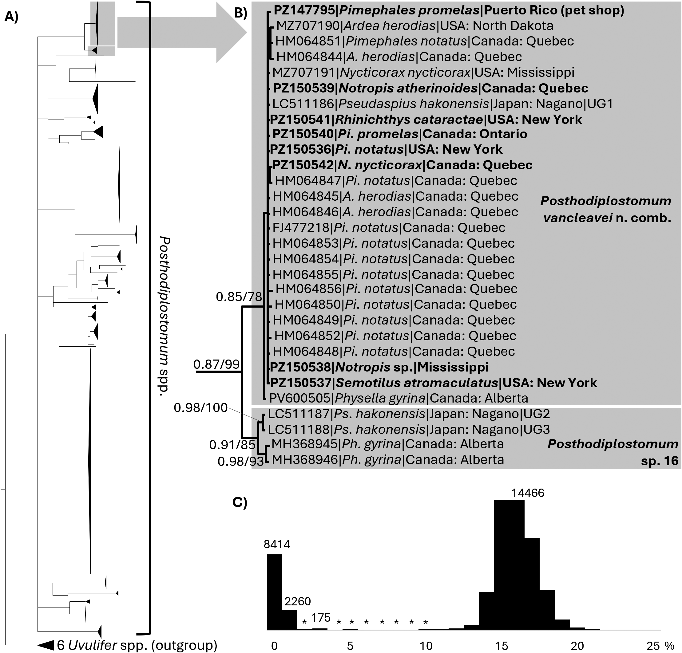
Partial CO1 sequences from metacercariae of P. vancleavei n. comb. from naturally infected leuciscids (Notropis sp., No. atherinoides, Pi. notatus, Pi. promelas, R. cataractae, and S. atromaculatus) and adults from naturally infected N. nycticorax in the present study formed a moderately supported clade (Fig. 4). This clade included published sequences from metacercariae of Posthodiplostomum spp. from leuciscids (Locke et al. Reference Locke, McLaughlin and Marcogliese2010; present study), cercariae from Physella gyrina (McPhail et al., 2025), as well as adult worms from naturally infected ardeids (A. herodias, N. nycticorax, Locke et al., Reference Locke, McLaughlin and Marcogliese2010; Achatz et al., Reference Achatz, Chermak, Martens, Pulis, Fecchio, Bell, Greiman, Cromwell, Brant, Kent and Tkach2021), all sampled in North America. This clade also contained one of three sequences from unidentified metacercariae from the big-scaled redfin (Pseudaspius hakonensis; Leuciscidae) in Japan (Komatsu et al. Reference Komatsu, Itoh and Ogawa2020). These sequences are here considered to be P. vancleavei n. comb., but are identified as P. minimum (Achatz et al., Reference Achatz, Chermak, Martens, Pulis, Fecchio, Bell, Greiman, Cromwell, Brant, Kent and Tkach2021; McPhail et al., 2025), Posthodiplostomum sp. 4 (Locke et al. Reference Locke, McLaughlin and Marcogliese2010), and Posthodiplostomum sp. UG1 (Komatsu et al., Reference Komatsu, Itoh and Ogawa2020).
A) Phylogenetic analysis of 351 partial CO1 sequences overlapping by at least 205 bp from Posthodiplostomum spp., with collapsed clades corresponding to different species (except for the Uvulifer clade, which comprises representatives of six species). The expanded inset B) shows Posthodiplostomum vancleavei n. comb. and Posthodiplostomum sp. 16. Sequences generated in this study are bold. Nodes of the Bayesian Inference topology shown are annotated with posterior probabilities/percent bootstrap support from 1000 replicates from a separate maximum likelihood analysis. The full tree is available as a supplementary file. C) Histogram of uncorrected P distances among the 351 CO1 sequences of Posthodiplostomum (* indicates non-zero values less than 100).

The most closely related species to P. vancleavei n. comb. in the CO1 phylogeny (Fig. 4) was Posthodiplostomum sp. 16 (provisional name of Achatz et al. Reference Achatz, Chermak, Martens, Pulis, Fecchio, Bell, Greiman, Cromwell, Brant, Kent and Tkach2021), a moderately supported clade comprising two sequences from metacercariae from Ps. hakonensis in Nagano, Japan (identified as Posthodiplostomum sp. UG2 and UG3, Komatsu et al. Reference Komatsu, Itoh and Ogawa2020) and two from cercariae from Phy. gyrina in Alberta, Canada (identified as Posthodiplostomum sp. 4, Gordy & Hanington, Reference Gordy and Hanington2019). In addition to the reciprocal monophyly indicating CO1 of Posthodiplostomum sp. 16 and P. vancleavei n. comb. are distinct evolutionary lineages (Fig.4B), the CO1 p-distances between P. vancleavei n. comb. and Posthodiplostomum sp. 16 averaged 5.28% (range 3.46–6.45%), which is greater than typical intraspecific variation when genetic distances within Posthodiplostomum are viewed as frequency distribution (Fig. 4C). Within these two species, the maximum uncorrected CO1 p-distances were 1.95% (P. vancleavei n. comb.) and 1.92% (Posthodiplostomum sp. 16). Other than Posthodiplostomum sp. 16, the CO1 of P. vancleavei n. comb. differed by at least 11.77% from all other species.
Posthodiplostomum Lin II and all other species sequenced by Pérez-Ponce de León et al. (Reference Pérez-Ponce de León, Sereno-Uribe, Pinacho-Pinacho and García-Varela2022) were not collected in the present study. The CO1 of Posthodiplostomum sp. Lin II from Pi. promelas in Durango, Mexico (Pérez-Ponce de León et al. Reference Pérez-Ponce de León, Sereno-Uribe, Pinacho-Pinacho and García-Varela2022), are from a different region of CO1 than that analysed in Fig. 4, and differed from the mitochondrial genome of P. vancleavei n. comb. by 14.9%. The CO1 sequences of four other lineages from Pérez-Ponce de León et al. (Reference Pérez-Ponce de León, Sereno-Uribe, Pinacho-Pinacho and García-Varela2022) differed from P. vancleavei n. comb. by 14.4–19.49%. This indicates that Posthodiplostomum Lin II, and all lineages from Pérez-Ponce de León et al. (Reference Pérez-Ponce de León, Sereno-Uribe, Pinacho-Pinacho and García-Varela2022), are different species from P. vancleavei n. comb.
The rDNA operon (PZ138402) of P. vancleavei n. comb. from Pi. promelas from the pet store in Puerto Rico was identical to all three 414-bp nuclear rDNA sequences from metacercariae from Ps. hakonensis in Japan (LC511183–5, including all of ITS2, Komatsu et al., Reference Komatsu, Itoh and Ogawa2020), which include representatives of both P. vancleavei n. comb. and Posthodiplostomum sp. 16. The ITS1-5.8S-ITS2 sequences from Pérez-Ponce de León et al. (Reference Pérez-Ponce de León, Sereno-Uribe, Pinacho-Pinacho and García-Varela2022) (OK315755–75, all 1,074 bp long) differ from the rDNA operon of P. vancleavei n. comb. by 2.7–9.2%. Intraspecific differences occurred at 2 out of 1,263 positions between the partial 28S sequence from P. vancleavei n. comb. generated in this study and two sequences identified as P. minimum by Achatz et al. (Reference Achatz, Chermak, Martens, Pulis, Fecchio, Bell, Greiman, Cromwell, Brant, Kent and Tkach2021) from great blue heron (MZ710961) and black-crowned night heron (MZ710962). We re-assessed the 28S chromatograms of Achatz et al. (Reference Achatz, Chermak, Martens, Pulis, Fecchio, Bell, Greiman, Cromwell, Brant, Kent and Tkach2021); in MZ710962, one of the two sites that differ from the new rDNA operon displayed a double peak (T/G). In the new rDNA operon this site is a Thymine (T).
The mt genome assembly of P. vancleavei n. comb. comprised 0.07% (36,137 out of 50,520,068 reads) of the read pool and was 13,490 bp in length with mean coverage of 357 (range 241–450) reads per site (quality-trimmed reads were mean: 143, SD: 8, range 50–150 bp long). The order and lengths of the 13 protein-coding, 20 tRNA, and 2 RNA subunit genes were similar to those observed in other digeneans (Supplementary Table 1). We were unable to unambiguously assemble the repeat-rich region between ND5 and CO3. The rDNA assembly comprised 0.01% (9,210 reads) of the read pool and was 7,235 bp in length with mean coverage of 166 (range 68–690) reads per site. The rDNA assembly comprised the 18S gene (1,978 bp in length), ITS1 (604 bp), 5.8S (157 bp), ITS2 (286 bp), and 28S (4,210 bp).
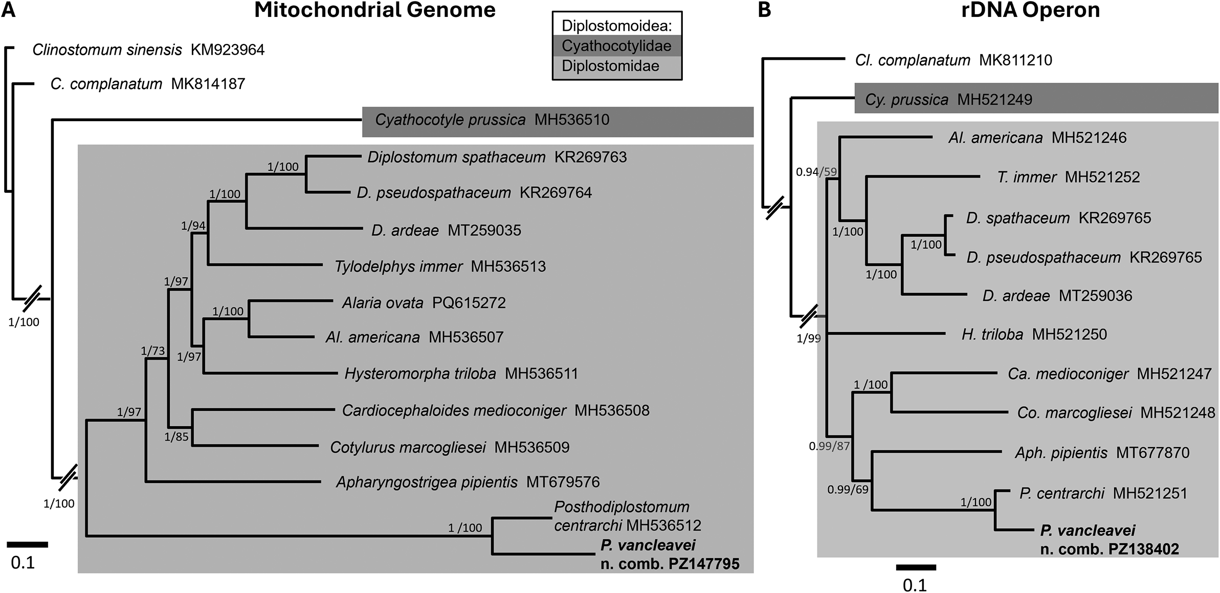
Phylogenetic analyses of both the mitochondrial genome and rDNA operon recovered Posthodiplostomum as a well-supported clade within the Diplostomoidea (Fig. 5), although topologies differed in some respects. In the mitochondrial genome tree (Fig. 5A), P. vancleavei n. comb. and P. centrarchi formed an early-diverging clade at the base of the Diplostomidae. In the rDNA operon tree (Fig. 5B), P. vancleavei n. comb. and P. centrarchi formed a clade distally nested within the Diplostomidae.
Phylogenetic analysis of A) mitochondrial genomes and B) rDNA operons from Posthodiplostomum vancleavei n. comb. and other members of the Diplostomoidea, rooted with Clinostomum (Schistosomatoidea). Nodes in the Bayesian Inference topology shown are annotated by posterior probabilities/percent bootstrap support from a separate Maximum Likelihood analysis with 1,000 replicates. Trees were generated under a GTR+G+I model of nucleotide substitution.

Discussion
Our study shows that the vast majority of records of P. minimum that have accumulated over the past century are misidentifications. The only confirmed specimens of this species are the adults that MacCallum (Reference MacCallum1921) obtained from a great blue heron in New York. Despite numerous records in diverse fishes, the second intermediate hosts of P. minimum are unknown. Under the previous concept of P. minimum, experimental (Hoffman Reference Hoffman1958) and molecular evidence (Locke et al. Reference Locke, McLaughlin and Marcogliese2010) indicated its metacercariae were specific to Nearctic leuciscid fishes, but here we show the adults described by MacCallum (Reference MacCallum1921) differ from adults of the species with metacercariae in minnows. For the latter species, we restore the name P. vancleavei n. comb., which applies to Posthodiplostomum sp. 4 of Locke et al. (Reference Locke, McLaughlin and Marcogliese2010), P. minimum of Achatz et al. (Reference Achatz, Chermak, Martens, Pulis, Fecchio, Bell, Greiman, Cromwell, Brant, Kent and Tkach2021), and the minnow line of Hoffman (Reference Hoffman1958), i.e., P. minimum minimum. We also show that Posthodiplostomum Lin II, which Pérez-Ponce de León et al. (Reference Pérez-Ponce de León, Sereno-Uribe, Pinacho-Pinacho and García-Varela2022) obtained from hosts in the Leuciscidae and other families, is a different species from P. vancleavei n. comb., and no evidence supports its identification as P. minimum. Below we discuss the historical basis and implications of our results for P. minimum, P. vancleavei n. comb., and other species of Posthodiplostomum.
History of Posthodiplostomum minimum revisited
The concept of P. minimum as a species with a wide range of second intermediate hosts owes much to Ferguson (Reference Ferguson1937), who obtained data in some respects similar to our own, but reached different conclusions. Ferguson (Reference Ferguson1937) re-examined the type specimens of MacCallum (Reference MacCallum1921) and considered them indistinguishable from worms he obtained from chicks fed leuciscids and centrarchids, as well as other specimens from naturally infected ardeids (A. herodias, N. nycticorax, green heron (Butorides virescens (L.)) and a gaviform (loon (Gavia immer (Brünnich))). Similarly, Miller (Reference Miller1954) examined the material of both MacCallum (Reference MacCallum1921) and Ferguson (Reference Ferguson1937) and considered them identical to experimentally obtained adults of what is now called P. centrarchi.
Where Ferguson (Reference Ferguson1937) and Miller (Reference Miller1954) saw one species, P. minimum, we see three (P. minimum, P. centrarchi, and P. vancleavei n. comb.) and attribute the discrepancy to two causes. First, early taxonomic decisions were made in a context of lower species diversity than is currently recognised in Posthodiplostomum. Ferguson (Reference Ferguson1937) worked when Dubois (Reference Dubois1936) had just established Posthodiplostomum, which initially contained five species and in North America was only represented by P. minimum. When Miller (Reference Miller1954) conducted his study, only one additional North American species was known (P. prosostomum). In contrast, even in the narrowest sense, i.e., excluding members of recent synonyms Mesoophorodiplostomum Dubois, Reference Dubois1936 and Ornithodiplostomum Dubois, Reference Dubois1936, at least 15 Posthodiplostomum spp. exist in North America, including seven with molecular and morphological support (P. centrarchi, P. dawnsherryae, P. erickgreenei, P. nanum, P. orchilongum, P. pacificum, and P. seminolense; Stoyanov et al., Reference Stoyanov, Georgieva, Pankov, Kudlai, Kostadinova and Georgiev2017; Achatz et al., Reference Achatz, Chermak, Martens, Pulis, Fecchio, Bell, Greiman, Cromwell, Brant, Kent and Tkach2021, 2025b) and eight known only by DNA sequences (Posthodiplostomum spp. 5, 8, 16–20, 23; Locke et al., Reference Locke, McLaughlin and Marcogliese2010; Achatz et al., Reference Achatz, Chermak, Martens, Pulis, Fecchio, Bell, Greiman, Cromwell, Brant, Kent and Tkach2021), some of which may correspond to four only known from morphology (P. boydae, P. minimum, P. opisthosicya, and P. prosostomum). Additional species diversity is likely to be revealed in future studies, because many of these species have only recently been recognized, and morphology alone is often inadequate for species-level work in this genus. For example, despite morphological characterization of all developmental stages, whether a species of Posthodiplostomum described by Ritossa et al. (Reference Ritossa, Flores and Viozzi2013) was new or corresponded to already named species could not be determined. Other examples include the morphologically similar, genetically distinct species recently described by Achatz et al. (Reference Achatz, Von Holten, Pritchard and Keel2025b).
The second important difference in current understanding, compared to earlier taxonomic studies of P. minimum by Ferguson (Reference Ferguson1936, Reference Ferguson1937) and others, is the recognition that metacercariae of some species of Posthodiplostomum are exclusive to narrow ranges of fish hosts. Second intermediate-host specificity is particularly well established for species with metacercariae in leuciscids and centrarchids of North America (Klak Reference Klak1940; Ferguson Reference Ferguson1943; Hoffman Reference Hoffman1958; Locke et al. Reference Locke, McLaughlin and Marcogliese2010; Boone et al. Reference Boone, Laursen, Colombo, Meiners, Romani and Keeney2018) and molecular surveys indicate it also applies to some (Díaz-Pernett et al. Reference Díaz-Pernett, Brant and Locke2022) but not all other systems (Pérez-Ponce de León et al. Reference Pérez-Ponce de León, Sereno-Uribe, Pinacho-Pinacho and García-Varela2022; Kudlai et al. Reference Kudlai, Binkienė, Rakauskas and Baker2026). Besides the implications for P. minimum and the minnow and sunfish lines of Hoffman (Reference Hoffman1958), these trends of high diversity and fish-host specificity suggest species-level taxonomic judgements in Posthodiplostomum should err on the side of splitting rather than lumping in considering morphological variation, especially given differences in second-intermediate-host usage. Indeed, both Ferguson (Reference Ferguson1937) and Miller (Reference Miller1954) acknowledged the possibility of two distinct species under the name P. minimum in light of second-intermediate-host specificity revealed in experiments.
Potential synonyms of P. minimum
As the oldest New-World species in the genus, P. minimum assuredly remains valid, and one of the subsequently described species in this region may be a junior synonym. We consider that the available morphological, host-use, and geographical data indicate four possible synonyms. Among species known from the Americas, the subspherical prosoma shape and body proportions of P. minimum are only shared with P. aztlanensis, P. dawnsherryae, P. nanum, and P. seminolense (Table 2). The subtlety of morphological variations among these four genetically distinct congeners (González-García et al. Reference González-García, López-Jiménez, Ortega-Olivares, Sereno-Uribe, de León G and García-Varela2024; Achatz et al. Reference Achatz, Von Holten, Pritchard and Keel2025b) makes it difficult to distinguish their relative potential as synonyms of P. minimum, considering the poor quality of the type material. However, the greater anterior extent of the vitellaria in P. aztlanensis (González-García et al., Reference González-García, López-Jiménez, Ortega-Olivares, Sereno-Uribe, de León G and García-Varela2024) may be significant.
Two of the four aforementioned species show evidence of host specificity exclusive of the type host of P. minimum, i.e., A. herodias. In the same locality in Georgia, P. dawnsherryae was found in four out of four great egrets (Ardea alba L.) studied, and P. seminolense in three out of three green herons (B. virescens) (Achatz et al. Reference Achatz, Von Holten, Pritchard and Keel2025b). These exclusive host associations were maintained in additional sampling in this area, including five great blue herons negative for these two species (TJA, personal observation). The possible definitive-host specificity of P. dawnsherryae and P. seminolense argues against their candidacy as synonyms of P. minimum, known only from great blue heron. However, additional sampling and experimental infection are needed to evaluate this possibility conclusively.
In contrast to this view from host specificity, from a geographical perspective, P. seminolense and P. dawnsherryae are the most likely candidates for synonyms of P. minimum because they occur in Georgia, closer to New York, where MacCallum (Reference MacCallum1921) collected P. minimum. However, geographic evidence has relatively little weight at this spatial scale because of the wide, overlapping ranges and mobility of many New-World ardeid definitive hosts (Kushlan and Hancock Reference Kushlan and Hancock2005). For example, intercontinental distributions have been demonstrated with molecular data in P. nanum (López-Hernández et al., Reference López-Hernández, Locke, de Melo, Rabelo and Pinto2018; Achatz et al. Reference Achatz, Chermak, Martens, Pulis, Fecchio, Bell, Greiman, Cromwell, Brant, Kent and Tkach2021) and other diplostomoideans with similar life cycles (Locke et al. Reference Locke, Al-Nasiri, Caffara, Drago, Kalbe, Lapierre, McLaughlin, Nie, Overstreet, Souza, Takemoto and Marcogliese2015; Hoogendoorn et al. Reference Hoogendoorn, Smit and Kudlai2020; Locke et al. Reference Locke, Drago, López-Hernández, Chibwana, Núñez, Van Dam, Achinelly, Johnson, de Assis, de Melo A and Pinto2021; Achatz et al. Reference Achatz, Martens, Kostadinova, Pulis, Orlofske, Bell, Fecchio, Oyarzún-Ruiz, Syrota and Tkach2022).
Geographic considerations also complicate the origins of P. minimum due to its description from a host in a major zoo. MacCallum (Reference MacCallum1921) did not mention whether the type host, A. herodias, was captive or wild caught, and this heron might have consumed exotic or imported fish housed in the zoo or its associated aquarium, with consequent exposure to nonnative parasites. For example, we found P. vancleavei n. comb., a North American species, in fish imported to the Caribbean, and Chibwana et al. (Reference Chibwana, Blasco-Costa, Georgieva, Hosea, Nkwengulila, Scholz and Kostadinova2013) found a diplostomid of African origin in fish imported to the Czech Republic from Nigeria. Ultimately, molecular data from the type material of MacCallum are needed to conclusively determine if a currently recognised species is a junior synonym of P. minimum. Unfortunately, the type specimens are old and were likely stored in formalin at some point (Anna Phillips, pers. comm.), both of which impede recovery and sequencing of DNA (Zimmermann et al. Reference Zimmermann, Hajibabaei, Blackburn, Hanken, Cantin, Posfai and Evans2008). Sequencing DNA of formalin-fixed archival material is possible (Hahn et al. Reference Hahn, Alexander, Grealy, Stiller, Gardiner and Holleley2021) but still not routine. Sequencing morphologically similar specimens newly collected from great blue heron in the type region may be the most practical way to address the identity of P. minimum.
Our data clarify several other recent identifications and records of P. minimum. Pérez-Ponce de León et al. (Reference Pérez-Ponce de León, Sereno-Uribe, Pinacho-Pinacho and García-Varela2022) considered Posthodiplostomum Lin II possibly conspecific with P. minimum, which they equated to the minnow-line of Hoffman (Reference Hoffman1958), herein renamed P. vancleavei n. comb. However, Posthodiplostomum Lin II differs from P. vancleavei n. comb. in both CO1 and ITS. The conclusion that Posthodiplostomum Lin II might be conspecific with the minnow line of P. minimum was based on its leucisid host, Pi. promelas, although Pérez-Ponce de León et al. (Reference Pérez-Ponce de León, Sereno-Uribe, Pinacho-Pinacho and García-Varela2022) sampled this fish at or even slightly beyond the southernmost limit of its reported natural range (Lee and Schute Reference Lee, Schute, Lee, Gilbert, Hocutt, Jenkins, McAllister and Stauffer1980). Pérez-Ponce de León et al. (Reference Pérez-Ponce de León, Sereno-Uribe, Pinacho-Pinacho and García-Varela2022) also found Posthodiplostomum Lin II in non-leuciscids, which they acknowledged conflicted with the defining feature of the minnow line of Hoffman (Reference Hoffman1958), namely its host specificity, which is also further supported in our expanded molecular survey of metacercariae from North American leuciscids. In addition, host associations of Posthodiplostomum Lin II are uncertain. For example, one specimen is associated with a mixture of fish hosts in Pérez-Ponce de León et al. (Reference Pérez-Ponce de León, Sereno-Uribe, Pinacho-Pinacho and García-Varela2022), who report it from olive skiffia (Skiffia lermae Meek) in their table I, and bulldog goodea (Alloophorus robustus (Bean)) and blackfin goodea (Goodea atripinnis Jordan) in GenBank records (OK315757, OK314910).
Other studies report P. minimum from bayad (Bagrus bajad (Forsskål)) (Siluriformes) in Egypt (Hamouda and Bazh Reference Hamouda and Bazh2019) and Nile tilapia (Oreochromis niloticus (L.)) (Cichliformes) in Nigeria (Vandi and Daniel Reference Vandi and Daniel2023). No support is provided for the unusual hosts and wide distributions that these records represent. On the contrary, figures in Hamouda and Bazh (Reference Hamouda and Bazh2019) show worms that are smaller and differ markedly in overall shape from metacercariae of P. vancleavei n. comb. and P. centrarchi and show little resemblance to adults of P. minimum of MacCallum (Reference MacCallum1921).
History of P. vancleavei n. comb. revisited
Hughes (Reference Hughes1928) argued that Agersborg (Reference Agersborg1926) probably misidentified M. anogenus, the reported type host of P. vancleavei n. comb., because this fish was uncommon as far south as Urbana, Illinois. However, Bailey (Reference Bailey1959) examined M. anogenus from Havana, Illinois, nearly as far south as the locality where Agersborg (Reference Agersborg1926) worked. Both Agersborg (Reference Agersborg1926) and Bailey (Reference Bailey1959) comment on the patchy and temporally variable distribution of this fish. We therefore accept M. anogenus as the type host. Our data show that a single Posthodiplostomum species predominates in leuciscids in the present and prior sequencing surveys (Locke et al. Reference Locke, McLaughlin and Marcogliese2010) encompassing the region where Agersborg (Reference Agersborg1926) worked. The metacercariae illustrated by Agersborg (Reference Agersborg1926) also resemble those studied herein, particularly specimens in wild-caught fish (Fig. 3B). Thus, the metacercariae from leuciscids in Agersborg (Reference Agersborg1926), Locke et al. (Reference Locke, McLaughlin and Marcogliese2010), and the present study can be confidently assigned to the same species.
Interestingly, the metacercariae of P. vancleavei n. comb. from Pi. promelas imported to a pet store in Puerto Rico were markedly smaller than those from wild-caught Pi. notatus in Quebec (Table 3, Fig. 3), although CO1 sequences indicate all these samples were conspecific (Fig. 4). We suspect that these size differences may reflect differences in worm age and density of infections. The mean length of Pi. promelas from the pet store was 34 mm (range 27–37 mm, n = 5), sizes which correspond to one year of age or less (Copp et al. Reference Copp, Godard, Verreycken, Davison, Kováč, Danylchuk, Spikmans and Britton2026; Danylchuk and Tonn Reference Danylchuk and Tonn2006), and infection levels in these small fish were high (100% prevalence, mean abundance 33.1 worms, range 14–51). The small size and high infection levels of Pi. promelas from the pet store are likely related to their origins in high-density aquaculture ponds in which harvested fish are as young as one month old (Stone et al. Reference Stone, Roy and Park2019). In P. centrarchi, the prosoma is notably shorter in younger worms (as seen here), even though these are of similar total length to metacercariae with characteristic, mature morphology, which occurs 27 days after infection (Hoffman Reference Hoffman1958), and Miller (Reference Miller1954) also describes effects of crowding on metacercarial morphology. Thus, the source of the Pi. promelas in the present study could affect metacercarial morphology through crowding of parasites that are more recently established within the host. In wild-caught fish, infections are often less intense and likely to be older. For example, here we studied paragenophores originating from the 111 Pi. notatus sampled by Locke et al. (Reference Locke, McLaughlin and Marcogliese2010, Reference Locke, McLaughlin and Marcogliese2013), which were larger (mean length 50 mm, range 37–78 mm), and therefore likely >1 year old (Westman Reference Westman1938), yet infection levels were much lower (prevalence 14%, mean abundance 1.1 worms, range 0–24). Similar or lower infection levels of Posthodiplostomum sp. occurred in leuciscids in non-aquacultural settings surveyed by Hughes (Reference Hughes1928), Muzzall and Peebles (Reference Muzzall and Peebles1987), Ruehle et al. (Reference Ruehle, Herrmann and Higgins2017), Marcogliese et al. (Reference Marcogliese, Gendron, Plante, Fournier and Cyr2006), and Hirtle et al. (Reference Hirtle, Ahn and Goater2023).
Sequences of CO1 reveal the presence of P. vancleavei n. comb. as far south as Mississippi, throughout northeastern North America, and as far northwest as central Alberta, Canada. The CO1 and ITS sequences of Komatsu et al. (Reference Komatsu, Itoh and Ogawa2020) indicate this species has also been introduced in Japan. The establishment of P. vancleavei n. comb. in Japan was probably facilitated by the introduction of Physa acuta, first intermediate host of P. vancleavei n. comb. in North America (Ferguson Reference Ferguson1937, Reference Ferguson1943). Physa acuta has been reported in Japan since the 1950s (Ansai et al. Reference Ansai, Nitta, Saito, Kojima and Waki2024). In the ponds of Nagano where Komatsu et al. (Reference Komatsu, Itoh and Ogawa2020) collected Ps. hakonensis infected with P. vancleavei n. comb., none of the 305 Ph. acuta examined harboured digeneans. However, low infection levels are typical of Ph. acuta in its non-native range (prevalence of 0.09% across 15 studies reviewed by Ebbs et al., Reference Ebbs, Loker and Brant2018), which suggests that larger sample sizes are needed to detect infected snails reliably.
We suspect that P. vancleavei n. comb. may also be present but still undetected in Europe. Michel et al. (Reference Michel, Faivre and Kinkelin1986) found metacercariae of Posthodiplostomum in a shipment of Pi. promelas imported to several western European countries, but perhaps due to the authors’ emphasis on a concomitant bacterial pathogen, this record has been overlooked. Pimephales promelas was first introduced to France 45 years ago and wild populations have subsequently been reported in England, Belgium, the Netherlands, and Germany (Anseeuw et al. Reference Anseeuw, Gaethofs and Louette2005; Copp et al. Reference Copp, Templeton and Gozlan2007, Reference Copp, Godard, Verreycken, Davison, Kováč, Danylchuk, Spikmans and Britton2026; Dümpelmann and Freyhof Reference Dümpelmann and Freyhof2015; Godard et al. Reference Godard, Almeida, Zięba and Copp2012; Keith and Allardi Reference Keith and Allardi1997; Michel et al. Reference Michel, Faivre and Kinkelin1986; Spikmans et al. Reference Spikmans, van Kessel, Dorenbosch, Kranenbarg, Bosveld and Leuven2010, 2011). In the USA, farmed Pi. promelas are typically raised in open, high-density ponds (Stone et al. Reference Stone, Roy and Park2019), conditions also favourable to P. vancleavei n. comb., and exported infections are probably not uncommon. For example, the imported fish acquired from a pet store in Puerto Rico in the present study were all small yet intensely infected, and other studies report even higher infection levels in farmed fish (e.g., Klak Reference Klak1940; Mitchell et al. Reference Mitchell, Smith and Hoffman1982). An internet search yielded several retailers in European countries selling Pi. promelas, along with other likely-imported North American leuciscids (e.g., Notropis and Cyprinella spp.), including a French vendor marketing Pi. promelas for outdoor ponds. If introduced in fish, P. vancleavei n. comb. would likely encounter necessary snail and bird hosts in many parts of Europe. Naturalised European populations of Ph. acuta are widespread and over two centuries old (Vinarski Reference Vinarski2017). The presence of P. vancleavei n. comb. in both N. nycticorax and A. herodias suggests this species could mature in European ardeid species. Finally, the recent introduction, establishment, and spread of two other Nearctic species in Europe serve as suggestive examples (P. centrarchi and Posthodiplostomum sp. 8, Kvach et al., Reference Kvach, Jurajda, Bryjová, Trichkova, Ribeiro, Přikrylová and Ondračková2017; Stoyanov et al., Reference Stoyanov, Georgieva, Pankov, Kudlai, Kostadinova and Georgiev2017; Čech et al., Reference Čech, Sándor, Molnár, Paulus, Papp, Preiszner, Vitál, Varga and Székely2020). These parasites likely faced a higher barrier to intercontinental dispersal than would P. vancleavei n. comb. because their metacercariae require members of the Centrarchidae, a family with a native range limited to the Nearctic. In contrast, the fish hosts in which P. vancleavei n. comb. has been confirmed with molecular or experimental work (Pimephales, Notropis, Rhinichthys, Semotilus, and Pseudaspius spp.) comprise distant lineages in the Leuciscidae, which is well represented in Europe (Schönhuth et al. Reference Schönhuth, Vukić, Šanda, Yang and Mayden2018). Therefore, the barrier to P. vancleavei n. comb. establishing in Europe seems comparatively low.
These considerations suggest that wild European populations of Pi. promelas should be screened for P. vancleavei n. comb. The presence and pathogenicity of this parasite should also be assessed in native European leuciscids, particularly those in current or historical sympatry with Pi. promelas. In England, for example, Pi. promelas were taken from the same small ponds as ide (Leuciscus idus (L.)) (Godard et al. Reference Godard, Almeida, Zięba and Copp2012). Not all leuciscids may be susceptible hosts. For example, golden shiner Notemigonus crysoleucas (Mitchill, 1814) were uninfected in surveys where sympatric Pimephales spp. were positive for P. vancleavei n. comb. (Klak Reference Klak1940; Locke et al. Reference Locke, McLaughlin and Marcogliese2013).
Molecular phylogeny and genetic variation of P. vancleavei n. comb.
Phylogenetic analysis of the whole mitochondrial genome and rDNA operon of P. vancleavei n. comb. yielded results broadly consistent with earlier work (Blasco-Costa and Locke Reference Blasco-Costa and Locke2017; Locke et al. Reference Locke, Van Dam, Caffara, Pinto, López-Hernández and Blanar2018; Locke et al. Reference Locke, Drago, Núñez, Souza and Takemoto2020; Achatz et al. Reference Achatz, Chermak, Martens, Pulis, Fecchio, Bell, Greiman, Cromwell, Brant, Kent and Tkach2021; Locke et al. Reference Locke, Drago, López-Hernández, Chibwana, Núñez, Van Dam, Achinelly, Johnson, de Assis, de Melo A and Pinto2021; Achatz and Tkach Reference Achatz and Tkach2025). Posthodiplostomum vancleavei n. comb. and P. centrarchi formed a well-supported clade. Sequencing of these markers and genome-wide approaches (e.g., Locke et al., Reference Locke, Van Dam, Caffara, Pinto, López-Hernández and Blanar2018) in additional taxa may help clarify higher relationships within the Diplostomoidea, and will likely provide greater resolution for exploring the interrelationships among Posthodiplostomum spp.
Only 0.2–0.7% CO1 variation exists between the mitochondrial genome sequence of P. vancleavei n. comb. in this study (PZ147795) and those of Achatz et al. (Reference Achatz, Chermak, Martens, Pulis, Fecchio, Bell, Greiman, Cromwell, Brant, Kent and Tkach2021) (MZ707190–1, identified as P. minimum), which strongly suggests conspecificity. However, two sites varied in the 28S sequences from the same specimens (PZ138402, MZ710961–2). Intraspecific variation in 28S is not often reported from diplostomoideans (e.g., Achatz et al. Reference Achatz, Chermak, Martens, Pulis, Fecchio, Bell, Greiman, Cromwell, Brant, Kent and Tkach2021; Young et al. Reference Young, Orlofske, Jadin, Tkach, Greiman, Locke, Fernandes, Brant, Bates, Michalski and Achatz2025), although low levels occur within species of other digenean taxa (Locke et al. Reference Locke, Calhoun, Cruz, Ebbs, Díaz Pernett, Tkach, Kinsella, Freeman, Blanar and PTJ2024; Díaz-González et al. Reference Díaz-González, López-Hernández, Tkach, Drago, Chibwana, Laidemitt, Blanar, Núñez, Dueñas-Díaz, Gomez-Puerta and Locke2025). Intraspecific variation in this conserved marker could reflect hybridization (e.g., Doanh et al. Reference Doanh, Guo, Nonaka, Horii and Nawa2013; Cribb et al. Reference Cribb, Barton, Blair, Bott, Bray, Corner, Cutmore, De Silva, Duong, Faltýnková, Gonchar, Hechinger, Herrmann, Huston, Johnson, Kremnev, Kuchta, Louvard, Luus-Powell, Martin, Miller, Pérez-Ponce de León, Smit, Tkach, Truter, Waki, Vermaak, Wee, Yong and Achatz2025) or polymorphism present within species or even the genomes of individual organisms. Examples of the latter can be found among the hundreds of copies of 28S in genomes of individual humans (Smirnov et al. Reference Smirnov, Chmúrčiaková, Liška, Bažantová and Cmarko2021).
Remarks on other Posthodiplostomum spp.
Stoyanov et al. (Reference Stoyanov, Georgieva, Pankov, Kudlai, Kostadinova and Georgiev2017) proposed that Posthodiplostomum sp. 3 of Locke et al. (Reference Locke, McLaughlin and Marcogliese2010) corresponds to the sunfish line of Hoffman (Reference Hoffman1958), P. minimum centrarchi, largely on the basis of host specificity of metacercariae for Lepomis in their samples from Bulgaria and Slovakia, and in those of Locke et al. (Reference Locke, McLaughlin and Marcogliese2010) in Canada. This host specificity has now been confirmed in numerous other studies (Kvach et al. Reference Kvach, Jurajda, Bryjová, Trichkova, Ribeiro, Přikrylová and Ondračková2017; Boone et al. Reference Boone, Laursen, Colombo, Meiners, Romani and Keeney2018; Locke et al. Reference Locke, Van Dam, Caffara, Pinto, López-Hernández and Blanar2018; Čech et al. Reference Čech, Sándor, Molnár, Paulus, Papp, Preiszner, Vitál, Varga and Székely2020; Achatz et al. Reference Achatz, Chermak, Martens, Pulis, Fecchio, Bell, Greiman, Cromwell, Brant, Kent and Tkach2021). Morphologically, however, the metacercariae of P. centrarchi studied by Stoyanov et al. (Reference Stoyanov, Georgieva, Pankov, Kudlai, Kostadinova and Georgiev2017) were substantially larger than those described by Hughes (Reference Hughes1928), to whom Hoffman (Reference Hoffman1958) referred for a description of the sunfish line. Stoyanov et al. (Reference Stoyanov, Georgieva, Pankov, Kudlai, Kostadinova and Georgiev2017) noted their specimens resembled P. minimum centrarchi of Davis and Miller (Reference Davis and Miller1989), but the latter study included specimens from crappies (Pomoxis spp.), a host genus that is refractive to infection by P. centrarchi (Hoffman, Reference Hoffman1958). This raises the question whether P. centrarchi might correspond to one of three other genetically distinguished species of Posthodiplostomum that also have metacercariae known from Lepomis: Posthodiplostomum cf. anterovarium (=Posthodiplostomum sp. 1 and 2, Locke et al. Reference Locke, McLaughlin and Marcogliese2010; Boone et al. Reference Boone, Laursen, Colombo, Meiners, Romani and Keeney2018), P. pricei (=Posthodiplostomum sp. 6, Locke et al. Reference Locke, McLaughlin and Marcogliese2010), and Posthodiplostomum sp. 5 (Locke et al. Reference Locke, McLaughlin and Marcogliese2010). However, we support the continued use of P. centrarchi as an identification for Posthodiplostomum sp. 3, with the implication that it is conspecific with the sunfish line of Hoffman (Reference Hoffman1958), for three reasons. First, molecular surveys show P. centrarchi is the most abundant and common form in Lepomis spp. in northeastern North America, where Hoffman (Reference Hoffman1958) collected his material; the other Posthodiplostomum spp. known from Lepomis spp. are comparatively uncommon (Locke et al. Reference Locke, McLaughlin and Marcogliese2010; Boone et al. Reference Boone, Laursen, Colombo, Meiners, Romani and Keeney2018). Second, P. pricei and Posthodiplostomum sp. 5 occur in the muscle of Lepomis spp., whereas P. centrarchi is known predominantly from visceral organs, as was the sunfish strain of Hoffman (Reference Hoffman1958). Finally, DNA sequence records suggest that P. cf. anterovarium, P. pricei, and Posthodiplostomum sp. 5 may also infect non-Lepomis centrarchids (e.g., Pomoxis spp.) and non-centrarchids. In contrast, the sunfish strain of Hoffman (Reference Hoffman1958) was unable to infect those hosts. Thus, despite the differences in morphology between the original description of metacercariae of P. centrarchi (Hughes, Reference Hughes1928) and the specimens of Stoyanov et al. (Reference Stoyanov, Georgieva, Pankov, Kudlai, Kostadinova and Georgiev2017), the current concept of P. centrarchi is well supported by its abundance in the region where Hoffman (Reference Hoffman1958) sampled, as well as host and tissue specificity consistent with Hoffman’s experiments.
Our phylogenetic results suggest an unidentified species (Posthodiplostomum sp. 16) closely related to P. vancleavei n. comb. occurs in Japan and North America. Similar to P. vancleavei n. comb., Posthodiplostomum sp. 16 also infects the leuciscid Ps. hakonensis in Japan. In North America, this species was encountered in two Ph. gyrina from Alberta, Canada (McPhail et al. Reference McPhail, Tomusiak, Veinot, Dodds and Hanington2026). Posthodiplostomum sp. 16 and P. vancleavei n. comb. share identical ITS and in partial CO1 their divergence is low to moderate (3.5–6.5%). Some authors might view these as lineages within a single species (e.g., up to 8% CO1 variation within species of Posthodiplostomum delineated by Pérez-Ponce de León et al. Reference Pérez-Ponce de León, Sereno-Uribe, Pinacho-Pinacho and García-Varela2022), as did two anonymous reviewers of this paper. However, P. vancleavei n. comb. and Posthodiplostomum sp. 16 were also thought to be more likely to be separate species by Komatsu et al. (2019) and Achatz et al. (Reference Achatz, Chermak, Martens, Pulis, Fecchio, Bell, Greiman, Cromwell, Brant, Kent and Tkach2021), and the bimodal distribution of CO1 distances in this genus (Fig. 4C) and others (e.g., Locke et al., Reference Locke, Al-Nasiri, Caffara, Drago, Kalbe, Lapierre, McLaughlin, Nie, Overstreet, Souza, Takemoto and Marcogliese2015) is more consistent with two species; the within- and between-species variation in CO1 seen here is also in line with previous studies on Posthodiplostomum and other diplostomoideans (Achatz et al. Reference Achatz, Chermak, Martens, Pulis, Fecchio, Bell, Greiman, Cromwell, Brant, Kent and Tkach2021, Reference Achatz, Martens, Kostadinova, Pulis, Orlofske, Bell, Fecchio, Oyarzún-Ruiz, Syrota and Tkach2022, Reference Achatz, Locke, Arrascaeta, Montes, Barneche, Fecchio, Bell, Oyarzún-Ruiz, Souza, Takemoto and Tkach2025a; Young et al. Reference Young, Orlofske, Jadin, Tkach, Greiman, Locke, Fernandes, Brant, Bates, Michalski and Achatz2025) and other taxa (Locke et al. Reference Locke, Caffara, Barčák, Sonko, Tedesco, Fioravanti and Li2019). Identical ITS, as seen between P. vancleavei n. comb. and Posthodiplostomum sp. 16, is not uncommon in closely related digeneans (Cribb et al. Reference Cribb, Barton, Blair, Bott, Bray, Corner, Cutmore, De Silva, Duong, Faltýnková, Gonchar, Hechinger, Herrmann, Huston, Johnson, Kremnev, Kuchta, Louvard, Luus-Powell, Martin, Miller, Pérez-Ponce de León, Smit, Tkach, Truter, Waki, Vermaak, Wee, Yong and Achatz2025). Notably, in this case CO1 variation cannot be attributed to population-level structure due to geography or spatial scale because P. vancleavei n. comb. and Posthodiplostomum sp. 16, which form reciprocally monophyletic lineages indicating long evolutionary separation, occur sympatrically and in the same hosts, in both Japan and in North America.
Concluding remarks
This study demonstrates long-standing misidentification of P. minimum and allowed the resurrection and transfer of P. vancleavei n. comb. In addition, we expand genetic resources for phylogenetic study of diplostomoids and highlight two directions for future work: the possibility that P. vancleavei n. comb. is established in Europe, and that a currently recognised species of Posthodiplostomum is junior synonym of P. minimum of MacCallum (Reference MacCallum1921).
Supplementary material
The supplementary material for this article can be found at http://doi.org/10.1017/S0022149X26101485.
Acknowledgements
We are very grateful to Amanda Robinson and Anna Phillips at the Smithsonian National Museum of Natural History for the loan of specimens of MacCallum (Reference MacCallum1921); to Sara V. Brant (Museum of Southwestern Biology) and Gabor Racz (Harold W. Manter Laboratory of Parasitology) for the loan of specimens of P. vancleavei n. comb.; to Susan Wylie and staff at Le Nichoir Wild Bird Conservation Centre (Hudson, Quebec) for access to salvaged bird carcasses; and to two anonymous reviewers for constructive suggestions that improved the manuscript. This work was supported by the National Science Foundation (DEB award 1845021) and the Clark Foundation.
Data availability
The newly generated sequences are deposited in the GenBank database under the accession numbers PZ147795, PZ138402, PZ150536-PZ150542. Morphological vouchers of parasites were deposited in the Smithsonian Institution, National Museum of Natural History (USNM 1771408-1771420) and the Museum of Southwestern Biology (MSB:Para:53289-53292).
Competing interest
The authors declare no conflict of interest, financial, or otherwise.
Ethical standard
Animals were collected and handled with permission from local agencies (e.g., the Department of Natural and Environmental Resources of Puerto Rico and the Institutional Animal Care and Use Committee of the University of Puerto Rico at Mayagüez).