Introduction
Bipolar disorder (BD) is a debilitating illness characterized by recurrent mood episodes with inter-episode remission (American Psychiatric Association, 2013). Yet, patients with BD are often misdiagnosed and the average time delay between clinical onset and diagnosis is 5–10 years (Baldessarini, Tondo, Baethge, Lepri, & Bratti, Reference Baldessarini, Tondo, Baethge, Lepri and Bratti2007). The identification of endophenotypes could increase our understanding of the underlying pathophysiology of BD and thereby improve diagnostic accuracy and guide treatment-selection. An illness endophenotype is a disease-associated trait present in both acute and remitted states of the disorder as well as in unaffected first-degree relatives (URs) to patients at a higher rate than in the general population (Gottesman & Gould, Reference Gottesman and Gould2003; Leboyer et al., Reference Leboyer, Bellivier, Nosten-Bertrand, Jouvent, Pauls and Mallet1998). Indeed, difficulty with emotion regulation is a key feature of BD that not only present during acute mood episodes and in remission (Townsend & Altshuler, Reference Townsend and Altshuler2012), but also in patients' URs, suggesting that emotion dysregulation represents a promising illness endophenotype (for a review, see Miskowiak et al., Reference Miskowiak, Kjærstad, Meluken, Petersen, Maciel, Köhler and Carvalho2017).
Efficient emotion regulation depends on the adaptive interaction between emotion-generating limbic regions (primarily the amygdala) and prefrontal cortical (PFC) regions involved in cognitive control (Ochsner, Bunge, Gross, & Gabrieli, Reference Ochsner, Bunge, Gross and Gabrieli2002). In healthy individuals, voluntary emotion down-regulation during the processing of unpleasant stimuli is associated with increased amygdala and PFC activation and negative amygdala-PFC functional connectivity (i.e. higher PFC activation coupled with lower amygdala activation; Banks, Eddy, Angstadt, Nathan, & Phan, Reference Banks, Eddy, Angstadt, Nathan and Phan2007; Blair et al., Reference Blair, Smith, Mitchell, Morton, Vythilingam, Pessoa and Drevets2007; Phan et al., Reference Phan, Fitzgerald, Nathan, Moore, Uhde and Tancer2005). Neuroimaging studies have provided consistent evidence for aberrant neural activation and connectivity during voluntary emotion regulation in patients with BD compared to healthy controls (HCs) (Miskowiak et al., Reference Miskowiak, Kjærstad, Meluken, Petersen, Maciel, Köhler and Carvalho2017; Picó-Pérez, Radua, Steward, Menchón, & Soriano-Mas, Reference Picó-Pérez, Radua, Steward, Menchón and Soriano-Mas2017; Townsend & Altshuler, Reference Townsend and Altshuler2012; Zilverstand, Parvaz, & Goldstein, Reference Zilverstand, Parvaz and Goldstein2017). Specifically, patients with BD show amygdala hyperactivation during voluntary down-regulation of negative emotional responses (Corbalán, Beaulieu, & Armony, Reference Corbalán, Beaulieu and Armony2015; Kanske, Schönfelder, Forneck, & Wessa, Reference Kanske, Schönfelder, Forneck and Wessa2015), coupled with aberrant activation within the dorsolateral and the ventrolateral prefrontal cortex (DLPFC, VLPFC). However, the direction of the case-control differences in PFC activation varies between studies (Morris, Sparks, Mitchell, Weickert, & Green, Reference Morris, Sparks, Mitchell, Weickert and Green2012; Rive et al., Reference Rive, Mocking, Koeter, van Wingen, de Wit, van den Heuvel and Schene2015; Sankar et al., Reference Sankar, Purves, Colic, Lippard, Millard, Fan and Constable2020; Townsend & Altshuler, Reference Townsend and Altshuler2012; Townsend et al., Reference Townsend, Torrisi, Lieberman, Sugar, Bookheimer and Altshuler2013; Zhang et al., Reference Zhang, Ai, Opmeer, Marsman, van der Meer, Ruhé and Van Tol2020), possibly due to neurobiological heterogeneity within the diagnosis (Njau et al., Reference Njau, Townsend, Wade, Hellemann, Bookheimer, Narr and Brooks2020). The amygdala-PFC connectivity during emotion regulation also shows divergence between studies and has been reported to be either absent (Morris et al., Reference Morris, Sparks, Mitchell, Weickert and Green2012), reduced (Townsend et al., Reference Townsend, Torrisi, Lieberman, Sugar, Bookheimer and Altshuler2013), or positive (Kanske et al., Reference Kanske, Schönfelder, Forneck and Wessa2015). These emerging findings imply that emotion dysregulation in BD may be associated with deficient prefrontal top-down control coupled with amygdala hyperactivity.
Only two studies have investigated neural responses during voluntary down-regulation of negative emotions to aversive images in adult URs of patients with BD; both reported reduced PFC engagement (Kanske et al., Reference Kanske, Schönfelder, Forneck and Wessa2015; Meluken et al., Reference Meluken, Ottesen, Phan, Goldin, Di Simplicio, Macoveanu and Miskowiak2018) and one showed aberrant positive amygdala-PFC connectivity (Kanske et al., Reference Kanske, Schönfelder, Forneck and Wessa2015). The sparsity of studies on the neural correlates of emotion regulation in URs represents a significant gap in the literature, particularly since examination of URs has the potential to identify neural correlates of familial risk or resilience. Specifically, brain-based measures found in patients with BD and UR relative to HC may reflect neural markers of risk, whereas regions where URs show differences in brain activity compared to BD patients and HC may reflect potential resilience, protective, or compensatory brain changes that may mitigate the effect of familial risk (Wiggins et al., Reference Wiggins, Brotman, Adleman, Kim, Wambach, Reynolds and Leibenluft2017). Furthermore, no study has assessed neural responses during emotion regulation in recently diagnosed remitted patients with BD. Consequently, there is a lack of knowledge of whether the reduced ability to down-regulate unpleasant emotion in BD is a trait deficit that is present already at illness onset or whether it represents a scar-like effect of recurrent episodes.
Here, we examined the neural responses during emotion regulation in a large sample of recently diagnosed patients with BD and their URs compared to HCs. We hypothesized (i) that patients with BD in full or partial remission and – to a lesser degree – their URs would exhibit aberrant PFC (DLPFC or VLPFC) activity during down-regulation of unpleasant emotions relative to HCs, (ii) that patients with BD and their URs would display aberrant fronto-amygdalar functional connectivity compared to HCs, and (iii) that at a behavioral level, patients and – to a lesser extent – their URs would exhibit difficulties with down-regulating unpleasant emotions.
Methods and materials
Study design and participants
The present study is a cross-sectional investigation of baseline data from the Bipolar Illness Onset (BIO) study, an ongoing longitudinal study that aims to identify biomarkers for BD (Kessing et al., Reference Kessing, Munkholm, Faurholt-Jepsen, Miskowiak, Nielsen, Frikke-Schmidt and Vinberg2017). The diagnostic status of all participants was assessed in a semi-structured interview based on the Schedules for Clinical Assessment in Neuropsychiatry (SCAN) (Wing et al., Reference Wing, Babor, Brugha, Burke, Cooper, Giel and Sartorius1990) and undertaken by MDs or MSc in psychology. Regardless of diagnostic group, individuals with a history of severe brain injury, neurological disorder (including dementia), current severe somatic illness, and/or substance abuse disorder were excluded. Patients with BD were recruited exclusively from the Copenhagen Affective Disorder Clinic, where they were diagnosed with BD within 2 years prior to study enrollment. In order to reflect the true heterogeneity of the disorder, all patients referred to the clinic between 18 and 65 years of age between June 2015 and November 2018 after having received a BD diagnosis were asked to participate in the study. Thus, the age of the included sample ranged between 15 and 65 years of age. Patients were diagnosed with BD according to the SCAN interview using International Classification of Diseases (ICD-10) criteria (World Health Organization, 1992), and were in full or partial remission according to their total score (⩽14) on both the Hamilton Depression Rating Scale (HDRS-17) (Hamilton, Reference Hamilton1967) and the Young Mania Rating Scale (YMRS) (Young, Biggs, Ziegler, & Meyer, Reference Young, Biggs, Ziegler and Meyer1978). The relatives’ sample comprised of siblings and offspring of patients, aged 15–40 years, that had no personal lifetime history of mental disorders or substance use and were recruited subsequent to patient consent. Unrelated individuals were recruited from the University Hospital, Rigshospitalet, Blood Bank as healthy controls. They had no personal or family (up to first-degree relatives) history of mental disorders or substance abuse. An estimate of verbal intelligence quotient (IQ) was obtained in all participants using the Danish version of the National Adult Reading Test (DART) (Nelson & O'Connell, Reference Nelson and O'Connell1978) while education achievement was measured in years in education. The authors assert that all procedures contributing to this work comply with the ethical standards of the relevant national and institutional committees on human experimentation and with the Helsinki Declaration of 1975, as revised in 2008. The study was approved by the Committee on Health Research Ethics of the Capital region of Denmark (protocol number: H-7-2014-007) and the Danish Data Protection Agency, Capital Region of Copenhagen (protocol number: RHP-2015-023). Informed consent was obtained for all participants prior to study participation.
Emotion regulation paradigm
During functional magnetic resonance imaging (fMRI) data acquisition, participants performed a well-established emotion regulation paradigm (Banks et al., Reference Banks, Eddy, Angstadt, Nathan and Phan2007; Phan et al., Reference Phan, Fitzgerald, Nathan, Moore, Uhde and Tancer2005) involving the presentation of 24 neutral and 48 unpleasant pictures from the International Affective Picture System (IAPS) (Lang, Bradley, & Cuthbert, Reference Lang, Bradley and Cuthbert1997) (online Supplementary Table S1). Participants were instructed to either simply view the images (‘passive view’ condition) or try to dampen their response to aversive images (‘dampen’ condition). The paradigm consisted of three conditions: passive view of neutral images (four images), passive view of unpleasant images (four images), and a voluntary down-regulation condition that involved only unpleasant images (four images). Different sets of unpleasant images were used in the passive view and dampen conditions but these were matched for valence (p = 0.54) and arousal (p = 0.56) according to the IAPS normative ratings (Lang et al., Reference Lang, Bradley and Cuthbert1997). Each of the three conditions was presented randomly six times, interleaved by a 16 s fixation cross on a blank screen. Each condition always included different set of pictures. The total time of the paradigm was 12 min. Each condition started with an instruction to ‘view’ or ‘dampen’ (4 s), was followed by the presentation of four corresponding images (4 s each), and concluded with a rating of unpleasantness (4 s) on a range from 1 (not at all unpleasant) to 5 (very unpleasant) (4 s), indicated by participants pressing a button with their right hand. No instructions were given to participants with respect to possible emotion regulation strategies during the ‘dampen’ condition to allow them to choose the strategy that they habitually employ in similar real-life situations. Information about the strategy they chose was collected after the scan. Details regarding the strategies used are presented in the online Supplementary material.
Analysis of fMRI data
Pre-processing and first-level analysis
Data pre-processing and first-level analysis were conducted using fMRI Expert Analysis Tool (FEAT) version 6.0 (Woolrich, Ripley, Brady, & Smith, Reference Woolrich, Ripley, Brady and Smith2001) from FMRIB Software (FSL; http://www.fmrib.ox.ac.uk/fsl). Pre-processing included brain extraction, image B0 field distortion correction using the acquired field map, linear, and nonlinear registration to structural space, spatial normalization to the Montreal Neurological Institute (MNI) standard space, motion correction, and spatial smoothing (Gaussian kernel full width half maximum = 5 mm). All participants' registrations were visually inspected to ascertain a good fit. The time series in each session were high pass-filtered (to max 0.008 Hz). The first-level analysis was conducted using a general linear model (GLM) with three conditions: ‘passive view neutral’, ‘passive view negative’, and ‘dampen negative’, modeled as blocks convolved with a canonical hemodynamic response function. For scans with head movement peaks that excided a mean displacement of 1 mm (n = 7), affected volumes in the time series were regressed out from the first-level GLMs. See online supplementary material for information on fMRI data acquisition.
Group-level analysis
Group-level analysis were conducted in FEAT using the FLAME estimation method (Woolrich, Behrens, Beckmann, Jenkinson, & Smith, Reference Woolrich, Behrens, Beckmann, Jenkinson and Smith2004) and included two first-level contrasts: dampen negative > passive view negative (emotion regulation) and passive view negative > passive view neutral (emotion reactivity). All GLM models included additional regressors to account for variance correlations within families (one regressor per family represented by a patient with BD and their URs) (Woolrich et al., Reference Woolrich, Ripley, Brady and Smith2001).
We first investigated our a priori hypothesis (I) that emotion regulation (dampen negative > passive view negative) would be associated with aberrant PFC activity in BD and URs compared to HCs using a PFC volume of interest. A mask of PFC was therefore constructed in FSLview by adding bilateral cortical regions anterior to the precentral sulcus: superior, middle, and inferior frontal gyri, the frontal medial cortex, ventrolateral, medial orbitofrontal, and subgenual cortices, the frontal poles, insula and anterior cingulate cortex. The PFC structures were based on the Howard-Oxford Cortical Structural Atlas (Desikan et al., Reference Desikan, Ségonne, Fischl, Quinn, Dickerson, Blacker and Hyman2006) thresholded at 25%. We also performed an exploratory whole-brain analysis to investigate potential group differences in neural activity within other brain regions during emotion regulation. For exploratory purposes, the same approach was used for the emotion reactivity contrast (passive view negative > passive view neutral). Significance level for clusters was p < 0.05 corrected for multiple comparisons using the Gaussian random field (GRF) theory following a cluster forming threshold of Z > 2.57 (uncorrected p < 0.005). The main effect of group (BD, UR, or HC) was assessed using an F-test. Upon significant F-test, pairwise comparisons between groups were conducted. We also conducted post-hoc FEAT analyses adjusting for subsyndromal depression and mania (HDRS-17 and YMRS scores). Peak activations were reported in MNI coordinates and the Brodmann area (BA) labels. Mean percent BOLD signal change within the suprathreshold clusters was extracted using the featquery tool in FSL for visual illustration of the direction of the effects. Extracted BOLD signal change from these clusters was also used for post-hoc assessment of the potential effects of childhood trauma and psychotropic medications. With regards to childhood trauma, we compared extracted BOLD signal change during emotion down-regulation (i) within significant F-test using linear mixed models analysis with group (BD v. UR v. HC) as fixed factor, familial relationship as random factor, and CTQ total score as covariate; and (ii) within significant pairwise clusters in BD v. HC using analyses of covariance with CTQ total score as covariate. With regards to the potential effects of medications, we compared BOLD signal change during emotion regulation (i) in BD v. HC with independent samples t tests while eliminating each medication group (i.e. patients taking antidepressants, anticonvulsants, antipsychotics, and lithium, respectively); (ii) in BD v. HC while controlling for medication status (1/0 for yes/no) of each class of medication (antidepressants, anticonvulsants, antipsychotics, lithium) as covariates in the same model; and (iii) in BD patients with no medication v. HC using independent samples t tests.
Functional connectivity analysis
Psychophysiological interaction (PPI) analyses were conducted to investigate hypothesis (II), that BD patients and URs exhibit aberrant functional connectivity from the amygdala to the PFC during emotion regulation. Functional masks of the left and right amygdalae were used as seed regions, in accordance with other studies on functional connectivity during emotion regulation in BD (e.g. Banks et al., Reference Banks, Eddy, Angstadt, Nathan and Phan2007; Kanske et al., Reference Kanske, Schönfelder, Forneck and Wessa2015; Morris et al., Reference Morris, Sparks, Mitchell, Weickert and Green2012; Townsend et al., Reference Townsend, Torrisi, Lieberman, Sugar, Bookheimer and Altshuler2013), given their key role in the processing of emotions (e.g. Ochsner, Silvers, & Buhle, Reference Ochsner, Silvers and Buhle2012; Phelps & LeDoux, Reference Phelps and LeDoux2005; Phillips, Ladouceur, & Drevets, Reference Phillips, Ladouceur and Drevets2008). The time-courses of activity was extracted from left and right amygdalae and added separately to the original first level GLM models together with the PPI term (interaction between time course and the ‘dampen negative’ event). Due to our hypothesis on negative coupling between amygdala and PFC (Banks et al., Reference Banks, Eddy, Angstadt, Nathan and Phan2007; Blair et al., Reference Blair, Smith, Mitchell, Morton, Vythilingam, Pessoa and Drevets2007; Phan et al., Reference Phan, Fitzgerald, Nathan, Moore, Uhde and Tancer2005), the second level analysis was performed on the negative PPI first level contrasts, otherwise using the same second level model as above including the familial relationship regressors.
Associations between neuronal activity, behavioral data, childhood trauma, and subsyndromal symptoms
We conducted exploratory Pearson' correlation analyses to assess the association between BOLD signal extracted from the regions showing significant group differences and participants' behavioral ratings during emotion regulation of aversive images, childhood trauma (CTQ total score), and subsyndromal mania and depressive symptoms (YMRS and HDRS scores, respectively) across the entire cohort. Findings were considered significant at p < 0.05 after subsequent correction for multiple comparisons using Benjamini–Hochberg procedure to control false discovery rate (FDR). We further considered uncorrected p < 0.05 as trend findings. Finally, to investigate whether childhood trauma modified the association between diagnosis and PFC hypoactivity and whether PFC hypoactivity moderated the association between CTQ and depressive symptoms, respectively, two exploratory post-hoc linear regression analyses were carried out with (i) activity in the PFC [i.e. mean of extracted signal change in the left dorsomedial prefrontal cortex (DMPFC), DLPFC, and VLPFC] as dependent variable and group (BD v. HC), CTQ, and the interaction between group and CTQ as predictor variables; and (ii) HDRS as dependent variable and group (BD v. HC), CTQ, extracted signal change in the PFC, and the interaction between CTQ and PFC activity as predictor variables. Descriptions of the statistical analysis of behavioral data and emotion regulation strategies used during fMRI are provided in the online supplementary material.
Results
Participants
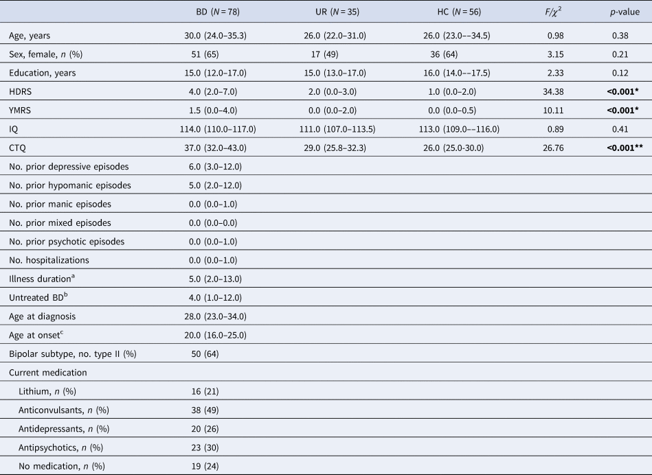
Participants were well-matched with respect to age, sex, years of education, and IQ (ps ⩾ 0.12). Although the burden of symptoms in this sample was very low, patients had higher HDRS-17 and YMRS scores than their URs and HCs (all ps ⩽ 0.002). Patients with BD reported significantly more childhood trauma (CTQ total score) compared to HC (p < 0.001), where URs reported intermediate levels of childhood trauma that significantly differed from their affected proband and HC (p < 0.001 and p = 0.04, respectively) (Table 1).
Demographic and clinical information patients with BD, their URs, and HCs

HAMD, Hamilton Depression Rating Scale; YMRS, Young Mania Rating Scale; CTQ, Childhood Trauma Questionnaire.
Continuous variables are presented as medians (interquartile range). Categorical variables are presented as n (%).
a Defined as difference between first hypomanic, manic, or mixed episode and time of study participation.
b Defined as interval between first (hypo)manic episode and diagnosis of BD.
c Age of patient at time of first hypomanic, manic, or mixed episode.
*HC < BD; UR < BD.
**HC < UR < BD.
Functional magnetic resonance imaging results
Emotion regulation
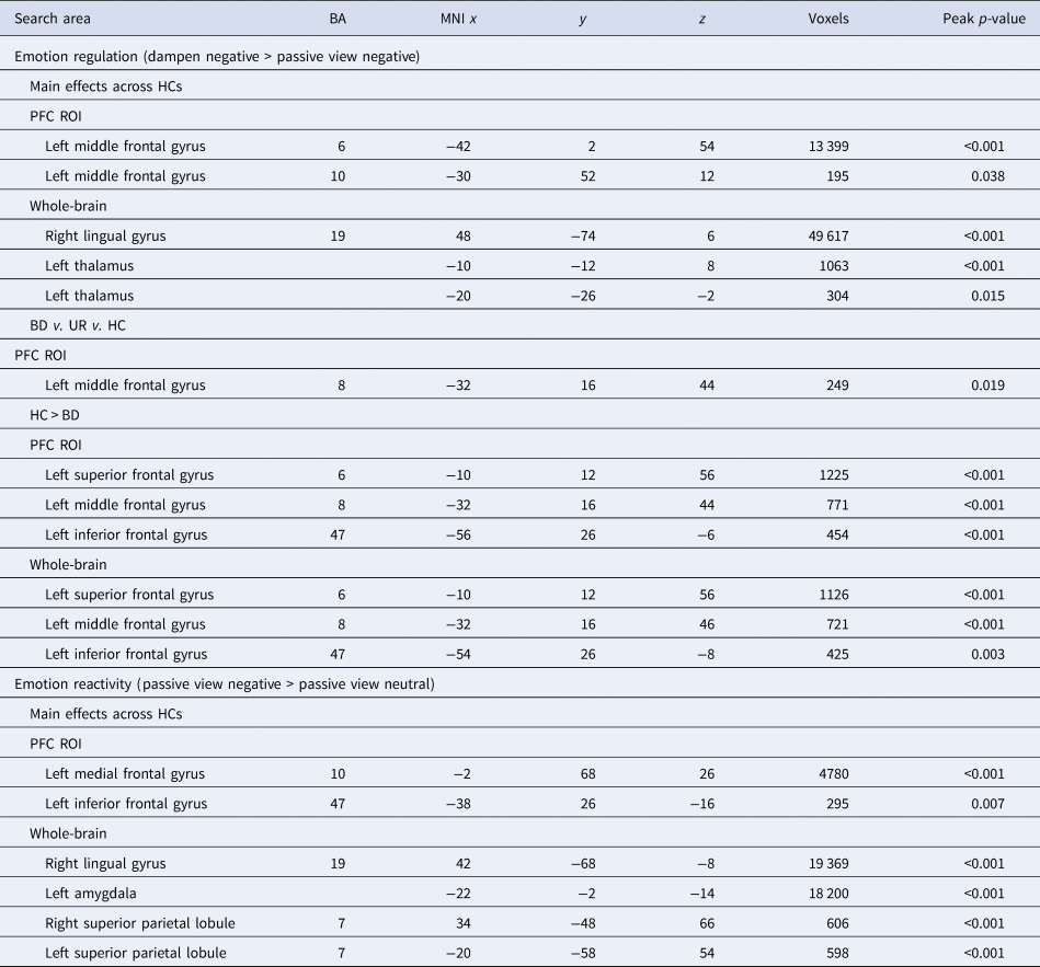
Main effect of task in healthy control participants: Emotion regulation (i.e. dampen negative > passive view negative contrast) activated the left supplemental motor area (BA6) and the DMPFC (BA10) within the PFC region of interest (ROI) (Table 2). Whole-brain analysis revealed that emotion regulation also activated the right occipital lingual gyrus (BA19) and the left thalamus (Table 2).
Main effects of task and group on brain activation in patients with BD, their URs, and HCs during emotion regulation and reactivity

Group comparisons of neural response during emotion regulation
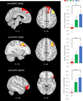
PFC analyses: We identified a main effect of group difference in a DLPFC (BA8) cluster in the left middle frontal gyrus (Table 2, Fig. 1). Pairwise tests revealed that compared to HCs, patients with BD had significantly lower activation in three clusters, located in the left DMPFC (BA6), left DLPFC (BA8), and the left VLPFC (BA47) (Table 2, Fig. 1). Relatives displayed intermediate levels of activity in these regions that did not differ significantly from the BD or HC groups. In post-hoc group level FEAT analyses covaried for subsyndromal depression and mania symptoms, the significant main effect of group disappeared but pairwise comparisons of the three groups showed that patients with BD had significantly lower activation in the left DMPFC (BA6) and the bilateral DLPFC (BA8/6) compared to HCs (Table S3).
Group comparisons of neural response during emotion regulation. Left: Decreased activity in left DMPFC, DLPFC, and VLPFC during emotion regulation of aversive images (dampen negative > passive view negative contrast) in patients with BD v. HCs (marked in red) and significant F-test in left DLPFC (marked in yellow). Right: Mean percent signal change within the left DMPFC, DLPFC, and VLPFC in patients with BD, their URs, and HCs; patients with BD exhibited significantly lower BOLD signal change during emotion regulation in the left DMPFC, DLPFC, and VLPFC compared to HCs. BOLD signal change in left DMPFC, DLPFC, and VLPFC did not significantly differ between URs and HCs. Error bars represent standard error of the mean. **p < 0.01. DMPFC, dorsomedial prefrontal cortex; DLPFC, dorsolateral prefrontal cortex; VLPFC, ventrolateral prefrontal cortex; BD, bipolar disorder; UR, unaffected first-degree relatives; HC, healthy controls.

We still found a significant main effect of group in the DLPFC (BA8), as well as lower BOLD signal in the left DLPFC (BA 8) in patients with BD compared to HC, after controlling for childhood trauma (p = 0.045 and p = 0.04, respectively). However, the reduced BOLD signal in the left DMPFC and VLPFC in BD v. HC rendered non-significant after adjusting for childhood trauma (ps ⩾ 0.10).
The reduced BOLD signal in the left DMPFC, DLPFC, and VLPFC in patients with BD compared to HCs remained significant after excluding patients taking antidepressants (ps ⩽ 0.004), antipsychotics (ps ⩽ 0.019), lithium (ps ⩽ 0.001), and anticonvulsants (ps ⩽ 0.016) with the exception of the left DMPFC which was reduced to a trend (p = 0.057) when excluding patients taking anticonvulsants. We still found reduced BOLD signal in the left DMPFC, DLPFC, and VLPFC (ps ⩽ 0.01) in BD v. HC after controlling for medication status (antidepressants, antipsychotics, lithium, anticonvulsants) as covariates in the model, and when the analyses were conducted after limiting the BD sample to non-medicated patients only v. HC (ps ⩽ 0.003), suggesting that medication did not change the brain reactivity.
Whole-brain analyses: The whole-brain analysis revealed no significant main effect of group difference (Table 2). Exploratory post-hoc pairwise comparisons showed lower activity in patients with BD than HCs in the left DMPFC (BA6), DLPFC (BA8) and VLPFC (BA47), with the URs exhibiting intermediate levels of activity that did not differ from BD or HC (Table 2). Post-hoc group-level analyses adjusting for symptom severity revealed no significant main effect of group, but the observed differences between BD and HC prevailed in the pairwise comparisons with highly overlapping regions (Table S3).
Emotion reactivity
Main effect of task in healthy control participants: Emotion reactivity (i.e. passive view negative > passive view neutral contrast) activated the left frontopolar cortex (BA10) and the left VLPFC (BA47) within the PFC ROI, and – as demonstrated in the whole-brain analysis – the right occipital lingual gyrus (BA19), left amygdala, and the bilateral superior parietal lobule (BA7) (Table 2).
Group comparisons of neural response during emotion reactivity: There were no group differences during emotion reactivity (passive view negative > passive view neutral) in the PFC ROI or in the exploratory whole-brain analysis. Given the absence of behavioral and brain activation evidence for a group effect, no further analyses on reactivity were undertaken.
Functional connectivity from the amygdala
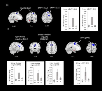
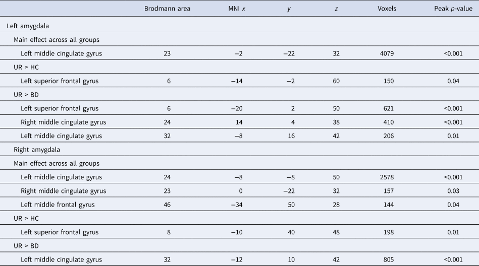
Across all groups, there was a negative coupling during dampening of emotions between both left and right amygdalae seed regions and an extensive dorsomedial region including the bilateral middle cingulate gyri (BA23/24) and the left superior frontal gyrus (BA8). Although there was no overall difference across the three groups in functional coupling, exploratory pairwise comparisons revealed a more negative functional coupling (i.e. stronger anti-correlations of activity over time) during dampening between the bilateral amygdalae and two DMPFC clusters in the left superior frontal gyrus (BA6/BA8) in URs compared to HCs during (Table 3, Fig. 2). URs also exhibited a more negative functional coupling than patients with BD during dampening between the right amygdala and a clusters in the left middle cingulate gyrus (BA32) that extended up to the DMPFC, as well as between left amygdala and three clusters spanning the bilateral middle cingulate gyri (BA24/BA32) and a DLPFC (BA6) cluster in the left superior frontal gyrus (Table 3, Fig. 2). There were no significant differences in functional coupling between patients with BD and HCs.
Functional connectivity analysis between the right and left amygdalae and prefrontal cortex during emotion regulation. When dampening emotions, (a) URs showed increased negative connectivity of the bilateral amygdalae (red) with two clusters in the DMPFC (blue) compared to HCs; and (b) URs showed increased negative connectivity of the left amygdala (red) with the bilateral middle cingulate and the DLPFC (blue) and the right amygdala (red) with the left middle cingulate (blue) compared to patients with BD. Error bars represent standard error of the mean.

Negative functional connectivity with left and right amygdalae during emotion regulation in patients with BD, their URs, and HCs

Note. Group differences in the PPI analyses could also be interpreted from a positive connectivity point of view.
Behavioral ratings of in-scanner emotion regulation and reactivity
For emotion regulation during fMRI, there was a statistically significant effect of group (F (2,104.5) = 3.63, p = 0.03), even after adjusting for HDRS and YMRS scores (p = 0.050). Follow-up independent samples t tests revealed that patients with BD and their URs were less successful at down-regulating their emotional response to aversive images compared to HCs (BD v. HC: t = 2.51, df = 132, p = 0.01; UR v. HC: t = 2.27, df = 88, p = 0.03) (Fig. S1). There were no significant differences between groups on emotional reactivity to aversive images (p = 0.21).
Distancing was the preferred strategy in all participants for down-regulating their response to the unpleasant images (see online supplement for details). Patients with BD were less likely to employ this strategy compared to HCs (p = 0.01).
Associations between BOLD fMRI, functional connectivity, emotion ratings, childhood trauma, and subsyndromal symptoms
Across the entire sample, childhood trauma was significantly associated with more hypo-activity in the left VLPFC during emotion down-regulation (r = −0.22, FDR-adjusted p = 0.048) (but not left DMPFC, left DLPFC, or amygdala-PFC functional connectivity: FDR adjusted p ⩾ 0.09). In contrast, emotion ratings or subsyndromal symptoms did not significantly correlate with BOLD response in the DMPFC, DLPFC and VLPFC (FDR adjusted p ⩾ 0.052) or amygdala-PFC functional connectivity (FDR adjusted p ⩾ 0.15) after adjusting for multiple comparisons (see Tables S4 and S5). For uncorrected p-values and post-hoc exploratory within-group correlation analyses, see the online supplementary material.
Linear regression analysis assessing whether the association between diagnosis and childhood trauma modified activity in PFC, revealed a model explaining a significant proportion of the variance in PFC activity during emotion down-regulation [adjusted R 2 = 0.06; F (3, 122) = 3.62, p = 0.02]. However, the group by childhood interaction did not significantly predict hypoactivity in the PFC during emotion down-regulation (p = 0.85). Linear regression analysis assessing whether CTQ and PFC activation during emotion-regulation moderated depressive symptom severity revealed that the model explained a significant proportion of the variance in depressive symptom severity [adjusted R 2 = 0.30, F (4, 121) = 14.15, p 0.001]. Childhood trauma significantly predicted depressive symptoms [β = 0.07, 95% CI (0.00–0.13), p = 0.046], but neither hypo-activity in the PFC nor the PFC activity by group interaction predicted depressive symptoms (ps < 0.71).
Discussion
This is the largest fMRI study to date that investigated the neural underpinnings of voluntary emotion regulation in 78 remitted patients recently diagnosed with BD, 35 URs and 56 HCs. Consistent with our hypothesis I, patients with BD exhibited hypo-activity in the left DMPFC, DLPFC, and VLPFC during down-regulation of unpleasant emotions compared to HCs, with URs displaying intermediate levels of activity that did not differ from either group. Importantly, hypo-activity during down-regulation prevailed in post-hoc analyses co-varied for mood symptoms and when excluding each medication group of patients. However, exploratory pair-wise comparisons revealed that URs exhibited more negative amygdala- DMPFC coupling than HCs and more negative amygdala-DLPFC-cingulate connectivity than BD patients. Patients with BD showed intermediate functional connectivity levels that did not differ from HCs (hypothesis II). However, we found no differences between patients with BD, their URs, and HCs in neural activity during negative emotional reactivity. At the behavioral level, patients with BD and their URs were less successful at down-regulating their negative emotional response to aversive images compared to HCs – also after adjusting for subsyndromal mood symptoms (hypothesis III).
The hypo-activity in dorsal and ventral PFC in recently diagnosed remitted patients with BD during emotion regulation is in accordance with prior research on patients with BD at later illness stages. Indeed, a majority of studies have identified aberrant activity in the DLPFC and VLPFC, although the direction of these abnormalities differ between studies (Morris et al., Reference Morris, Sparks, Mitchell, Weickert and Green2012; Rive et al., Reference Rive, Mocking, Koeter, van Wingen, de Wit, van den Heuvel and Schene2015; Sankar et al., Reference Sankar, Purves, Colic, Lippard, Millard, Fan and Constable2020; Townsend & Altshuler, Reference Townsend and Altshuler2012; Townsend et al., Reference Townsend, Torrisi, Lieberman, Sugar, Bookheimer and Altshuler2013; Zhang et al., Reference Zhang, Ai, Opmeer, Marsman, van der Meer, Ruhé and Van Tol2020). The DLPFC and VLPFC are implicated in voluntary emotion regulation; specifically, the DLPFC/VLPFC is thought to be involved in the effortful inhibition of the amygdala, mediated by feedback from the ventromedial PFC (Phillips et al., Reference Phillips, Ladouceur and Drevets2008). Our study provides further evidence for the involvement of the VLPFC in BD, another region known to be involved in contextual response selection across a number of tasks (Chen, Suckling, Lennox, Ooi, & Bullmore, Reference Chen, Suckling, Lennox, Ooi and Bullmore2011; Delvecchio, Sugranyes, & Frangou, Reference Delvecchio, Sugranyes and Frangou2013). The present demonstration of hypo-activity in these regions therefore points to trait-related deficits in top-down regulation already at early in the course of the illness.
Relatives were comparable to HCs in their neural activity during emotion regulation despite their poorer ability to down-regulate negative emotions to the aversive images. Although these behavioral data are in line with previous reports, the brain imaging findings differ from two previous studies that found lower prefrontal (including medial frontal gyrus, posterior dorsal anterior cingulate cortex, and the left superior frontal gyrus) (Meluken et al., Reference Meluken, Ottesen, Phan, Goldin, Di Simplicio, Macoveanu and Miskowiak2018) and increased amygdala (Kanske et al., Reference Kanske, Schönfelder, Forneck and Wessa2015) activity during reappraisal of aversive images. This divergence may be due to the use of different emotion regulation strategies. Specifically, participants in both previous studies were explicitly instructed to use cognitive reappraisal to down-regulate their emotional response to unpleasant images (i.e. reinterpret the meaning of the pictures, e.g. thinking of the people in the aversive images as actors). In contrast, participants in our study were allowed to use their habitual responses. As a result, they reported using distancing (i.e. viewing the aversive image from a detached perspective) more frequently than cognitive reappraisal. In keeping with this, prior studies identified no significant differences between URs and HCs when they were instructed to use distraction (Kanske et al., Reference Kanske, Schönfelder, Forneck and Wessa2015) or mental imagery (Meluken et al., Reference Meluken, Ottesen, Phan, Goldin, Di Simplicio, Macoveanu and Miskowiak2018) instead of reappraisal.
The lack of differences between patients with BD and HCs in functional coupling between the right amygdala and prefrontal areas was unexpected. In fact, previous studies found that patients with BD fail to exhibit a normal negative amygdala-PFC coupling during emotion regulation to aversive images (Kanske et al., Reference Kanske, Schönfelder, Forneck and Wessa2015; Morris et al., Reference Morris, Sparks, Mitchell, Weickert and Green2012; Townsend et al., Reference Townsend, Torrisi, Lieberman, Sugar, Bookheimer and Altshuler2013) that has been interpreted as inefficient top-down regulation of limbic responsivity. Our results, on the other hand, suggest that patients who are recently diagnosed with BD have intact fronto-amygdalar connectivity, but that hypoactivity in the DMPFC, DLPFC, and VLPFC prevents patients from successfully recruiting prefrontal areas essential for successful emotion regulation. Furthermore, given that fronto-amygdalar functional connectivity was normal in recently diagnosed patients in the current study, but previously found to be abnormal in BD patients at later illness stages (Phillips et al., Reference Phillips, Ladouceur and Drevets2008; Townsend & Altshuler, Reference Townsend and Altshuler2012), this difference may reflect illness-related ‘scarring’, whereby fronto-amygdalar functional connectivity during emotion regulation deteriorates with repeated mood episodes (Goodwin, Martinez-Aran, Glahn, & Vieta, Reference Goodwin, Martinez-Aran, Glahn and Vieta2008). Although this is in line with a neuroprogressive account of neural changes over time, we cannot exclude the possibility of a neurodevelopmental origin of emotion dysregulation in BD given the cross-sectional study design. Indeed, studies investigating children and adolescents with BD have also found evidence of aberrant fronto-amygdalar connectivity during processing of negative faces (e.g. Ladouceur et al., Reference Ladouceur, Farchione, Diwadkar, Pruitt, Radwan, Axelson and Phillips2011; Passarotti, Ellis, Wegbreit, Stevens, & Pavuluri, Reference Passarotti, Ellis, Wegbreit, Stevens and Pavuluri2012), suggesting that neurodevelopmental abnormalities in fronto-amygdalar connectivity could play a role in the pathophysiology of BD.
Our demonstration of greater negative amygdala-PFC functional connectivity in URs compared to HCs contrasts with the study of Kanske and colleagues (Kanske et al., Reference Kanske, Schönfelder, Forneck and Wessa2015) who reported positive amygdala-PFC connectivity in URs compared to HCs during emotion regulation. We note that the URs in our sample were younger than URs in the sample by Kanske et al. (mean age: 27 ± 6 years v. 37 ± 13 years, respectively). Since BD onset typically occurs in the mid-20s (Baldessarini et al., Reference Baldessarini, Tondo, Vázquez, Undurraga, Bolzani, Yildiz and Lolich2012), it is possible that the current sample had not passed all age-related risk periods and therefore had a greater projected lifetime risk of mood disorder compared to UR samples in previous studies. This, coupled with BD patients and URs being less successful than HCs at down-regulating their emotions at the behavioral level, would suggest that the enhanced negative fronto-amygdalar coupling in our UR sample may reflect a risk-marker of BD (Wiggins et al., Reference Wiggins, Brotman, Adleman, Kim, Wambach, Reynolds and Leibenluft2017). Given that the ‘normative’ negative fronto-amygdalar connectivity was enhanced in URs, one might argue that our findings reflect a compensatory mechanism of resilience. Nevertheless, URs in this study were still less successful than HCs at down-regulating their unpleasant emotions, suggesting that their greater negative fronto-amygdalar functional connectivity reflected unsuccessful compensatory attempts to down-regulate unpleasant emotions. Whether increased negative fronto-amygdalar connectivity represents a risk marker (i.e. predicting illness onset in URs) or a resilience marker (i.e. relatives remain unaffected) will be examined in our ongoing longitudinal part of the cohort study (Kessing et al., Reference Kessing, Munkholm, Faurholt-Jepsen, Miskowiak, Nielsen, Frikke-Schmidt and Vinberg2017).
Our finding that childhood trauma was more prevalent in patients and their URs than in HCs is in accordance with previous studies suggesting an association between childhood trauma and BD susceptibility and severity (Aas et al., Reference Aas, Henry, Andreassen, Bellivier, Melle and Etain2016; Coello, Munkholm, Nielsen, Vinberg, & Kessing, Reference Coello, Munkholm, Nielsen, Vinberg and Kessing2019; Ottesen et al., Reference Ottesen, Meluken, Scheike, Kessing, Miskowiak and Vinberg2018). Interestingly, childhood trauma was associated with more hypo-activity in the left VLPFC during down-regulation of emotions to aversive images (as seen in patients with BD). However, post-hoc regression analyses suggested that childhood trauma did not influence the association between diagnosis and PFC hypoactivity, suggesting that patients – whether with or without childhood maltreatment – display PFC hypo-activity during emotion down-regulation. Childhood trauma in BD has previously been associated with affective lability (Aas et al., Reference Aas, Henry, Bellivier, Lajnef, Gard, Kahn and Leboyer2017) and negative effects on the limbic network, including decreased amygdala volume and frontolimbic coupling (Souza-Queiroz et al., Reference Souza-Queiroz, Boisgontier, Etain, Poupon, Duclap, d'Albis and Delavest2016). Taken together, this may indicate that emotion dysregulation moderates the relationship between childhood trauma and adverse clinical outcomes. Specifically, traumatic experiences during childhood may contribute to development of maladaptive coping mechanisms and less effective down-regulate aversive emotions, which, at the neural level, may be exhibited by failure to recruit higher-order prefrontal resources for top-down control of emotions. Such difficulties with emotion regulation may again lead to more stress vulnerability thereby increased risk of recurrent mood episodes and worsening clinical course of BD (Daruy-Filho, Brietzke, Lafer, & Grassi-Oliveira, Reference Daruy-Filho, Brietzke, Lafer and Grassi-Oliveira2011; Dodd, Lockwood, Mansell, & Palmier-Claus, Reference Dodd, Lockwood, Mansell and Palmier-Claus2019). However, we found no moderating effect of PFC hypoactivity on the association between childhood trauma and depressive symptoms in our analyses. This may suggest that while childhood maltreatment is indeed associated with more depressive symptoms, this is not affected by participants' ability to activate their DMPFC, DLPFC, and VLPFC when required to down-regulate.
Strengths of the study included the large sample of well-defined patients with recently diagnosed BD, URs, and HCs. Also, URs and HCs were free of any psychiatric illness and psychotropics, thereby enabling direct comparisons between these two groups to elucidate risk markers that are specific to BD without the confounding effect of psychopathology and medication. Indeed, ‘asymmetric screening’ (i.e. stricter inclusion criteria for HCs than URs) would likely results in overestimated between-group differences (Snitz, Reference Snitz2005). Given that the UR sample was generally young (years of age, mean ± s.d.: 27.2 ± 6.4), they may in the future have onset of psychiatric illness. A limitation was that, although patients were recently diagnosed with BD, their average delay in diagnosis was 4 years. This reflects the difficulties in recruiting patients in the early course of the illness as the average delay between onset and diagnosis in BD is 5–10 years (Baldessarini et al., Reference Baldessarini, Tondo, Baethge, Lepri and Bratti2007; Fritz et al., Reference Fritz, Russell, Allwang, Kuiper, Lampe and Malhi2017). Patients also had to reach clinical remission before being included in the study, further adding to the delay. Hence, we cannot exclude the possibility that the current findings are a result of pathology related to clinical progression. Nevertheless, the patients in the current study had a substantially shorter illness duration compared to patients in other studies (mean 8.7 years in the current study v. 18.0 years in previous studies). Notably, the median age of patients in the study was 30 (interquartile range: 24, 35) where only one patient was over the age of 60 and one patient over the age of 50. This was done intentionally as we wanted our patient sample to reflect the true heterogeneity of the disorder, thereby increasing the generalizability of our study results to a larger BD population. Including participants with late onset BD may reflect a different phenotype of BD. Nevertheless, group differences prevailed also after excluding participants ⩾50 years of age. The relatively small UR sample hampered comparison of functional connectivity between URs with and without subsyndromal symptoms and emotional regulation difficulties to clarify whether enhanced negative functional amygdala-PFC connectivity reflect resilience or risk. A limitation of PPI functional connectivity analysis is that it cannot be used for causal inferences about the direction of the relationship (O'Reilly, Woolrich, Behrens, Smith, & Johansen-Berg, Reference O'Reilly, Woolrich, Behrens, Smith and Johansen-Berg2012).
This first large fMRI study in recently diagnosed BD patients and their URs revealed evidence that unsuccessful down-regulation of negative emotion in response to unpleasant visual stimuli is a familial dimension of BD. Our findings further point to deficient prefrontal top-down regulation already at BD illness onset. The findings warrant prospective follow-up of our BD cohort and URs to clarify whether the observed PFC hypo-activity and enhanced negative functional coupling during regulation of unpleasant emotions are predictive of an adverse illness course and risk of onset, respectively.
Supplementary material
The supplementary material for this article can be found at https://doi.org/10.1017/S0033291721002737.
Acknowledgements
KWM holds a 5-year Lundbeck Foundation Fellowship (R215-2015-4121).
Financial support
The study is funded by grants from the Mental Health Services, Capital Region of Denmark, The Danish Council for Independent Research, Medical Sciences (DFF-4183-00570), Markedsmodningsfonden (the Market Development Fund 2015-310), Gangstedfonden (A29594), Helsefonden (16-B-0063), Innovation Fund Denmark (the Innovation Fund, Denmark, 5164-00001B), Copenhagen Center for Health Technology (CACHET), EU H2020 ITN (EU project 722561), and Augustinusfonden (16-0083).
Conflict of interest
MV has received consultancy fees from Lundbeck in the past 3 years. LVK has within the preceding 3 years been a consultant for Lundbeck. KWM has received consultancy fees from Lundbeck and Janssen-Cilag in the past 3 years. The remaining authors declare no conflicts of interest.