Tetralogy of Fallot with alleged absence of the pulmonary valve is characterised by absence or presence of only a rudimentary ridge of pulmonary valve tissue, resulting in severe pulmonary regurgitation with subsequent aneurysmal dilation of the pulmonary trunk and the right and left pulmonary arteries.Reference Roche, Greenway and Redington 1 Severe respiratory problems due to bronchial obstruction by dilated pulmonary arteries frequently occur. We describe a case of a neonate who developed obstruction of the superior caval vein by an aneurysmal right pulmonary artery with absent pulmonary valve.
Case report
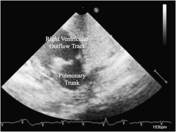
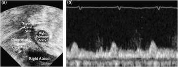
In our male patient, double outlet right ventricle, tetralogy of Fallot type, with absent pulmonary valve was diagnosed prenatally. After birth, the baby was immediately intubated for ventilatory assistance. Post-natal two-dimensional echocardiography revealed a relatively large ridge at the level where the pulmonary valve leaflets would be expected (Fig 1). The pulmonary trunk and right and left pulmonary arteries were extraordinarily enlarged – diameter of pulmonary trunk was 17 mm and of right and left pulmonary arteries was 12 mm. The superior caval vein was compressed by an aneurysmal right pulmonary artery, and the upstream superior caval vein distal to the obstruction was dilated (Fig 2a). Doppler echocardiogram of the blood flow from the superior caval vein to the right atrium demonstrated an uncharacteristic pulsatile pattern due to pulsation of the adjacent aneurysmal right pulmonary artery (Fig 2b). Because of severe hypoxia, partial pressure of oxygen in the arterial blood <30 Torr, despite appropriate ventilatory support and persistent facial oedema, surgical intervention was planned on day 1. We opted for a palliative approach – ligation of the pulmonary trunk and placement of a modified Blalock–Taussig shunt – because of the patient’s severe hypoxia, systemic oedema, anuria, and relatively low birth weight (2530 g). He was extubated 33 days postoperatively and was discharged 73 days after surgery because of neurological complications – agenesis of the corpus callosum.
Echocardiogram of the right ventricular outflow tract demonstrates the large rudimentary ridge.

( a ) Echocardiogram demonstrates that the superior caval vein is compressed by aneurysmal right pulmonary artery and the distal portion of the superior vena cava is dilated. ( b ) Pulse Doppler echocardiogram of blood flow from superior caval to right atrium. It demonstrates diastolic dominant pulsatile pattern, which is influenced by pulsation of the adjacent right pulmonary artery.

Discussion
In patients with tetralogy of Fallot with absent pulmonary valve syndrome, the pulmonary trunk and right and left pulmonary arteries dilate because of severe pulmonary regurgitation. The aneurysmal pulmonary arteries can potentially compress the tracheobronchial tree and this compression can result in severe tracheobronchial obstruction. It has also been reported that the dilated pulmonary artery can obstruct the left coronary artery.Reference Sengupta, Saxena and Rajani 2 In our patient, the unique feature is the compression of the superior caval vein by the aneurysmally dilated right pulmonary artery. This abnormality has not been reported. We speculate that the relatively large rudimentary ridge at the level of pulmonary valve leaflets may have contributed to the severity of the dilation of the pulmonary artery (Fig 1). This syndrome is named “absent” pulmonary valve; however, the pulmonary trunk usually has a small ridge at the ventriculoarterial junction. In our case, the ridge was large and stenotic as well as regurgitant. Therefore, it augmented the turbulent flow in the pulmonary arterial tree. As a result, the pulmonary trunk and the right and left pulmonary arteries were extraordinary dilated and compressed, not only the tracheobronchial tree, but also the superior caval vein.
Acknowledgement
None.
Financial Support
This research received no specific grant from any funding agency, commercial or not-for-profit sectors.
Conflicts of Interest
None.