In the field of developmental psychopathology, behaviors and symptoms experienced by children have their origins in disruptions to the biology of the brain. In order to improve the diagnosis and treatment of psychiatric disorders in children, we must understand not only the neurobiology associated with emotional and behavioral problems as they arise but also the developmental origins of any neurobiological abnormalities. Identifying the developmental origins of psychopathology will improve our understanding of the underlying mechanisms, because the symptomatology that arises is often a consequence of disruptions in achieving common biological milestones. For all forms of mental illness, but particularly those that emerge during the dynamic developmental periods of infancy and childhood, early events in the development of neuronal cells and the subsequent changes to the overall layout of central nervous system connectivity are of particular interest. Embryonic and early postnatal developmental most certainly contribute substantially to the origins of childhood psychiatric illness; converging evidence for this principle for disorders such as autism, Tourette syndrome, and mood disorders comes from studies implicating genes with significant roles during early phases of neurogenesis and synaptic formation and work showing neurobiological risk phenotypes prior to the onset of any illness (Sowell et al., Reference Sowell, Kan, Yoshii, Thompson, Bansal and Xu2008; Stevens, Smith, Rash, & Vaccarino, Reference Stevens, Smith, Rash and Vaccarino2010).
As interesting as the events of early human growth and development are, obtaining precise information about them has been off limits to our technical ability to investigate the human brain. Studies of cellular and molecular changes that occur during development in nonhuman animals have elucidated many components of normal brain development. Human neuroscience has not been able to make the same gains: electrophysiology and neuroimaging have spatial and temporal resolution limitations and postmortem analysis of human brain is limited by variability from multiple sources, a small supply of well-characterized human brain tissue, and inherent difficulties in manipulating gene and protein expression in postmortem material.
However, new approaches have emerged that make it possible to examine both human neurodevelopment in vitro and the genetic and epigenetic factors dynamically controlling this process. We can deprogram fibroblasts obtained from a skin biopsy of any individual into human-induced pluripotent stem cells (hiPSCs) that closely resemble multipotent stem cells isolated from human embryos (Takahashi et al., Reference Takahashi, Tanabe, Ohnuki, Narita, Ichisaka and Tomoda2007; Vaccarino et al., Reference Yu, Vodyanik, Smuga-Otto, Antosiewicz-Bourget, Frane and Tian2011; Yu et al., Reference Yu, Vodyanik, Smuga-Otto, Antosiewicz-Bourget, Frane and Tian2007). The hiPSCs can be differentiated into neuronal progenitors and more mature neurons in vitro, allowing the study of neuronal development and its possible alterations in the human population, including patients with neuropsychiatric disorders. Because hiPSCs retain the unique genetic signature of the individual from which they are derived, they can be used to understand the cellular and molecular consequences of individual genetic abnormalities, as well as how gene expression across an individual's entire genome at different stages may be related to disease risk (Vaccarino, Stevens, et al., Reference Vaccarino, Stevens, Kocabas, Palejev, Szekely and Grigorenko2011).
Induced pluripotent stem cells (iPSCs) are important not only for understanding human development and looking directly at the neurobiology associated with some psychiatric disorders but also for advancing research on the genetic basis of developmental psychopathology. There are at least three areas where the hiPSC technology can deliver crucial new understanding in human genetics and psychopathology. First, it can elucidate the relationships between specific gene mutations/variants and epigenetic modification as well as changes in the expression of that gene in developing neural cells. One can also elucidate relationships between gene variants and genome-wide gene expression, and thus advance our understanding of gene–gene interactions (see the Integrative Analyses Using the hiPSC Model Section). Second, hiPSC can help understand the effect of perturbing gene expression on the typical development of human neural cells. This is the first time that gene structure and gene expression can be related to the biology of brain development. Third, this unique model can potentially elucidate the relationships between gene splice variants uniquely expressed in human brain development and psychopathology. As genomic studies uncover risk genes and single gene etiologies, hiPSCs can provide an appropriate context for further developments in genomic research by which we can understand how these genes affect the expression of other genes in an individual's genome, as well as their neuronal cells, multicellular systems and, perhaps, the functioning of the brain across development. The in vitro study of human neurodevelopment has the potential to reveal specific roles for known and unknown genes and their structural variants at different time points and in different neuronal precursors. At present, much of our conclusions about findings from genome-wide association studies and of rare variants comes from our knowledge about nonhuman biological systems. In addition, much of our background knowledge driving the examination of neurobiological human samples is driven by this same knowledge about nonhuman biological systems. New insights about human neurodevelopment and its unique features can come from integrating known genes of importance from clinical populations with a framework for these genes' functioning taken from nonhuman model systems and utilizing these combined data to examine hiSPCs.
The hiPSC Approach
The notion that studying development as it happens is useful for understanding the human brain is not a new one. Stem cells derived from embryos were historically used to understand this process in vitro because these cells have the ability to develop into all of the other cell types of the organism, specifically of interest here, the brain. During typical development and growth of an individual, the fate potential of cells becomes progressively restricted as cells become specialized into different subtypes. Embryonic stem cells are pluripotent, without fate restriction and therefore can be used to understand genes and developmental processes driving fate commitment. Work with these cell types, however, has been limited to only a few established stem cell lines due to ethical concerns. These few available lines were a poor representation of overall human genetic diversity and could not demonstrate the developmental events that might lead to psychiatric illness. Alternatives to embryonic stem cells are stem cells specific to brain regions of interest in psychiatric research, the so-called adult neural stem cells present in the subventricular and subgranular zones of the brain ventricles and hippocampus. These cells in children and adults demonstrate the universal principle of development in their fate restriction: they only produce cells of the olfactory bulb and dentate gyrus and have limited abilities to become neurons of different types, even upon transplantation into different central nervous system regions (Alvarez-Buylla, Herrera, & Wichterle, Reference Alvarez-Buylla, Herrera and Wichterle2000; Lim, Flames, Collado, & Herrera, Reference Lim, Flames, Collado and Herrera2002; Milosevic, Noctor, Martinez-Cerdeno, Kriegstein, & Goldman, Reference Milosevic, Noctor, Martinez-Cerdeno, Kriegstein and Goldman2008; Vaccarino, Ganat, Zhang, & Zheng, Reference Vaccarino, Ganat, Zhang and Zheng2001). These stem cells also appear to be limited in their capacity to divide, which restricts their ability to recapitulate the highly neurogenic period of development during embryogenesis.
By studying stem cells from multiple species, we have learned about their characteristics and neural fate potential. Stem cells from bone marrow and other mesenchymal tissues are thought to be able to produce neural stem cells when cultured in the right milieu containing important neuronal support cells like astrocytes (Fricker-Gates & Gates, Reference Fricker-Gates and Gates2010; Jiang et al., Reference Jiang, Henderson, Blackstad, Chen, Miller and Verfaillie2003; Trzaska et al., Reference Trzaska, King, Li, Kuzhikandathil, Nowycky and Ye2009) or when induced to express genes important for differentiation and maintenance of neural precursors (Dezawa et al., Reference Dezawa, Kanno, Hoshino, Cho, Matsumoto and Itokazu2004). Although the capacity of these cells to divide and recapitulate early development is limited, they have been used in mice and other species as sources of factors that stimulate neuronal repair in neurodegenerative and inflammatory conditions (Constantin et al., Reference Constantin, Marconi, Rossi, Angiari, Calderan and Anghileri2009; Kranz et al., Reference Kranz, Wagner, Kamprad, Scholz, Schmidt and Nitzsche2010; Lee et al., Reference Lee, Lee, Lee, Carter, Chang and Oh2012; Momin, Mohyeldin, Zaidi, Vela, & Quinones-Hinojosa, Reference Momin, Mohyeldin, Zaidi, Vela and Quinones-Hinojosa2010; Wang et al., Reference Wang, Yasuhara, Shingo, Kameda, Tajiri and Yuan2010; Xia et al., Reference Xia, Hong, Chen, Lan, Zhang and Liao2010).
The restriction in cell division and fate potential that occurs in all human cells through development is realized through regulation of gene expression by transcription factors that bind and activate specific promoters as well as through epigenetic modifications on the chromatin and the genomic DNA sequence itself. The epigenetic processes that determine fate restriction were thought to be irreversible until recently. Two major advances in the past 15 years have pushed the technology of stem cell research to a new level, which is past the limitation that stem cells only produced a handful of cell lineages or cell growth mediators. First, in 1997, a sheep named “Dolly” was the first animal cloned from an adult somatic cell (Wilmut, Schnieke, McWhir, Kind, & Campbell, Reference Wilmut, Schnieke, McWhir, Kind and Campbell1997). Second, in 2007, capitalizing on prior research showing that egg cells have the potential to reprogram the chromatin of differentiated nuclei (Gurdon, Reference Gurdon1987, Reference Gurdon1988), Takahashi et al. (Reference Takahashi, Tanabe, Ohnuki, Narita, Ichisaka and Tomoda2007) discovered that four gene products encoding transcription factors (Oct4, c-Myc, Sox2, and Klf4) were sufficient to reprogram somatic cells into virtually immortal stem-cell-like cells called iPSCs. Other articles independently reported similar findings (Meissner, Wernig, & Jaenisch, Reference Meissner, Wernig and Jaenisch2007; Wernig et al., Reference Wernig, Meissner, Foreman, Brambrink, Ku and Hochedlinger2007; Yu et al., Reference Yu, Vodyanik, Smuga-Otto, Antosiewicz-Bourget, Frane and Tian2007) and determined that these iPSCs were remarkably similar to embryo-derived stem cells with respect to gene expression profile, epigenetic marks, and fate potential (Mikkelsen et al., Reference Mikkelsen, Ku, Jaffe, Issac, Lieberman and Giannoukos2007). The iPSCs can be derived from different types of somatic cells, including skin, hair follicle cells, and blood (Aasen et al., Reference Aasen, Raya, Barrero, Garreta, Consiglio and Gonzalez2008; Maherali & Hochedlinger, Reference Maherali and Hochedlinger2008; Meng et al., Reference Meng, Neises, Su, Payne, Ritter and Gridley2012; Staerk et al., Reference Staerk, Dawlaty, Gao, Maetzel, Hanna and Sommer2010). In recent years iPSCs, which are most commonly derived from skin fibroblasts, have been an intense focus of investigation and scrutiny. Despite significant efforts, their derivation is still inefficient (although recent methods seem more promising; Fusaki, Ban, Nishiyama, Saeki, & Hasegawa, Reference Fusaki, Ban, Nishiyama, Saeki and Hasegawa2009; Warren et al., Reference Warren, Manos, Ahfeldt, Loh, Li and Lau2010) and the characterization of “truly” reprogrammed cells versus those that are only partially reprogrammed or unstable has proven difficult. Recent derivation of iPSC using technologies that avoid permanent integration of foreign DNA into the genome are also rapidly evolving (Fusaki et al., Reference Fusaki, Ban, Nishiyama, Saeki and Hasegawa2009; Okita et al., Reference Okita, Matsumura, Sato, Okada, Morizane and Okamoto2011; Si-Tayeb et al., Reference Si-Tayeb, Noto, Sepac, Sedlic, Bosnjak and Lough2010), which are of particular importance for the therapeutic applications of these cells.
Deriving iPSCs from human beings (hiPSC) has advantages compared to embryonic and other types of stem cells for research involving human psychopathology: hiPSC are derived from a living person of any age and can be used for prospective studies as well as in diagnostics. Because hiPSC maintain one person's genetic constitution, the fact that these cells can be used to produce differentiated cells (e.g., pancreatic, cardiac, neural cells) genetically compatible with the person of origin is significant for potential cell replacement therapies.
Developmental Neuroscience and hiPSCs
Research in model systems such as mice can and has revealed significant information about common aspects of mammalian development. However, in the case of some organs and for the nervous system in particular, there are substantial differences between species. Gene expression in the brain has many layers of complexity, and, at the earliest stages of brain development, there is an especially pronounced divergence among species (Clowry, Molnar, & Rakic, Reference Clowry, Molnar and Rakic2010; Colantuoni et al., Reference Colantuoni, Lipska, Ye, Hyde, Tao and Leek2011; Geschwind, Reference Geschwind2011; Johnson et al., Reference Johnson, Kawasawa, Mason, Krsnik, Coppola and Bogdanovic2009; Kang et al., Reference Kang, Kawasawa, Cheng, Zhu, Xu and Li2011; Rakic, Reference Rakic2009). In simply looking at the structure of the mature brain, differences between mouse and humans are very apparent. For example, in humans as compared to lower mammalian species such as the mouse, the cerebral cortex is 1,000 times larger in surface area, has many more glial cells proportionally, and contains a more complex range of neuronal subtypes (Rakic, Reference Rakic1995). Upper cortical layers are much more developed in primates and humans than in mice. During development, the human cortex encompasses a much thicker subventricular zone layer, which is closely associated with blood vessels and that generates the larger number of neurons destined to populate superficial cortical layers (Anstrom et al., Reference Anstrom, Thore, Moody, Challa, Block and Brown2005; Kriegstein, Noctor, & Martinez-Cerdeno, Reference Kriegstein, Noctor and Martinez-Cerdeno2006; Martinez-Cerdeno, Noctor, & Kriegstein, Reference Martinez-Cerdeno, Noctor and Kriegstein2006). Cells of the subventricular zone comprise “outer radial glial cells,” a type of neuronal progenitor that is found at much higher frequency in humans than in mice, and it has been hypothesized to be crucial for the morphogenesis of the characteristic convolutions (gyri and sulci) of the primate cortex (Dehay & Kennedy, Reference Dehay and Kennedy2007; Lui, Hansen, & Kriegstein, Reference Lui, Hansen and Kriegstein2011; Molnar et al., Reference Molnar, Metin, Stoykova, Tarabykin, Price and Francis2006). How the human cortex generates gyri and sulci and how it becomes regionalized are matters of current debate among neurobiologists, because these processes are difficult to model in lower mammalian species. The neuronal cells found in mouse and human brains are different in their structure and organization and probably play a different role in the mature neural circuitry. Therefore, hiPSCs can demonstrate not only the precise series of events necessary for normal human brain development but also what aspects of development are uniquely human, shedding light on the neurodevelopmental origins of the expansion of the cortex and the underlying biology of many other behavioral features of human nature.
At the heart of what hiPSCs will be able to reveal about uniquely human biology are the genetics that are specific to the human brain. From the earliest time in brain development, the human brain diverges from animals such as mice, and these differences in biological structure and function are a result of gene expression differences. These differences arise from a number of aspects of genetic regulation and functioning. Different timing of the expression of common genes of early brain development can lead, for example, to a more prolonged neurogenic period in a human brain than in a mouse brain; different splicing of gene transcripts can produce not only distinct gene products but also more or fewer isoforms from a single gene's transcription; and, of course, there are some genes that are entirely unique to each species. Quantitative mapping of the genes and their level of transcription across brain regions at different time points of development is ongoing, promising a “developmental transcriptome” that can document some of these species differences (Kang et al., Reference Kang, Kawasawa, Cheng, Zhu, Xu and Li2011).
The use of hiPSCs may provide a clear focus for the integration of work on the developmental transcriptome in early human development, and on the precise molecular mechanisms being mapped out during early development in mouse. The hiPSC approach, basing investigations on known significant factors from the developmental transcriptome, can evaluate the dynamic molecular interactions occurring in early brain development much as has been done in mouse model systems for decades. With hiPSCs, the expression of the entire genome can be captured for specific cell types, just as they enter or exit specific developmental stages. As described in detail below, our ability to analyze not just single genes but whole genome gene expression at each point in time and/or type of cell in humans, combined with our knowledge from other sources, will give great insight into what components of the human genome are critical for neuroscience.
Applications of hiPSCs
A unique feature of the hiPSC model is that it permits the study of alterations in cellular and molecular pathways while maintaining the genomic structure of the individuals from which the cells originate. Some investigators have already made use of this feature of hiPSCs to study neurodevelopmental disorders such as Rett and Timothy syndromes and even psychiatric disorders with complex inheritance such as schizophrenia (Brennand et al., Reference Brennand, Simone, Jou, Gelboin-Burkhart, Tran and Sangar2011; Marchetto et al., Reference Marchetto, Carromeu, Acab, Yu, Yeo and Mu2010; Pasca et al., Reference Pasca, Portmann, Voineagu, Yazawa, Shcheglovitov and Pasca2011; Pedrosa et al., Reference Pedrosa, Sandler, Shah, Carroll, Chang and Rockowitz2011). The ability to study each individual patient allows for a more thorough understanding of the genetic heterogeneity of psychiatric disorders and how each disorder may still converge upon specific molecular pathways and a gene network whose function is altered during neurodevelopment. For example, in the study of autism spectrum disorders (ASDs), various mutations have been documented as an underlying cause of syndromes that include social deficits (Campbell, Li, Sutcliffe, Persico, & Levitt, Reference Campbell, Li, Sutcliffe, Persico and Levitt2008; Sbacchi, Acquadro, Calo, Cali, & Romano, Reference Sbacchi, Acquadro, Calo, Cali and Romano2010; Yaspan et al., Reference Yaspan, Bush, Torstenson, Ma, Pericak-Vance and Ritchie2011). Across many of these mutations, a few molecular pathways or gene networks are repeatedly represented, including those systems involved in synaptic development, implicated by the fragile X mutuation, and the Rett syndrome gene (MECP2), as well as the regulation of neural stem cells, implicated by PTEN and the TSC1 and TSC2 genes, which form the tuberous sclerosis complex (D'Hulst & Kooy, Reference D'Hulst and Kooy2009; Ehninger & Silva, Reference Ehninger and Silva2011; Shepherd & Katz, Reference Shepherd and Katz2011; Zhou & Parada, Reference Zhou and Parada2012). The convergence of these previous findings not only gives direction to investigations of hiPSCs in patients with autism, but it also demonstrates how hiPSC may provide data that can identify additional gene networks critical in autism. This approach is particularly revealing for patients that have not been shown to have an obvious single-gene mutation. In such groups of patients, genetic studies would be targeted at revealing a responsible genomic alteration. The hiSPC approach will look at a general sequence of the genome and, by looking at whole genome expression, will examine molecular pathways in action during early development and determine if these patient groups share alterations of the same gene networks.
For example, the gene pathways that mediate the increase in brain size found in children with ASDs are unknown. This phenotype was one of the first physical features described in patients with autism by Leo Kanner (Kanner & Eisenberg, Reference Kanner and Eisenberg1957). Since those initial reports, increased growth in head circumference has been shown to occur in about 20%–40% of patients with ASDs early in development, mainly in the first year of life (Chawarska et al., Reference Chawarska, Campbell, Chen, Shic, Klin and Chang2011; Courchesne, Carper, & Akshoomoff, Reference Courchesne, Carper and Akshoomoff2003; Courchesne, Redcay, & Kennedy, Reference Courchesne, Redcay and Kennedy2004; Davidovitch, Patterson, & Gartside, Reference Davidovitch, Patterson and Gartside1996; Miles, Hadden, Takahashi, & Hillman, Reference Miles, Hadden, Takahashi and Hillman2000; Woodhouse et al., Reference Woodhouse, Bailey, Rutter, Bolton, Baird and Le Couteur1996). An enlargement of underlying brain volume was confirmed by postmortem brain examination in patients with ASDs (Palmen, van Engeland, Hof, & Schmitz, Reference Palmen, van Engeland, Hof and Schmitz2004) and structural neuroimaging (Hazlett, Poe, Gerig, Smith, & Piven, Reference Hazlett, Poe, Gerig, Smith and Piven2006; Palmen et al., Reference Palmen, Hulshoff Pol, Kemner, Schnack, Durston and Lahuis2005; Palmen & van Engeland, Reference Palmen and van Engeland2004). Not only has an increased growth of head and brain size been shown to be more pronounced in early stages of development, but it has also been shown to be more pronounced in both gray and white matter components of the neocortex and basal ganglia, particularly those in frontal and medial cortical regions (Boddaert et al., Reference Boddaert, Chabane, Gervais, Good, Bourgeois and Plumet2004; Carper & Courchesne, Reference Carper and Courchesne2005; Courchesne et al., Reference Courchesne, Carper and Akshoomoff2003; Herbert et al., Reference Herbert, Ziegler, Deutsch, O'Brien, Lange and Bakardjiev2003; Langen et al., Reference Langen, Schnack, Nederveen, Bos, Lahuis and de Jonge2009). Important hypotheses about the cellular changes that may occur in autism may be deduced from this specific information about gray matter versus white matter changes in the brain. For example, the increase of gray matter may be caused by an increased number of cells and/or their synapses, with increased number of axonal fibers projecting to subcortical regions or to other cortical areas underlying the expanded white matter. Neuroimaging has demonstrated increased surface area without an increase in cortical thickness in patients with ASDs (Hazlett et al., Reference Hazlett, Poe, Gerig, Styner, Chappell and Smith2011) and postmortem studies have shown that the prefrontal cortex in patients with ASDs has an increased number of neurons (Courchesne et al., Reference Courchesne, Mouton, Calhoun, Semendeferi, Ahrens-Barbeau and Hallet2011) and more closely packed mini-column structures (Casanova, Buxhoeveden, Switala, & Roy, Reference Casanova, Buxhoeveden, Switala and Roy2002). All of these findings, taken together, suggest that volume changes may arise more from the number of cells generated, particularly those contributed by the uniquely human process that accounts for a greatly increased cortical surface area (Hill et al., Reference Hill, Inder, Neil, Dierker, Harwell and Van Essen2010; Rash, Lim, Breunig, & Vaccarino, Reference Rash, Lim, Breunig and Vaccarino2011). Together with studies suggesting abnormal early volume growth and neuron number in amygdala (Schumann & Amaral, Reference Schumann and Amaral2006; Schumann et al., Reference Schumann, Hamstra, Goodlin-Jones, Lotspeich, Kwon and Buonocore2004) and hippocampus (Lawrence, Kemper, Bauman, & Blatt, Reference Lawrence, Kemper, Bauman and Blatt2010), converging evidence now supports the hypothesis that ASDs may involve an early alteration in neurogenesis (Vaccarino, Grigorenko, Smith, & Stevens, Reference Vaccarino, Grigorenko, Smith and Stevens2009), or, at the very least, a change in the regulation of early developmental events in the telencephalon. What genes and pathways may be involved in these processes can only be identified by observing early development and only in patients that show this phenotype. Therefore, analysis of hiPSCs from patients with ASD and abnormal head growth offer us the chance to link our observations of behavior, overall growth, and cellular events.
In the future, this analysis will permit a more in-depth stratification of patients' clinical characteristics with reference to the genetic–cellular–molecular network and with specific alterations in cortical regions, areas, or systems inferred from neuroimaging data. Associating data sets pertaining to cellular events during development with those pertaining to gene expression and with clinical data, particularly genomics, may give us an indication of aspects of the phenotype that may be adaptive or maladaptive, and the associated prognostic indications.
Modeling Early Cortical Development With hiPSC
Regional specification of neural cell types within the brain and the underlying differences in gene expression appear within progenitors at early stages of development, well before neurons are born (Ayoub et al., Reference Ayoub, Oh, Xie, Leng, Cotney and Dominguez2011; Rubenstein, Martinez, Shimamura, & Puelles, Reference Rubenstein, Martinez, Shimamura and Puelles1994; Simeone, Acampora, Gulisano, Stornaiuolo, & Boncinelli, Reference Simeone, Acampora, Gulisano, Stornaiuolo and Boncinelli1992). Likewise, recent studies of the human transcriptome revealed that brain regions are regionally distinct with respect to the genes they express already at the onset of the formation of the human central nervous system, 4–8 weeks postconception (Colantuoni et al., Reference Colantuoni, Lipska, Ye, Hyde, Tao and Leek2011; Kang et al., Reference Kang, Kawasawa, Cheng, Zhu, Xu and Li2011). This is universally true in all vertebrates; fundamental differences exist in the differentiation programs that govern the development of brain and spinal cord at the progenitor stage during gastrulation (Acampora et al., Reference Acampora, Mazan, Lallemand, Avantaggiato, Maury and Simeone1995; Tam, Reference Tam1989; Withington, Beddington, & Cooke, Reference Withington, Beddington and Cooke2001).
Although neurons derived from hiPSCs usually express some regional markers (e.g., forebrain or spinal cord; Hu et al., Reference Hu, Weick, Yu, Ma, Zhang and Thomson2010; Kim et al., Reference Kim, O'Sullivan, Sanchez, Hwang, Israel and Brennand2011) and manifest excitatory and inhibitory properties (Hu et al., Reference Hu, Weick, Yu, Ma, Zhang and Thomson2010; Marchetto et al., Reference Marchetto, Carromeu, Acab, Yu, Yeo and Mu2010), it is important to ascertain if they achieve true regional specification in vitro and for what specification this signature might exist. For example, hiPSCs should recapitulate the transcriptional program that gives rise to the mammalian telencephalon if they are to model neuropsychiatric disorders of higher cognitive functions such as social and language disabilities (Hansen, Rubenstein, & Kriegstein, Reference Hansen, Rubenstein and Kriegstein2011).
Using a recently described free-floating method for neural differentiation that relies on formation of embryoid body-like aggregates (Eiraku et al., Reference Eiraku, Watanabe, Matsuo-Takasaki, Kawada, Yonemura and Matsumura2008), we have been successful in differentiating hiPSCs into three-dimensional (3D), forebrain-like structures, which recapitulates the gene expression profile found in the embryonic human neocortex (Mariani et al., Reference Mariani, Simonini, Palejev, Tomasini, Coppola and Szekely2012) and permits a resolution of the stages and types of developmental abnormalities found in childhood disorders.
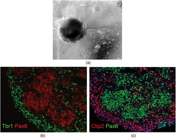
This 3D aggregation protocol for neuronal differentiation of hiPSCs gives rise to multilayered structures, characterized by apicobasal polarity mimicking the early aspects of corticogenesis in forebrain. As it develops in vitro, each hiPSC-derived aggregate spontaneously segregates into discrete layers (Figure 1): (a) a layer of radial glial cells with their apical end-feet facing a central lumen; (b) a layer of intermediate neuronal precursors with characteristics of human subventricular zone, found superficially to the radial glial layer; and (c) a layer of more mature excitatory-type neurons outside the subventricular zone. Moreover, these neuronal progenitors within the aggregates express transcription factors that specify layer-specific cortical neurons similar to those that arise during normal mammalian development. A remarkable aspect of this is that transcription factors for both lower and upper layer identity coexist in each aggregate in a nonoverlapping manner (Figure 1), even if their proportion and spatial organization does not fully recapitulate that of the developing human cortex. In addition, these multilayered structures contain GABAergic precursor cells as well as more mature inhibitory GABAergic neurons, which often segregate in a separate area from the excitatory neurons, suggesting that both excitatory and inhibitory neurons differentiate and mature in this preparation. Extensive synaptic differentiation is also achieved after 50–70 days in culture, with both pre- and postsynaptic markers expressed by the differentiating neurons and morphologically identifiable by electron microscopy (Mariani et al., Reference Mariani, Simonini, Palejev, Tomasini, Coppola and Szekely2012).
Human iPSCs-derived aggregates spontaneously organize in a multilayered fashion that recapitulates the architecture of normal human cerebral cortex during development. (a) A low power image of a three-dimensional aggregate after 50 days of neuronal differentiation in vitro. (b, c) Cryosections and immunostaining of these structures show that each aggregate contain layers of radial glial cells expressing (b, c) the transcription factor PAX6 and, more superficially, a layer of more mature neurons expressing transcription factors (b) such as TBR1, expressed by layers IV–V excitatory cortical neurons and other earlier cortical neurons, and (c) CTIP2, that is, specifically expressed by layer V cortical excitatory neurons. [A color version of this figure can be viewed online at http://journals.cambridge.org/dpp]

Global gene expression profiles using gene microarrays revealed that our hiPSC-derived 3D multilayered structures have a pattern of gene expression more typical of dorsal forebrain than ventral forebrain, and excluded the expression of genes of more posterior and ventral regions of the brain and spinal cord. To determine how the gene expression profile of these hiPSC-derived multilayered aggregates matched with the patterns of gene expression of the developing human brain, we performed a correlation analysis between whole genome transcripts expressed by our hiPSC-derived cells and whole genome gene expression data sets of human postmortem brain tissue samples derived from different regions and including several developmental stages (Kang et al., Reference Kang, Kawasawa, Cheng, Zhu, Xu and Li2011). The highest correlation was found to be with the human cerebral cortex at 4–10 weeks postconception, with enrichment in the associations for 10 postconceptual weeks and frontal regions of the developing human brain (Mariani et al., Reference Mariani, Simonini, Palejev, Tomasini, Coppola and Szekely2012).
The overall indication of this work is that hiPSCs can be differentiated into forebrain-like structures recapitulating the unique gene expression profile found in the embryonic human neocortex (Mariani et al., 2012). Gene expression in precursor and mature neural cells derived from hiPSC can be also examined with high throughput mRNA sequencing (RNA-Seq), which allows for a more quantitative study of the dynamics of gene expression and splice variants in neuronal development.
In principle, this should permit the resolution of stages and types of abnormalities found in developmental disorders, including ASD. Although genetic studies have found a number of polymorphism, gene sequence or copy number variant associations with specific disorders such as autism, obsessive compulsive disorder, or attention-deficit/hyperactivity disorder, these associations are often inconclusive because of the large number of genes/variants involved and the extreme degree of genetic polymorphism of human populations. The hiPSC approach may theoretically be able to identify more robust genomic association by extending analyses to a different level, that is, differences in amount of gene expression in neural cells implicated in these developmental disorders. As explained in the following section, these associations of gene expression differences with disorders and with their biological endophenotypes may be independent and complementary to studies of structural gene variation, localizing instead to regulation at the level of transcription or mRNA and leading to the identification of many other cellular processes and regulatory events critical for developmental psychopathology.
Integrative Analyses Using the hiPSC Model
The hiPSCs model offers a cell-centric approach, giving relatively easy access to genetic and epigenetic data, the associated cellular phenotypes, clinical data, and possibly structural and functional imaging from the individuals that donated the cells. In this way, a large set of heterogeneous data is generated, which warrants the implementation of integrative data analytic approaches, to account for the hierarchical/multiscale nature of the data. Genetics of gene expression (Montgomery et al., Reference Montgomery, Sammeth, Gutierrez-Arcelus, Lach, Ingle and Nisbett2010; Myers et al., Reference Myers, Gibbs, Webster, Rohrer, Zhao and Marlowe2007) and imaging genomics (Hariri & Weinberger, Reference Hariri and Weinberger2003) are two examples of integrative approaches in this direction, respectively integrating gene expression with individual/genotype and organ/tissue morphometry with individual/genotype. The large number of variables that arises from each individual's structural genomic variants makes the analysis of such data sets challenging. Because of this, both of these approaches have required large sample sizes of individual patients to achieve statistically significant results. Alternative approaches are now emerging, exploiting pathway enrichment analysis (Schoof, Iles, Bishop, Newton-Bishop, & Barrett, Reference Schoof, Iles, Bishop, Newton-Bishop and Barrett2011; Simonson, Wills, Keller, & McQueen, Reference Simonson, Wills, Keller and McQueen2011; Weng et al., Reference Weng, Macciardi, Subramanian, Guffanti, Potkin and Yu2011) to address these limitations Such analyses consider pathways rather than individual genes, implicitly accounting, to a limited extent, for linkage disequilibrium.
Where gene expression data are available, two types of analysis are feasible: one knowledge-driven method is pathway analysis, which relies on past findings about the biological roles of genes and their relatedness within previously delineated functional pathways (Hicks, Asfour, Pannuti, & Miele, Reference Hicks, Asfour, Pannuti and Miele2011; Xiong, Ancona, Hauser, Mukherjee, & Furey, Reference Xiong, Ancona, Hauser, Mukherjee and Furey2012; Zhong, Yang, Kaplan, Molony, & Schadt, Reference Zhong, Yang, Kaplan, Molony and Schadt2010). The second, data-driven, method is gene coexpression network analysis, which examines the relatedness of genes in their actual patterns of coexpression, within the tissues or cells that are being investigated (Ben-David & Shifman, Reference Ben-David and Shifman2012). Both of these approaches have proved to be powerful in uncovering important factors that were missed by the standard gene-by-gene analysis. Specifically, gene networks and pathways are a way to represent genes according to their coexpression and, probably, common function. Summarizing genes, which could number in the thousands, by the many fewer pathways/networks they participate in has the advantage of dramatically reducing the number of comparisons to be made, and, therefore, increase statistical power of the analysis.
In the context of the hiPSC model, data-driven gene coexpression network analysis allows for sets of coexpressed transcripts (“modules”) to be determined. By analyzing gene expression within these sets, it is possible to identify the module(s) correlating with specific diagnoses and/or their biological endophenotypes, that is, specific alterations in cellular functions as their development unfolds in vitro. For example, by using the cellular data from the hiPSCs, it can be determined whether certain modules correlate with altered differentiation of neuronal subtypes, changes in synapse numbers, neurite length, and other cellular alterations.
Modules associated with cellular alterations may also be examined to identify correlations with genotype and neuroimaging phenotypes across patients and typically developing controls. The convergence of network analyses on the neurodevelopmental transcripts with imaging, clinical, and genotype data obtained in the same individuals is a powerful approach leading to the identification of functionally related genes and pathways that may underlie altered neuronal development and may provide biological underpinnings of structural and functional alterations of the brain in neurodevelopmental disorders. In the future, where the availability of high throughput epigenomic, proteomic, and metabolomic data from the same individual becomes feasible, along with more detailed cellular characterization, multimodality imaging data (e.g., magnetic resonance imaging, functional magnetic resonance imagining, positron emission tomography, EEG), as well as clinical and environmental data, these biologically driven data reduction approaches, like the coexpression network, may become the key to any integrative analysis.
In conclusion, new techniques that can be used to closely examine the neurobiology and genetics of childhood psychiatric disorders promise to uncover mechanisms that may explain many behavioral and neurological observations. Each individual's etiology of illness may be identifiable, and common etiologies may be discovered. In patients with ASDs, the significant number of individuals who share a specific phenotype and developmental process of increased brain size may be heterogeneous with respect to their susceptibility genes. This would be expected because, during development, genes and their products often interact with each other in unexpected ways, that is, as transactivating transcription and epigenetic factors. The hiPSC approach can provide us with a model to investigate how disparate genomic loci can exert common functional effects. These commonalities can be revealed by common functions in cellular aspects of development (i.e., cell proliferation) and the underlying shared patterns of gene expression across developmental times and cell types. With an extensive knowledge of the cellular mechanisms of disease, our ability to diagnose and treat childhood psychiatric illness will significantly improve. In particular, the uniting of neurobiological and genomic data promises dramatic changes in our understanding of these disorders and may allow for a notable shift in the global outlook on childhood psychopathology.