Introduction
Originally described by Gilmore (Reference Gilmore1933) based on a specimen from the Djadokhta Formation at Bayan Zag, Pinacosaurus is one of the most commonly encountered dinosaur taxa in the Gobi Desert (Watabe and Suzuki, Reference Watabe and Suzuki2000; Currie et al., Reference Currie, Badamgarav, Koppelhus, Sissons and Vickaryous2011). It has become one of the best known ankylosaur in terms of the number and preservational quality of specimens. In addition to Bayan Zag, the Djadokhta-age beds at Bayan Mandahu in Inner Mongolia have produced more than a dozen articulated juvenile partial skeletons (Burns et al., Reference Burns, Currie, Sissons and Arbour2011; Currie et al., Reference Currie, Badamgarav, Koppelhus, Sissons and Vickaryous2011). From this site, the Chinese-Belgian expeditions (Godefroit et al., Reference Godefroit, Suberbiola, Li and Dong1999) recovered a specimen identified as representative of a distinct species, Pinacosaurus mephistocephalus Godefroit, Pereda-Suberbiola, Li, and Dong, 1999, although Pinacosaurus grangeri Gilmore, Reference Gilmore1933 can also be found at that site (Burns et al., Reference Burns, Currie, Sissons and Arbour2011).
A third Djadokhta-age site is Alag Teeg, originally discovered in 1969 by the Soviet-Mongolian Paleontological Expedition and excavated in 1969 and 1970 (Tverdochlebov and Zybin, Reference Tverdochlebov and Zybin1974). Several sources report that the expedition found several specimens of P. grangeri in 1969 (Tverdochlebov and Zybin, Reference Tverdochlebov and Zybin1974; Maryańska, Reference Maryańska1977; Fastovsky and Watabe, Reference Fastovsky and Watabe2000; Currie et al., Reference Currie, Badamgarav, Koppelhus, Sissons and Vickaryous2011), using a bulldozer to remove overburden and expose the fossil-bearing level. Although most specimens collected were represented by isolated, disarticulated postcranial elements, some of the postcrania recovered were articulated. The Mongolian-Japanese Joint Paleontological Expedition also collected from Alag Teeg from 1993 to 1998, reporting about 30 individuals, although many were damaged or removed by fossil poachers (Watabe and Suzuki, Reference Watabe and Suzuki2000). Many specimens were preserved upright in life positions in the lower mudstone section at Alag Teeg, interpreted as the floodplain of a braided system (Fastovsky, Reference Fastovsky2000). Their description is in preparation elsewhere. In 2001 and between 2003 and 2007, the “Dinosaurs of the Gobi” (Nomadic Expeditions) found remains of over 40 Pinacosaurus specimens. These are housed in the Paleontological Center in Ulaan Baatar, and many were described by Currie et al. (Reference Currie, Badamgarav, Koppelhus, Sissons and Vickaryous2011), who suggested that the Alag Teeg specimens might represent a different species than those from nearby sites like Bayan Zag because of their stratigraphically lower position.
Despite the attention given to the Alag Teeg locality by several institutions and expeditions for over forty years, a detailed description of the fossil material, which was restricted largely to manual and pedal elements, has only recently been published (Currie et al., Reference Currie, Badamgarav, Koppelhus, Sissons and Vickaryous2011). The original material, first discovered by the Soviet-Mongolian Paleontological Expedition in 1969–1970, has escaped the attention of specialists and remained formally undescribed because of a general focus on cranial morphology over the years in ankylosaur research. This contribution remedies this by describing these specimens for the first time. The occurrence of many conspecifics in the same assemblage also allows us to address postcranial variability within Pinacosaurus.
Systematic paleontology
Institutional Abbreviations
HMNS, Center for Paleobiological Research, Hayashibara Biochemical Laboratories, Inc., Okayama, Japan; INBR, Victor Valley Museum, Apple Valley, California, USA; IVPP, Institute for Vertebrate Paleontology and Paleoanthropology, Beijing, China; MPC, Paleontological Center of the Mongolian Academy of Sciences, Ulaan Baatar, Mongolia; PIN, Palaeontological Institute, Russian Academy of Sciences, Moscow, Russia; ZPAL, Zoological Institute of Paleobiology, Polish Academy of Sciences, Warsaw, Poland.
Other Abbreviations
SD, standard deviation of the mean; α, allometric coefficient.
Remarks
Ankylosaur material at the PIN was collected from the Late Cretaceous beds of the Alagteeg Formation at Alag Teeg, Mongolia (Fig. 1), by the Soviet-Mongolian Paleontological Expedition in 1969 and 1970. The collection is composed of approximately 285 specimens, including ~160 vertebrae (disarticulated, except for three articulated series, including one of four cervicals, one of seven caudals, and another of 21 caudals), over 20 rib fragments, ~20 girdle elements, ~30 complete and fragmentary limb bones, ~30 isolated manual and pedal bones, and ~15 cervical half-ring fragments. This material, all of which is prepared, was examined by the first and second authors via measurements, observations, and photographs. Only the most informative specimens (83 in total) were catalogued and form the basis of this manuscript.
Additional comparative material referable to P. grangeri from Alag Teeg, Bayan Zag, and Baya Mandahu was examined by the first and third authors at the IVPP, MPC, and ZPAL, and by the first author at the HMNS. Positional and orientation terminology for limb elements follows Currie et al. (2011). Statistical calculations and regression analyses were performed in Microsoft Excel. Uncorrected p values were obtained in PAST (Hammer et al., 2001). Elements of the manus and pes have been described in detail from the collections of the MPC by Currie et al. (2011). MPC-D 100/1305 was recently referred by Carpenter et al. (2011) to Saichania chulsanensis Maryańska, 1977; however, its assignment to any ankylosaurid taxon is presently equivocal (Arbour and Currie, 2013) and the specimen number is used here without a taxonomic referral. References to Saichania here refer only to the holotype (MPC-D 100/151). In addition to examining the robustness of each element, specimens were compared with other ankylosaur taxa to see if ontogenetic trends in growth can be separated from interspecific differences. Measurement data for all specimens used in regression and statistical analyses are available as Supplemental Data.
Holotype
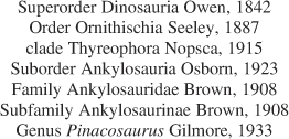
AMNH 6523, skull and mandible of an adult specimen, atlas, axis, and several associated osteoderms. Material is dorsoventrally crushed and has undergone expanding matrix distortion.

Occurrence
?upper Santonian to upper Campanian of Mongolia and PRC. Material is from the Alagteeg and Djadokhta formations (Campanian, Upper Cretaceous). The type locality is Bayan Zag, Gobi Desert, Mongolia.
Material
PIN 3144, basicranium, articulated, associated, and disarticulated postcranial elements, disarticulated cervical half ring segments.
Cranial description
Basicranium
Although this description focuses on postcranial elements, the PIN collection includes one partial basicranium (Fig. 2) from Alag Teeg. The occipital condyle is 2.4 times wider than it is high, more similar to the condition in Saichania (MPC-D 100/151) than ZPAL MgD II/1, although this may be partially influenced by dorsoventral crushing. It incorporates the basioccipital and exoccipitals; the latter contribute to the dorsolateral corners of the occiput and form 75% of its dorsal width. The sutures separating the exoccipitals from the basioccipital are prominent as in ZPAL MgD II/1. Conversely, the exoccipitals are excluded from the occipital condyle in Euoplocephalus tutus (Lambe, Reference Lambe1902; Vickaryous and Russell, Reference Vickaryous and Russell2003). There is no median basioccipital foramen on the ventral surface of the basioccipital between the basitubera and occiput. This foramen is absent in Minotaurasaurus ramachandrani Miles and Miles, 2009. It has not been reported for Ankylosaurus magniventris Brown, 1908 (Carpenter, Reference Carpenter2004) or Euoplocephalus tutus (Vickaryous and Russell, Reference Vickaryous and Russell2003). It is present, however, in Saichania (Maryańska, Reference Maryańska1977). The basisphenoid bifurcates anteriorly into bilateral, anterolaterally directed basipterygoid processes. As preserved, the sutural contact between the basioccipital and basisphenoid is not visible.
Locality map showing productive Mongolian ankylosaur localities, including Alag Teeg in south central Mongolia.

Basicranium of juvenile Pinacosaurus grangeri, Gilmore Reference Gilmore1933, in, (1), dorsal and, (2), ventral views. Anterior is up.
Axial skeleton description
Cervical vertebrae
Four articulated cervicals (PIN 3144/49/1–4; Fig. 3) were collected. Although the neural arches are attached to the centra, the neurocentral sutures are clearly visible. The amphiplatyan centra are consistently wider than long (by an average ratio of 1:0.88) as in Ankylosaurus (Coombs, 1986; Carpenter, Reference Carpenter2004) and Euoplocephalus (juvenile and adult); in contrast, the centra in MPC-D 100/1305 become longer than wide posteriorly in the series (Carpenter et al., Reference Carpenter, Hayashi, Kobayashi, Maryańska, Barsbold, Sato and Obata2011). Ventrally, each has a median longitudinal ridge for attachment of the longitudinal ligament. The articular faces of any single centrum are not parallel in lateral view, but give the centrum a trapezoidal shape to accommodate the sigmoidal shape of the neck as in other ankylosaurs (Coombs, 1986; Carpenter et al., Reference Carpenter, Hayashi, Kobayashi, Maryańska, Barsbold, Sato and Obata2011). There are no horizontal bony laminae in the neural canals like those reported for Saichania (MPC-D 100/151) by Maryańska (Reference Maryańska1977). Each diapophysis is elliptical in cross-section and projects laterally from the base of the prezygapophysis. The prezygapophyses are relatively as large as those figured for MPC-D 100/1305 (Carpenter et al., Reference Carpenter, Hayashi, Kobayashi, Maryańska, Barsbold, Sato and Obata2011, fig. 12). The neural spines are damaged in all four vertebrae, and there is no evidence to suggest the cervical ribs were fused with any of the vertebrae.
Articulated cervical vertebrae (?c3–c6) of juvenile Pinacosaurus grangeri in, (1), dorsal; (2), ventral; (3), left lateral; and, (4), right lateral views. Anterior is to the right in (1), (2), and (4) and to the left in (3). Abbreviations: dps=diapophysis; ncs=neurocentral suture; poz=postzygapophysis; prz=prezygapophysis; sp=neural spine; tp=transverse process; vmr=ventral median ridge.
Dorsal vertebrae
The amphiplatyan anterior and posterior faces of each dorsal centrum (Fig. 4) are equal in width and height. The centra are constricted at midlength (to an average of 72% the widths of the anterior and posterior faces) into an hourglass shape when viewed ventrally. There are no horizontal grooves on the lateral surfaces of the centra as reported in MPC-D 100/1305 (Carpenter et al., Reference Carpenter, Hayashi, Kobayashi, Maryańska, Barsbold, Sato and Obata2011; Arbour and Currie, Reference Arbour and Currie2013). Such depressions are also absent in Euoplocephalus tutus and Talarurus plicatospineus Maleev, 1952 (Arbour and Currie, Reference Arbour and Currie2013). Although dorsal neural arches are found at Alag Teeg, none are fused to the centra. Their sutures extend the entire length of the dorsal surface of a centrum. The prezygapophyses form a single median sagittal U-shaped structure (concave dorsally) similar to those of most ankylosaurs (Carpenter, Reference Carpenter2004; Carpenter et al., Reference Carpenter, Hayashi, Kobayashi, Maryańska, Barsbold, Sato and Obata2011). The neural spines are vertically oriented plates that are roughly rectangular in shape when viewed laterally. Like MPC-D 100/1305 (Carpenter et al., Reference Carpenter, Hayashi, Kobayashi, Maryańska, Barsbold, Sato and Obata2011), they do not show transversely expanded distal ends as they do in Ankylosaurus (Carpenter, Reference Carpenter2004). Each transverse process projects dorsolaterally, angles anteriorly, and is supported by two ventrally diverging struts that terminate on the parapophysis immediately dorsal to its articular surface. There is no evidence that dorsal ribs were fused to any of the neural spines. There is also nothing to suggest that ossified tendons were associated with the dorsals as in MPC-D 100/1305 (Carpenter et al., Reference Carpenter, Hayashi, Kobayashi, Maryańska, Barsbold, Sato and Obata2011).
Dorsal centrum of juvenile Pinacosaurus grangeri in, (1), dorsal; (2), ventral; (3), anterior or posterior, and, (4), (5), lateral views. Orientation uncertain. Abbreviations: nas=neural arch sutural surface; nc=neural canal; np=notochordal prominence; pd=pedicle.
Sacral vertebrae
All sacral vertebrae (Fig. 5) are separate from each other, even in the largest Alag Teeg specimen (MPC-D 100/1333). Ventrally, each sacral centrum has a median longitudinal ridge but lacks a ventral groove, the latter of which is known in nodosaurs (e.g., Edmontonia, Peloroplites) and Cedarpelta (Carpenter et al., 2008). The neural canal excavates the dorsal surface of the centrum. The intervertebral articulations of the centra range from semi lunar to heart-shaped when viewed anteriorly or posteriorly. Lack of fusion of the synsacrum in these juvenile specimens makes it difficult to identify and differentiate dorsosacrals from dorsals, and sacrocaudals from free caudals.
Sacral centra of juvenile Pinacosaurus grangeri in, (1), dorsal; and, (2), ventral views. Anterior is to the right. Abbreviations: nas=neural arch sutural surface; nc=neural canal; s1, s2=first, second sacral.
Caudal vertebrae
In ankylosaurines, the caudal series is divided into the free (= anterior) caudals and those (posterior) forming the handle of the tail club, although none from the latter series were identified in the PIN collection. The free centra (Fig. 6) in P. grangeri are amphiplatyan, and their heights, widths and lengths are roughly equidimensional. The neural arches are not fused to the centra in any of the specimens collected from Alag Teeg. An anteroposteriorly elongate median groove occupies the ventral surface of each centrum. The chevrons (= haemal arches) are not fused to the centra, although their articular facets are visible on the posteroventral corners of the centra. Ossified tendons are not present on the free caudals.
Articulated free caudal centra of juvenile Pinacosaurus grangeri in (1), dorsal; (2), ventral; (3), left lateral; and (4), right lateral views. Anteriormost centrum in (5a, b), anterior; and (5c, d), posterior views. Posterior-most centrum in (6a, b), anterior and (6c, d), posterior views. Anterior is to the right in (1) and (4) and to the left in (2) and (3). Dorsal is up in (5) and (6). Abbreviations: cha=chevron (haemal spine) articulation; nas=neural arch sutural surface; np=notochordal prominence; pd=pedicle; tp=transverse process; vmg=ventral median groove.
Dorsal ribs
Smaller dorsal ribs from Alag Teeg have distinct tubercula and capitulae, demonstrating that they were not fused to the vertebrae in young animals. Those ribs (PIN 3144/68 and 69) associated with a larger individual are medially broken at these articular surfaces, suggesting that they were fused to the vertebrae. Immediately distal to the articular surfaces, the ribs have T-shaped cross sections as in other derived ankylosaurs (Coombs, Reference Coombs1971). This cross-sectional shape changes distally along the rib until it is triangular and eventually elliptical.
Appendicular skeleton description
Scapula
The scapula (Fig. 7.2) is medially concave to conform to the shape of the thorax. The constricted, neck-like region is poorly developed as is characteristic for ankylosaurines, but in contrast with the distinct necks of derived nodosaurid scapulae. The acromion extends on average 40% the length of the scapula (although the range is from 30% to 50% with a standard deviation of 7.4% due to the ambiguities in determining the exact posterior-most extent of the acromion). In MPC-D 100/1305, the acromion extends one-third the length of the scapula (Carpenter et al., Reference Carpenter, Hayashi, Kobayashi, Maryańska, Barsbold, Sato and Obata2011). It is formed by a lateral deflection of the dorsal border of the scapula but does not curve ventrally as in Ahshislepelta minor Burns and Sullivan, Reference Burns and Sullivan2011.
Left shoulder girdle elements of juvenile Pinacosaurus grangeri. Coracoid in, (1a,b), lateral; and (1c, d), medial views. Scapula in (2a, b), lateral and (2c, d), medial views. Anterior is to the right in (1a, b) and (2c, d) and to the left in (1c, d) and (2a, b). Abbreviations: acr=acromion process; adp=anterodorsal process; ca= coracoid articulation; cof= coracoid foramen; gfs= glenoid fossa; mb= medial brace; n= scapular neck; sa= scapular articulation.
Coracoid
None of the coracoids (Fig. 7.1) from Alag Teeg are coossified to scapulae. Posteroventrally, the coracoid contributes to half of the glenoid fossa. As is characteristic for most ankylosaurines (Carpenter et al., Reference Carpenter, Hayashi, Kobayashi, Maryańska, Barsbold, Sato and Obata2011), a ridge extends from the glenoid fossa to the anterodorsal process, giving the process a dorsolateral projection and imparting a sigmoidal curve to the anterior border of the coracoid. The glenoid foramen is situated anterior to the scapular contact dorsal to the glenoid fossa. The rounded anterior edges of the coracoids distinguishes them from those of MPC 100/1305, which have flat anterior edges (Arbour and Currie, Reference Arbour and Currie2013), although it is unknown whether this difference is taxonomic, ontogenetic, and/or individually variable.
Humerus
Proximally, each humerus (Fig. 8) is expanded mediolaterally to an average of 2.5 times the width of the shaft, forming a deltopectoral crest and medial process flanking the head. Distally, the humerus is expanded into the medial and lateral condyles to an average of 2.4 times the width of the shaft.
Left partial humerus of juvenile Pinacosaurus grangeri in (1), anterior and (2), posterior views. Note that the deltopectoral crest is largely broken off. Proximal is up. Abbreviations: dp=deltopectoral crest; hh=humeral head; it=internal (medial) tuberosity; lc=lateral condyle; lepc=lateral epicondyle; mc=medial condyle; mepc=medial epicondyle.
Godefroit et al. (Reference Godefroit, Suberbiola, Li and Dong1999) considered that the humeri were diagnostic for the two species of Pinacosaurus in that P. mephistocephalus had a better-developed deltopectoral crest that terminated distal to humeral mid-length. This is more similar to the condition in Saichania chulsanensis and Shanxia tianzhenensis Barrett, You, Upchurch, and Burton, Reference Barrett, You, Upchurch and Burton1998, according to Godefroit et al. (Reference Godefroit, Suberbiola, Li and Dong1999); however, the humeral shaft has a more rounded cross-section in P. mephistocephalus. Currie et al. (Reference Currie, Badamgarav, Koppelhus, Sissons and Vickaryous2011) noted that the deltopectoral crest in two humeri (MPC-D 100/1344 and MPC-D 100/1346) from Alag Teeg exhibited the condition reported for P. mephistocephalus. The character was considered non-diagnostic at the species level by Burns et al. (Reference Burns, Currie, Sissons and Arbour2011), who suggested it was possibly size-dependent.
Ulna
Proximally, the medial process of the ulna (Fig. 9), against which the radius articulates, is prominent as in other ankylosaurines (Maryańska, Reference Maryańska1977; Carpenter et al., Reference Carpenter, Hayashi, Kobayashi, Maryańska, Barsbold, Sato and Obata2011). The olecranon process is low and blunt as in MPC-D 100/1305 (Carpenter et al., Reference Carpenter, Hayashi, Kobayashi, Maryańska, Barsbold, Sato and Obata2011). The proximal articular surface comprises 66% of the total length of the ulna in Saichania and 50% in ZPAL MgD II/1, although the olecranon is more developed in PIN 614 than it is in ZPAL MgD II/1 (Maryańska, Reference Maryańska1977). The shaft tapers distally to the radial (circumferential) articulation. This articulation conforms to the ulnar notch of the radius, and forms an anteroposteriorly elongate oval in distal view.
Right forelimb zeugopodial elements of juvenile Pinacosaurus grangeri in anterior (1), posterior (2), proximal (3a), and distal (3b) views. Proximal is up in (1) and (2). Anterior is up in (3). Abbreviations: op=olecranon process; r=radius; rg=radial groove of the ulna; rh=radial head; stp=styloid process; u=ulna; unr=ulnar notch of the radius.
Radius
The straight radius (Fig. 9) has an anteroposteriorly elongate, oval (in proximal view) head with a cupped fovea for articulation with the humeral capitulum. The styloid process is underdeveloped as a blunt projection on the lateral side of the distal end of the radius. The distal articular surface is convex. The ulnar notch is a proximodistally elongate groove on the medial surface of the distal end of the radial shaft.
Ilium
Godefroit et al. (Reference Godefroit, Suberbiola, Li and Dong1999) considered the ilium (Fig. 10) diagnostic at the species level in Pinacosaurus; it forms a horizontal shelf dorsal to the acetabulum in P. mephistocephalus but overhangs the acetabulum laterally in P. grangeri. Burns et al. (Reference Burns, Currie, Sissons and Arbour2011) tentatively retained the character because their revision focused on cranial characters. The Alag Teeg specimens match the morphology observed in ZPAL MgD II/1, although there is no published lateral figure of the ilium of P. mephistocephalus for comparison with that taxon. The alae of the ilia from the Alag Teeg specimens show no such ventral deflection lateral to the acetabulum. In fact, well-preserved specimens (PIN 3144/5 and 3144/9) each has a dorsal deflection at the posterolateral corner of the ilium, leaving the acetabulum visible in lateral view. The same is observable on the ilium of PIN 614.
Right partial ilium of juvenile Pinacosaurus grangeri in (1), dorsal; (2), ventral; (3), lateral; and (4), medial views. Anterior is to the right in (1), (2), and (4) and to the left in (3). Abbreviations: ac=acetabulum; isped=ischial peduncle; poap=postacetabular process; prap=preacetabular process.
Ischium
Proximally, the ischium is expanded and forms the entire medial surface of the acetabulum, seen as a lateral concavity on the ischium. There is no evidence that the sacral ribs were fused to the ischium as in Ankylosaurus (Carpenter, Reference Carpenter2004). The distal end is anteroposteriorly expanded to 131% the anteroposterior diameter of the shaft.
Femur
The minimum circumference to length ratio (49%) in the Alag Teeg femora (Fig. 11) is similar to that reported for MPC-D 100/1305 (47%) by Carpenter et al. (Reference Carpenter, Hayashi, Kobayashi, Maryańska, Barsbold, Sato and Obata2011). The articular surfaces and muscle attachment sites are not as rugose as they are in larger ankylosaur femora (e.g., MPC-D 100/1305; Carpenter et al., Reference Carpenter, Hayashi, Kobayashi, Maryańska, Barsbold, Sato and Obata2011). The greater trochanter is prominent along the lateral edge of the proximal end of the femur. It extends 23% the length of the femur as in other ankylosaurines (Carpenter et al., Reference Carpenter, Hayashi, Kobayashi, Maryańska, Barsbold, Sato and Obata2011). The shaft is anteroposteriorly compressed to a ratio of 1:0.45 (transverse/anteroposterior), although this is variable (SD=0.19). The fourth trochanter is visible on the posteromedial side of the shaft. The distal condyles are anteriorly separated by an intercondylar notch. The lateral condyle extends farther distally than the medial condyle, although the medial condyle is larger.
Left femur of juvenile Pinacosaurus grangeri in (1), anterior; (2), posterior; (3), lateral; (4), medial; and (5), distal views. Proximal is up in (1–4). Anterior is up in (5). Abbreviations: grt=greater trochanter; hf=femoral head; icn=anterior intercondylar notch; lc=lateral condyle; mc=medial condyle; pf=popliteal fossa.
Tibiotarsus
Distally, the tibial shaft (Fig. 12) flares into lateral and medial condyles separated anteriorly by a sulcus. Astragali are preserved in articulation with the posteroventral, distal surfaces of two tibiae (PIN 3144/38 and 39). Their morphologies and positions are similar to those described for P. grangeri specimens from the MPC (Currie et al., Reference Currie, Badamgarav, Koppelhus, Sissons and Vickaryous2011) and a juvenile North American ankylosaurine (Coombs, 1986). They articulate but are not fused to the tibiae, unlike in adult ankylosaurines (Coombs, Reference Coombs1979). No fibulae were recovered from Alag Teeg by the Soviet-Mongolian Expedition, but those in the collections of the MPC have been described by Currie et al. (Reference Currie, Badamgarav, Koppelhus, Sissons and Vickaryous2011).
Left tibia of juvenile Pinacosaurus grangeri in (1), anterior; (2), posterior; (3), lateral; (4), medial; and (5), distal views. Proximal is up in (1–4). Anterior is to the right in (5). Abbreviations: a=astragalus; iocr=interosseous crest; lc=lateral condyle; mc=medial condyle; tbp=tibial plateau.
Dermal skeleton description
Cervical half rings
All cervical half rings from Alag Teeg are incomplete due to an apparent lack of complete mineralization at early ontogenetic stages. The preserved portions represent pieces of the transverse bands of the cervical half rings and have crenulated anterior and posterior edges. Distal portions of the bands (Fig. 13) have smooth edges that transition into rounded anterior corners. The posterior corner is angular, and separates a crenulated edge to the distal end of the band. Osteodermal projections extend from the external surface of the bands and have rugose, pitted textures contrasting with the smooth textures of the bands. The thickness of each band varies, being relatively thinner deep to the osteodermal projections, and thicker between them. This gives the band an undulating appearance in anterior or posterior views. The interosteodermal portions of the band are truncated by crenulated surfaces perpendicular to the band. Here, two portions of the band may have contacted one another in interdigitating sutures (as can be seen in other ankylosaurine cervical half rings) or interfaced with nonmineralized dermis. In two distal specimens (PIN 1344/74 and 75), the osteodermal projections have more rugose surface textures than other specimens, although this may be ontogenetically affected. These distal projections do not cover the distal end of the band as in other (more skeletally mature) ankylosaurs (Penkalski, Reference Penkalski2001; Arbour et al., Reference Arbour, Burns and Sissons2009), but they do extend with distal inclinations.
Right distal cervical half ring segment of juvenile Pinacosaurus grangeri in (1), external; (2), basal; and (3), distal views, and right lateral view of skull and cervical half ring of P. grangeri (IVPP V16854) from the Bayan Mandahu locality, Inner Mongolia, People’s Republic of China, showing the position on the body of the cervical half rings (modified from Burns et al., Reference Burns, Currie, Sissons and Arbour2011; fig. 6B′). Anterior is up in (1), (2), and (4). External is up in (3). Abbreviations: anc=anterior corner of distal edge of band; ang=angular; db=smooth distal edge of band; lac=lacrimal; lom=lateral osteodermal mass covering narial region; ms=medial suture; osq=osteoderm covering squamosal; poc=posterior corner of distal edge of band; pmx=premaxilla; prf=prefrontal; rp=rugose osteodermal projection; so=supraorbital ossification; vc=vascular canal foramina.
Bivariate log-log plots for postcranial dimensions in juvenile Pinacosaurus grangeri from Alag Teeg for forelimb (1), humerus; (2), ulna; (3), radius; and hindlimb (4), femur; (5), tibia; (6), fibula elements. Element length is plotted against maximum proximal width (diamonds), maximum midshaft width (squares), maximum distal width (triangles), and midshaft circumference (Xs). Slope, intercepts, and fit statistics are presented in Table 1. Data used to create plots are available as Supplemental Data.
Appendicular allometry
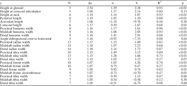
Scapulae from the PIN Alag Teeg collection (Table 1) become allometrically dorsoventrally taller as their lengths increase. Height at the glenoid process has the highest coefficient of allometry (α=1.29, R2=0.93, p<0.01) followed by the articular surface for contact of the coracoid (α=1.17, R2=0.80, p=0.13), neck height (α=1.14, R2=0.92, p<0.01), and posterior height (α=1.02, R2=0.88, p<0.01). The acromion grows with negatively allometry (α=−1.14, R2=0.49, p=0.26) when compared with scapular length, although this trend is not significant. Coracoid height is negatively allometric (α=0.59, R2=0.68, p=0.01) to its length. Comparison of scapular blade length (y) with humerus length (Table 2) suggests that the scapular growth was negatively allometric within Pinacosaurus (α=0.87, R2=0.94, p<0.01), whereas the lengths of these two elements stay the same (are isometric) in specimens of all sizes but different taxa (α=0.95, R2=0.89, p<0.01). The results of comparisons between humerus length and scapular blade width of the neck are more equivocal because of variability (both within Pinacosaurus and between all taxa), but do confirm that shaft robustness is increasing in ontogenetic (but not interspecific) series.
Analyses of size-related differences in the postcrania of juvenile Pinacosaurus grangeri Gilmore, Reference Gilmore1933 from Alag Teeg, Gobi Desert, Mongolia
The x-axis represents element length in all cases. Measurement data for all specimens used in regression and statistical analyses is available as Supplemental_Data_1.xls. Only analyses with N≥5 are included here.
Abbreviations: α=allometric coefficient (slope of regression line); b=regression line y-intercept; Δx=ratio of maximum to minimum x for samples used in analysis; N=number of samples.
Analyses of size-related differences in the postcrania of Pinacosaurus and other ankylosaur specimens of various taxa and body sizes

The x and y values represent element length in all cases Measurement data for all specimens used in regression and statistical analyses is available as Supplemental_Data_1.xls.
Abbreviations: α=allometric coefficient (slope of regression line); b=regression line y-intercept; Δx=ratio of maximum to minimum x for samples used in analysis; N=number of samples.
Among the PIN humeri from Alag Teeg (Figure 13), proximal and midshaft widths show size-dependence with respect to element length. Proximal width increases at a greater rate (α=1.17, R2=0.96, p<0.01) relative to humeral length than does midshaft width (α=1.08, R2=0.93, p<0.01). Distal humeral shows greater positive allometry (α=1.41, R2=0.88, p<0.01) than the proximal end. The deltopectoral crest shows no significant length-dependent trends with respect to crest length (α=1.15, R2=0.23, p=0.85) or its angle to the long axis of the humerus (α=0.15, R2=0.01, p=0.64). Few ankylosaur specimens have both femur and humerus, but comparison of humerus length with femoral length suggests that the humeral growth was negatively allometric within specimens of all sizes but different taxa (α=0.91, R2=0.95, p<0.01).
The radius and ulna of PIN Alag Teeg specimens show significant allometric trends. In radii, the distal end has higher positive allometry (α=1.14, R2=0.87, p<0.01) than the proximal end (α=1.12, R2=0.83, p<0.01). The shaft has lower positive allometry than both ends (α=1.07, R2=0.68, p<0.01). Positive allometry in the ulna is strongest proximally (α=1.49, R2=0.89, p<0.01), due likely to the development of the olecranon process. The ulnar shaft shows higher positive allometry (α=1.28, R2=0.73, p<0.01) than the distal end (α=1.03, R2=0.27, p=0.07), although this latter relationship is not significant. Comparison of radius length with humerus length suggests that the radial growth was negatively allometric within Pinacosaurus (α=0.89, R2=0.90, p=0.05), whereas the lengths of these two elements stay the same (are isometric) in specimens of all sizes but different taxa (α=0.99, R2=0.95, p<0.01).
In PIN Alag Teeg specimens, hind limb element widths do not show strong length-dependent correlations like those of the forelimb. Both proximal (α=1.03, R2=0.76, p<0.01) and distal ends (α=0.92, R2=0.63, p=0.01) of the femur are roughly isometric. The femoral shaft diameter (α=0.75, R2=0.27, p<0.01) and circumference (α=0.73, R2=0.47, p=0.01) are both negatively allometric. In the tibia, there are no significant correlations between length and proximal (α=0.59, R2=0.07, p=0.60), midshaft (α=0.54, R2=0.12, p=0.38), or distal (α=−0.51, R2=0.08, p=0.60) widths. The same is true for fibular proximal (α=−1.08, R2=0.18, p=0.54), midshaft (α=−0.68, R2=0.17, p=0.54), and distal (α=−1.70, R2=0.37, p=0.35) widths.
Comparison of femur length with tibia length suggests that the tibial growth was negatively allometric in specimens of all sizes but different taxa (α=0.86, R2=0.96, p<0.01). Unfortunately, there are only four specimens in which both elements are preserved. Comparison of femur length with the length of the third metatarsal suggests that metatarsus growth was isometric in specimens of all sizes but different taxa (α=1.07, R2=0.97, p<0.01).
Discussion
Taxonomic referral
Assignment of the Soviet-Mongolian ankylosaurine material described here is based on its occurrence in the bonebed at Alag Teeg. Several complete skulls collected from the same locality by the HMNS allow unequivocal referral to P. grangeri based on the current diagnosis from Burns et al. (Reference Burns, Currie, Sissons and Arbour2011). A description of the HMNS P. grangeri material from Alag Teeg is in preparation.
Currie et al. (Reference Currie, Badamgarav, Koppelhus, Sissons and Vickaryous2011) have suggested that due to their lower stratigraphic occurrence (Dashzeveg et al., Reference Dashzeveg, Dingus, Loope, Swisher, Dulam and Sweeney2005), the ankylosaurine specimens from Alag Teeg may represent a taxon distinct from those found at Bayan Zag. A problem when comparing P. grangeri specimens from different assemblages is the discrepancy in mean body size, which makes it difficult to distinguish between ontogenetic and possible taxonomically significant morphological variation. To date, there is little published data on the postcranial material from P. grangeri. Nevertheless, ZPAL MgD II/1 falls within the range of size variation of the Alag Teeg material. The large PIN 614 falls significantly outside of it. In addition, some material from Alag Teeg (possibly representing a single individual) is significantly larger than the remaining elements from the site.
Based on discrete cranial characters (Burns et al., Reference Burns, Currie, Sissons and Arbour2011) all of the ankylosaurine material from Alag Teeg and ZPAL MgD II/1 is referable to P. grangeri.
Size range of Alag Teeg P. grangeri
Arbour and Currie (Reference Arbour and Currie2011) noted that two humeri and a scapula (found together and presumably constituting one individual) were larger than other homologous elements in the MPC Alag Teeg collection. The scapula and humeri (MPC-D 100/1333) are significantly larger than all others from Alag Teeg (at least 1.4 times larger). The same is true for the large coracoid (PIN 3144/23). Because the humeri are left and right, and the scapula and coracoid are from the same side (left), this lends support to these large elements represent a single individual. Despite their size, the scapula and coracoid are unfused, as are several large neural spines in the PIN.
If all of the significantly larger elements from Alag Teeg do represent a single individual, then it would provide some of the first information on the timing of osteological fusion events in ankylosaurs. It is interesting that the neural arches are only preserved in articulation in the cervical vertebrae (although they are not fully coossified to the centra). There are no fully-fused neurocentral sutures on any of the dorsal, sacral, or free caudal vertebrae from Alag Teeg, although isolated arches have been recovered and demonstrate that their isolation is caused by lack of fusion and not breakage. In contrast, the neural arches of a juvenile North American ankylosaurid, referred to Euoplocephalus by Coombs (1986) were not found in association with the cervical centra. This might suggest that neurocentral closure began in the cervical series for P. grangeri and progressed in an anterior to posterior sequence. However, there are also examples of large unfused (not broken) neural arches from the cervical (PIN 3144/64), dorsal (PIN 3144/62), and caudal (PIN 3144/63) series. Evidence suggests the dorsal ribs and vertebrae were among the first elements to fully coosify during ontogeny. If most of the coosification is delayed ontogenetically, this would add support to the validity of taxa that may be relatively small in terms of body size, but show a skeletally mature pattern of coosification such as Ahshislepelta (Burns and Sullivan, Reference Burns and Sullivan2011).
Growth in ankylosaurs
The length of the deltopectoral crest varies among specimens referred to P. grangeri and does not appear to exhibit any pattern that would warrant considering it entirely dependent on humeral length. As stated by Currie et al. (Reference Currie, Badamgarav, Koppelhus, Sissons and Vickaryous2011) and supported here, the deltopectoral crest in the Alag Teeg specimens is on average 51%±2% the length of the humerus. In ZPAL MgD II-1, it actually extends 64% the length of the humerus. Burns and Sullivan (Reference Burns and Sullivan2011) demonstrate that proximal humeral width is linked to humeral size across a wide range of taxa. In that case, only Saichania diverged noticeably from the overall trend. This, however, may be influenced by taphonomic crushing of the humerus in Saichania, which causes the deltopectoral crest to extend more laterally than anteriorly according to Carpenter et al. (Reference Carpenter, Hayashi, Kobayashi, Maryańska, Barsbold, Sato and Obata2011). Perceived variability in deltopectoral crest length and angle may be due, in part, to difficulty measuring these features. Distally, the crest does not have a definite terminus but rather grades into the humeral shaft. Whatever the cause for this variation, humeral characters should be restricted to width measurements.
Overall, the widths of elements in the forelimbs in young P. grangeri show positive allometry and are strongly correlated with length. This suggests that forelimb characters can be useful for taxonomy and phylogenetic analyses for ankylosaurs because forms that diverge considerably from this overall trend should be readily apparent (e.g., proximal humeral width for Saichania). Because similarly strong correlations are not seen in hind limb elements, taxonomic characters should be restricted to those of the forelimb. At least in the early stages of growth, the forelimbs became more robust. In the zeugopodial elements, the allometric coefficient increases from proximal to distal in the radius but decreases proximal to distal in the ulna. This corresponds to an increase in robustness of the larger articulations in both elements (i.e., the olecranon of the ulna and distal end of the radius).
Although a digging function had been proposed (Maleev, Reference Maleev1954) to account for the robust structure of the ankylosaurian forelimb, Coombs (Reference Coombs1978a) and Maryańska (Reference Maryańska1977) recognize that the manus is unsuitable for such an activity. Maryańska (Reference Maryańska1977) proposed, instead, a scenario of burrowing whereas Coombs (Reference Coombs1978a) favoured a simple weight-bearing explanation. The forelimbs are shorter than the columnar hind limbs, and they are held in a flexed position. As a result, the entire weight of the presacral part of the body would have been supported by the forelimbs. It is possible, then, that the allometry observed in the robust forelimbs is simply a response to increasing body mass as the animal grew, supporting the hypothesis of Coombs (Reference Coombs1978a). The development of osteoderms on the forelimbs of other specimens, like those seen in MPC-D 100/1305, may exacerbate this trend in ontogenetically older individuals.
The articular surfaces of limb bones in juvenile P. grangeri are generally smooth and convex. For example, the proximal end of the radius is convex; the olecranon process is blunt, whereas it is a sharp, distinct projection in an adult ankylosaurine. Unlike mammals, sauropsid bones do not grow via secondary epiphyseal ossification centers (Chinsamy–Turan, Reference Chinsamy-Turan2005). The articular ends of developing long bones in dinosaurs are composed of uncalcified cartilage, which does not readily fossilize and is not preserved in the Alag Teeg specimens. The apparent articular ends of these immature bones represent the interface between calcified and uncalcified cartilage.
Ankylosaurid cervical half rings are usually interpreted as bands of fused, paired, smooth dermal elements with osteoderms fused to the external surface (Penkalski, Reference Penkalski2001; Arbour et al., Reference Arbour, Burns and Sissons2009). In the immature cervical half rings from Alag Teeg, however, osteodermal projections apparently emanate from the underlying band, becoming more rugose with an increase in their basal-apical height. Two distinct developmental processes (intramembranous ossification of the basal band and metaplastic mineralization of the dermis into the external osteoderms) have been hypothesized (Hayashi et al., Reference Hayashi, Burns, Watabe and Carpenter2012), but this will need to be tested histologically. Evidence from juvenile P. grangeri implies that development of the cervical half rings is dependent upon an intimate interaction between these two processes. A similar combination of processes is responsible for the development of cranial ornamentation in ankylosaurs (Vickaryous et al., Reference Vickaryous, Russell, Currie and Xhao2001).
Conclusions
All ankylosaurine material from Alag Teeg and ZPAL MgD II/1 are referable to P. grangeri. The assignment of PIN 614 to Pinacosaurus is equivocal, currently based on provenance, and requires the collection of adult postcranial material definitively referable to the genus. One individual from Alag Teeg has a significantly larger body size than the rest of the population. The large specimen indicates a delayed onset of postcranial fusion. This adds support to the validity of ankylosaur taxa erected using specimens of relatively small body size, but whose postcrania show a high degree of skeletal fusion. Relative to other ankylosaurines, the average body size of P. grangeri at Alag Teeg is small, although their exact ages will require histological data.
At least in early stages of growth, the element widths of the shoulder girdle and forelimb are strongly correlated with lengths, suggesting a weight-bearing origin for their robust morphology. The scapula becomes allometrically taller as its length increases, with the highest allometric coefficients occurring proximally. Humeral width shows the highest positive allometry at its distal end. Although zeugopodial elements exhibit positive allometry along their lengths, the allometric coefficient increases proximally for the ulna and distally for the radius. The hind limb does not show similar significant allometric trends.
Acknowledgments
We thank E.N. Kurochkin and V.P. Tverdochlebov who began research at Alag Teeg and for collecting the material described in this paper. We also thank V.R. Alifanov (PIN RAS), M. Borsuk-Białynicka (ZPAL), Zheng F. (IVPP), K. Tsogtbaatar (MPC), and M. Watabe (HMNS) for access to specimens at their respective institutions. E. Koppelhus and all members of the University of Alberta Laboratory for Vertebrate Palaeontology provided advice and support. V. Arbour read over an early version of this manuscript. Reviews by K. Carpenter and J. Kirkland greatly improved the quality of this manuscript. Funding to MEB was provided by the University of Alberta Department of Biological Sciences, Dinosaur Research Institute, and Jurassic Foundation.
Accessibility of supplemental data
Supplemental data deposited at Dryad: doi:10.5061/dryad.d0k6q