Introduction
Parapharyngeal space tumours are very challenging in terms of diagnosis and treatment. They represent only 0.5 per cent of all head and neck tumours.Reference Singh, Gupta and Singla1,Reference Ijichi and Murakami2 There is an extensive diversity of histological tumour types arising from parapharyngeal space tumours, but reports show that overall 80 per cent of them are benign and 20 per cent are malignant.Reference Riffat, Dwivedi, Palme, Fish and Jani3 Salivary gland neoplasms are considered to be the most frequent, followed by neurogenic tumours.Reference Ijichi and Murakami2 The pleomorphic adenoma is the most common tumour arising from the parapharyngeal space, accounting for 34 per cent of all cases.Reference Riffat, Dwivedi, Palme, Fish and Jani3,Reference Chen, Sun, Tang and Hu4
The parapharyngeal space is described as an inverted pyramid-like space extending from the skull base to the greater cornu of the hyoid bone.Reference Horowitz, Ben-Ari, Wasserzug, Weizman, Yehuda and Fliss5,Reference Bozza, Vigili, Ruscito, Marzetti and Marzetti6 The sphenoid bone and the petrous portion of the temporal bone define its superior limit (base). The junction of the greater cornu of hyoid bone with the posterior belly of digastric muscle constitutes its inferior limit (apex). The pterygomandibular raphe defines the anterior boundary, and the posterior aspect of the carotid sheath and the prevertebral fascia constitute the posterior limit. The medial boundary is formed by the buccopharyngeal fascia, which covers the superior pharyngeal constrictor muscle and the pharyngobasilar fascia plane. The medial pterygoid muscle and the condyle of the mandible define the lateral border.Reference Bozza, Vigili, Ruscito, Marzetti and Marzetti6–Reference Olsen8
The parapharyngeal space is separated into pre-styloid and post-styloid compartments by the tensor veli palatine muscle and styloid process.Reference Ijichi and Murakami2,Reference Metgudmath, Metgudmath, Malur, Metgudmath and Das9 Parapharyngeal space tumours can be primary or metastatic and arise from any structure of the parapharyngeal space itself or adjacent structures. Pre-styloid space lesions mainly originate from the retromandibular portion of the parotid gland (deep lobe) and its surrounding adipose tissue or lymph nodes. As for post-styloid space, tumours may arise from the internal carotid artery, jugular vein, cranial nerves IX, X, XI or XII, sympathetic chain, and lymph nodes draining the oral cavity, oropharynx, paranasal sinuses and thyroid gland.Reference Bozza, Vigili, Ruscito, Marzetti and Marzetti6 Tumours arising from the pre-styloid space are more frequent (59 per cent) compared with the post-styloid space (26 per cent). The rest (15 per cent) consist of lesions with indeterminate origin of both spaces.Reference Riffat, Dwivedi, Palme, Fish and Jani3,Reference López, Suárez, Vander Poorten, Mäkitie, Nixon and Strojan7 The clinical presentation varies depending on the involved structures, but the most frequent presentations are neck mass and oropharyngeal bulge in the soft palate, which pushes the tonsil posteriorly.Reference Singh, Gupta and Singla1,Reference Ijichi and Murakami2,Reference Bozza, Vigili, Ruscito, Marzetti and Marzetti6
Pre-operative investigation includes computed tomography (CT) and magnetic resonance imaging (MRI); both are sufficient to identify tumour size, limits, extension, location and its relation to nearby structures.Reference Ijichi and Murakami2,Reference Bozza, Vigili, Ruscito, Marzetti and Marzetti6,Reference Metgudmath, Metgudmath, Malur, Metgudmath and Das9 The surgical approach usually depends on the involved structures and the tumour accessibility. The most common approaches are transmandibular-transcervical, transparotid-transcervical, conventional transoral and transoral robotic surgery.Reference Ijichi and Murakami2,Reference Bozza, Vigili, Ruscito, Marzetti and Marzetti6
The literature regarding parapharyngeal space tumours is rich, but the heterogeneity of approaches and histology limits the level of evidence. The objective of the current research is to study the outcomes of the surgical treatment of pleomorphic adenoma of the parapharyngeal space in relation to the surgical approach.
Materials and methods
This single centre retrospective study was conducted from January 2008 to December 2020 at Toulouse University Hospital. We included all consecutive patients treated surgically for a pleomorphic adenoma occupying the parapharyngeal space. We only studied pleomorphic adenoma because it is the most common histopathology and to have a better consistency in terms of clinical outcome comparison.
Patient charts were reviewed retrospectively regarding patient characteristics, clinical presentation, diagnostic investigation, surgical approach, intra-operative data, post-operative complications, histopathology and follow up. Computed tomography and/or MRI scans were reviewed for eligibility. The surgical approach was chosen according to the tumour size, location and extension.
The surgical approaches were categorised into several groups (i.e. transparotid-transcervical, conventional transoral (without robotic or endoscopic assistance) and transoral robotic surgery). Transparotid-transcervical approach included a parotidectomy with facial nerve dissection, along with a cervical extension for a better exposure of the parapharyngeal space.
Results
A total of 21 patients (11 males and 10 females) were included over a period of 13 years. The mean age at diagnosis was 52.6 years (range, 24–75). In the majority of patients (66.7 per cent, n = 14), the tumour was incidentally found following a radiological investigation in the area of the head and neck, with either a normal clinical examination (47.6 per cent) or an oropharyngeal bulge (47.6 per cent) in the majority of patients (Table 1).
Symptoms at presentation and examination findings

In terms of clinical investigation, MRI was conducted in all cases for diagnosis and pre-operative evaluation for the surgical approach. A CT scan was performed in 12 patients (57.1 per cent). The radiological findings of all tumours were consistent with a pre-styloid space lesion. The lesions were in contact with the skull base in 12 patients (57.1 per cent). The oropharyngeal bulge was noted radiologically in 14 patients (66.7 per cent).
Fine-needle aspiration cytology (FNAC) was performed transorally (n = 9) or transcervically (n = 5) without radiological guidance in 14 patients (66.7 per cent) who had a clinically accessible mass. The result suggested pleomorphic adenoma in all cases. Overall, the pre-operative investigation was suggestive of pleomorphic adenoma in all patients.
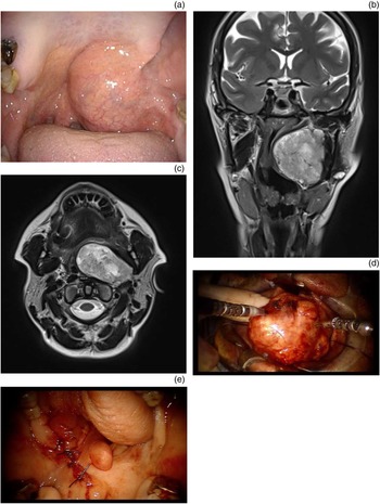
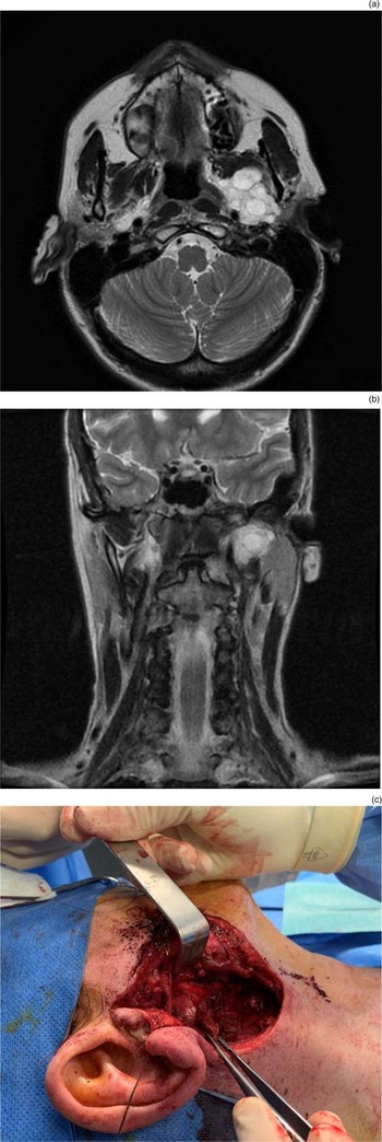
The surgical approaches used were the transparotid-transcervical approach (52.4 per cent, n = 11), transoral robotic surgery (28.6 per cent, n = 6) or the conventional transoral approach (19 per cent, n = 4) (Figure 1 and 2). No mandibular swing or combined approaches were required. The en bloc tumour excision was achieved with no intra-operative macroscopic capsular rupture in all but 3 patients (14.3 per cent). The first patient operated on with the transparotid-transcervical approach had a mass in contact with the skull base, for which the access was limited, leading to capsular rupture. The other two cases underwent transoral robotic surgery and had focal capsular spread. The fact that both tumours had prior biopsy breaching the capsule may be a predisposing factor for intra-operative capsular rupture. However, the excision was deemed macroscopically complete in all three patients, and no residual tumour was seen on the MRI conducted at one month and one year post-operatively.
Pleomorphic adenoma of the left parapharyngeal space in a 52-year-old woman. (a) Clinical examination showing an oropharyngeal bulge, (b) pre-operative coronal plane magnetic resonance imaging (MRI) scan, (c) pre-operative axial plane MRI scan showing a 5 cm mass occupying the parapharyngeal space, (d) intra-operative view of final dissection and delivery of the tumour with the finger and (e) intra-operative view of primary closure of the oropharynx.

Pleomorphic adenoma of the upper portion of the left parapharyngeal space in a 52-year-old woman, discovered incidentally on magnetic resonance imaging (MRI). (a) Pre-operative axial plane MRI slice, (b) pre-operative coronal MRI slice showing a mass limited to the upper portion of the parapharyngeal space, extending to the skull base and (c) intra-operative view after facial nerve dissection and tumour excision.

The mean operating time of each approach was as follows: transparotid-transcervical, 246.6 minutes; transoral robotic surgery, 190.8 minutes; and transoral surgery, 108.8 minutes. The transparotid-transcervical approach was significantly longer than the conventional transoral approach and transoral robotic surgery (p < 0.01). In addition, transoral robotic surgery was longer than the conventional transoral approach (p = 0.0004). There was no documentation of significant blood loss intra-operatively in our series. The length of hospitalisation showed no significant difference between the three groups, with a median of five days in all three surgical approaches.
The final histopathology was confirmed as pleomorphic adenoma in all patients. Most tumour subtypes (87.5 per cent) were stroma rich or myxoid. Adverse pathological features were seen in only two patients who presented with an incomplete capsule alongside either pseudopodia (n = 1) or satellite nodules (n = 1). Clear margins could not be confirmed in only the 3 patients with a capsular rupture (14.3 per cent).
The most common complication was facial nerve palsy (42.9 per cent, n = 9) in the transparotid-transcervical group, which was limited to the marginal mandibular nerve (33.3 per cent, n = 7) or was diffused for two patients (9.5 per cent) with House–Brackmann grade IV facial paralysis. The weakness was transient and resolved during follow up of up to nine months in all cases in the transparotid-transcervical group. It was attributed to prolonged retraction of the nerve during the dissection in the parapharyngeal space. Transient limitation of mouth opening was seen in 2 patients (9.5 per cent) of the conventional transoral group. It was likely related to prolonged use of the mouth gag. It was noted that no instances of first-bite syndrome occurred. Two patients (9.5 per cent) presented with Frey's syndrome. Both patients underwent a transparotid-transcervical approach for a tumour involving the superficial lobe, without possibility of preserving the superficial musculoaponeurotic system. In the transoral robotic surgery group, one patient presented with wound dehiscence at day seven, which required a transoral revision surgery for wound closure.
After a mean follow up of 21 months (range, 1–60), no patient experienced recurrence. Our protocol of follow up included a clinical examination one month and one year after surgery and an MRI at one year post-operatively. In case of intra-operative capsular rupture, an MRI was performed at one month to confirm the completeness of excision. Most patients were then referred to their primary care physician for further follow up.
Discussion
The clinical presentation in our series was consistent with what is found in the literature.Reference Singh, Gupta and Singla1,Reference Riffat, Dwivedi, Palme, Fish and Jani3,Reference Kuet, Kasbekar, Masterson and Jani10 The majority of our patients were asymptomatic at the time of diagnosis, which is a strong indicator that parapharyngeal space lesions are underdiagnosed. In terms of clinical investigation, MRI and FNAC were sufficient for an adequate clinical diagnosis and to determine the preferential surgical approach. Magnetic resonance imaging is the preferred imaging as it provides sufficient information concerning the tumour origin, nature, size and its relation to nearby vital structures. Moreover, it is well documented that MRI is superior to CT in diagnosis and assessment of pleomorphic adenoma of the parapharyngeal space.Reference Som, Sacher, Stollman, Biller and Lawson11,Reference Basaran, Polat, Unsaler, Ulusan, Aslan and Hafiz12 Therefore, we can suppose that MRI is sufficient to suggest the diagnosis of pleomorphic adenoma pre-operatively. However, we believe that FNAC can still provide a significant input, despite limited access to the parapharyngeal space. It largely relies on the experience of the cytologist and the surgeon or radiologist taking the sample.Reference Matsuki, Miura, Tada, Masubuchi, Fushimi and Kanno13–Reference Arnason, Hart, Taylor, Trites, Nasser and Bullock15
Understanding the anatomy of parapharyngeal space is crucial for the correct diagnosis and, most importantly, to decide how to approach the parapharyngeal space for the tumour excision. The majority of parapharyngeal space tumours are of salivary origin and are located in the pre-styloid compartment.Reference Riffat, Dwivedi, Palme, Fish and Jani3,Reference Chen, Sun, Tang and Hu4,Reference Kuet, Kasbekar, Masterson and Jani10 There are several factors determining the surgical approach to a parapharyngeal space tumour (e.g. its size, location and histopathology). Nevertheless, the decision is always subjective and depends on the surgeon's preference and comfort level.Reference Boyce, Curry, Luginbuhl and Cognetti16 An attempt to standardise surgical approach decision-making was made by Kanzaki and Nameki before the advent of transoral robotic surgery and relying on the pre-operative imaging and the division of the parapharyngeal space into six compartments.Reference Kanzaki and Nameki17
The transoral approach was first described by Harry Ehrlich in 1950 and is the most debated surgical approach in the literature.Reference Markou, Blioskas, Krommydas, Psillas and Karkos18,Reference Ehrlich19 It is associated with minimal access and poor vision of the operating field,Reference De Virgilio, Costantino, Mercante, Di Maio, Iocca and Spriano20 and it was only used during the first years of our study period, prior to the use of transoral robotic surgery in our centre. Transoral robotic surgery is a relatively new approach, with the first documented use in 2007.Reference O'Malley and Weinstein21 The Da Vinci® robotic system has the advantages of a magnified three-dimensional visualisation, tremor-reduction technology and wristed instruments with seven degrees of freedom.Reference Boyce, Curry, Luginbuhl and Cognetti16 Some studies suggest these advances have reduced the risk for tumour rupture, incomplete excision and uncontrollable bleeding intra-operatively.Reference De Virgilio, Costantino, Mercante, Di Maio, Iocca and Spriano20 For centres where transoral robotic surgery is not available, an endoscope-assisted transoral approach seems to provide satisfactory access.Reference Liu, Yu and Zhen22 The pre-operative investigation is crucial before making the decision of a transoral approach. The evaluation of the mouth opening is the most important aspect to anticipate the quality of both exposure and working space. Pre-operative imaging is paramount to rule out carotid artery encasement and bony erosion of the skull base. The main contraindications for transoral approaches are trismus, macroglossia and maxilla-mandibular defects.Reference Maglione, Guida, Pavone, Longo, Aversa and Villano23,Reference Arora, Kotecha, Acharya, Garas, Darzi and Davies24 Other studies advised against transoral robotic surgery for tumours bigger than 6 cm.Reference Maglione, Guida, Pavone, Longo, Aversa and Villano23 Another advantage of transoral robotic surgery is to limit the need for a transpalatal approach. The entrance door to the parapharyngeal space is made by a linear mucosal incision around the tonsillar fossa, preserving the palatoglossus, palatopharyngeus and superior constrictor muscles. Dividing the pterygoid muscles can improve the mouth opening and transoral exposure. There is usually no need for a mucosal reconstruction as a primary closure can be achieved.
The main risks for all transoral approaches are neurovascular injury, tumour spillage or implantation, incomplete excision, and surgical site infection.Reference Markou, Blioskas, Krommydas, Psillas and Karkos18,Reference Chan, Tsang, Eisele and Richmon25 In our series, two patients out of six from the transoral robotic surgery group presented with intra-operative capsular rupture. Both patients had undergone transoral incisional biopsies prior to the surgery, which was likely to induce a capsular fragility. A history of biopsy should be considered as an argument in favour of a transoral approach, in order to avoid seeding along the transcervical incision and to obtain a safe mucosal margin intra-orally. Moreover, follow-up imaging did not show any evidence of residual or recurrent disease. Some authors claim that tumour spillage during a transoral approach allows for a thorough washing and prevents tumour dissemination in the neck.Reference Sethi, Dale, Vidhyadharan, Krishnan, Foreman and Hodge26
The transparotid-transcervical approach was the most common in our series and is preferred by many for the good access and visualisation it provides.Reference Cohen, Burkey and Netterville27–Reference Zhi, Ren, Zhou, Wen and Zhang29 The keystone for this surgery is to preserve the facial nerve and limit the retraction injury. In order to improve the exposure, the authors recommend a nasotracheal intubation for a complete jaw closure during the surgery and an extension of the classical Blair's incision in the submandibular area to provide a better exposure of the major blood vessels and cranial nerves. The posterior belly of digastric muscle, stylohyoid muscle and stylomandibular ligament are commonly divided. The submandibular gland is retracted anteriorly or excised, and the mandible is protracted to identify the major neurovascular structures of the post-styloid space.Reference Olsen8,Reference Vallabhaneni, Mandakulutur, Vallabhaneni, Prabha and Banavara30 A parotidectomy is performed, as completely as possible posteriorly and in the deep lobe. Some studies advocate for preserving the superficial lobe of the parotid gland whenever feasible, with the aim of reducing the risk of Frey's syndrome and for a better cosmetic outcome.Reference van Hees, van Weert, Witte and René Leemans31 The main indication for this approach is a mass reaching the skull base and/or merging laterally within the deep lobe of parotid gland with ill-defined contour on radiological investigations.Reference Chen, Sun, Tang and Hu4,Reference Horowitz, Ben-Ari, Wasserzug, Weizman, Yehuda and Fliss5
The transcervical approach is the most frequently used for excision of parapharyngeal space mass of all origins, most notably those arising from the post-styloid compartment.Reference Markou, Blioskas, Krommydas, Psillas and Karkos18 The parapharyngeal space is accessed through a submandibular incision at the level of the hyoid bone, which can be further extended to the submental area for mandibulotomy with lip splitting. Many surgical steps are similar to those of the transparotid-transcervical approach (i.e. retraction of the submandibular gland, division of the posterior belly of the digastric muscle, the stylohyoid muscle and stylomandibular ligament and protraction of the mandible). The facial nerve is not identified. The mylohyoid muscle can be transected for further exposure of parapharyngeal space.Reference Basaran, Polat, Unsaler, Ulusan, Aslan and Hafiz12 However, none of our cases underwent an exclusively transcervical approach because all tumours originated from the deep lobe of the parotid gland, which justified a parotidectomy in our opinion. Some studies suggest the transcervical approach should be avoided for masses bigger than 4 cm because the mandible is a barrier for tumour manipulation and extraction.Reference Riffat, Dwivedi, Palme, Fish and Jani3,Reference Bozza, Vigili, Ruscito, Marzetti and Marzetti6,Reference Markou, Blioskas, Krommydas, Psillas and Karkos18,Reference Cohen, Burkey and Netterville27,Reference Papadogeorgakis, Petsinis, Goutzanis, Kostakis and Alexandridis32 Furthermore, it is not recommended for tumours with a long vertical dimension and with radiological suspicion of cranial foramen invasion as they are associated with a high risk of internal carotid artery laceration during blunt dissection.Reference Basaran, Polat, Unsaler, Ulusan, Aslan and Hafiz12 Overall, many authors support the transcervical approach because it can provide good control of the lesion while avoiding dissection of the facial nerve, but it is mainly reserved for tumours with limited attachment to the deep lobe of the parotid.Reference Horowitz, Ben-Ari, Wasserzug, Weizman, Yehuda and Fliss5,Reference Malone, Agrawal and Schuller33
Mandibulotomy is essentially indicated for malignant tumours, recurrent neoplasms, large benign tumours and highly vascular neoplasms with a need for vascular control.Reference Basaran, Polat, Unsaler, Ulusan, Aslan and Hafiz12 There are three ways of conducting the osteotomy: through the body (lateral mandibulotomy), and midline and paramedian approaches. Following the osteotomy, the mandible is swung and the floor of the mouth is stretched. An incision in the floor of the mouth is made 1 cm medial to the gingiva across the mucosa, soft tissues and muscles of mouth floor and extended up to the anterior tonsillar pillar. It carries a possible risk of inferior alveolar nerve anaesthesia, malocclusion, teeth loss, malunion or non-union of the mandible, and in some cases it might require a tracheostomy.Reference Vallabhaneni, Mandakulutur, Vallabhaneni, Prabha and Banavara30,Reference Patel34 No mandibulotomy was performed in our series, although it is reported in 2.0 to 20.5 per cent of cases in the literature.Reference Olsen8,Reference Cohen, Burkey and Netterville27,Reference Malone, Agrawal and Schuller33,Reference Hughes, Olsen and McCaffrey35,Reference Miller, Wanamaker, Lavertu and Wood36 From our standpoint, mandibulotomy is now unnecessary for the vast majority of pleomorphic adenomas of the parapharyngeal space. Additionally, no combined approach was necessary in our series for a complete tumour manipulation and excision. As a result, the morbidity and complications of the mandibular split were avoided, and the length of hospitalisation was limited.
• Parapharyngeal space tumours can be excised with various surgical approaches
• Accessibility and involved structures, shown on magnetic resonance imaging and computed tomography, are the main determining factors for surgical approach
• The transparotid-transcervical approach is chosen when there is no oropharyngeal bulge or there is intra-parotid location or lateral extension behind the pterygoid process or inner ramus of mandible
• The chosen approach must provide adequate tumour visualisation in order to achieve clear excision margins
The histopathological subtypes of pleomorphic adenoma are divided into three groups based on their epithelial element: mucoid, myxoid or chondroid.Reference Seifert, Brocheriou, Cardesa and Eveson37 The most common is the myxoid subtype, which is documented to have a higher rate of recurrence and is associated with incomplete and thin capsules.Reference Dulguerov, Todic, Pusztaszeri and Alotaibi38 However, there are a number of pathological differences between a pleomorphic adenoma originating from the superficial or deep lobe of the parotid gland. Deep lobe pleomorphic adenomas tend to be bigger, have a thicker capsule and there is less invasion of the capsule by the tumour.Reference Harney, Murphy, Hone, Toner and Timon39 The rate of recurrence documented in the literature ranges between 0 and 10.5 per cent.Reference Horowitz, Ben-Ari, Wasserzug, Weizman, Yehuda and Fliss5,Reference Basaran, Polat, Unsaler, Ulusan, Aslan and Hafiz12,Reference Cohen, Burkey and Netterville27,Reference van Hees, van Weert, Witte and René Leemans31,Reference Shahab, Heliwell and Jones40,Reference Lim, Park, Kang, Kim, Choi and Kim41 Our rate of complete surgical excision and the absence of recurrence during the follow up seem to validate our management. Indeed, a concession can be made on the surgical exposure in order to avoid the morbidity of a systematic transcervical approach or a mandibulotomy.
The complications were dominated by facial nerve palsy in our series because of the prolonged retraction of the nerve during tumour dissection in the parapharyngeal space. This occurs from 5.2 to 30.8 per cent of cases in the literature. Permanent facial nerve palsy has been reported after up to 7.7 per cent of procedures.Reference Ijichi and Murakami2,Reference Chen, Sun, Tang and Hu4–Reference Bozza, Vigili, Ruscito, Marzetti and Marzetti6,Reference Cohen, Burkey and Netterville27,Reference van Hees, van Weert, Witte and René Leemans31
The limits of our study are its retrospective design and the small number of patients, which prevented statistical comparison between groups and a short follow-up period. Indeed, these tumours evolve slowly and late recurrences can still occur.
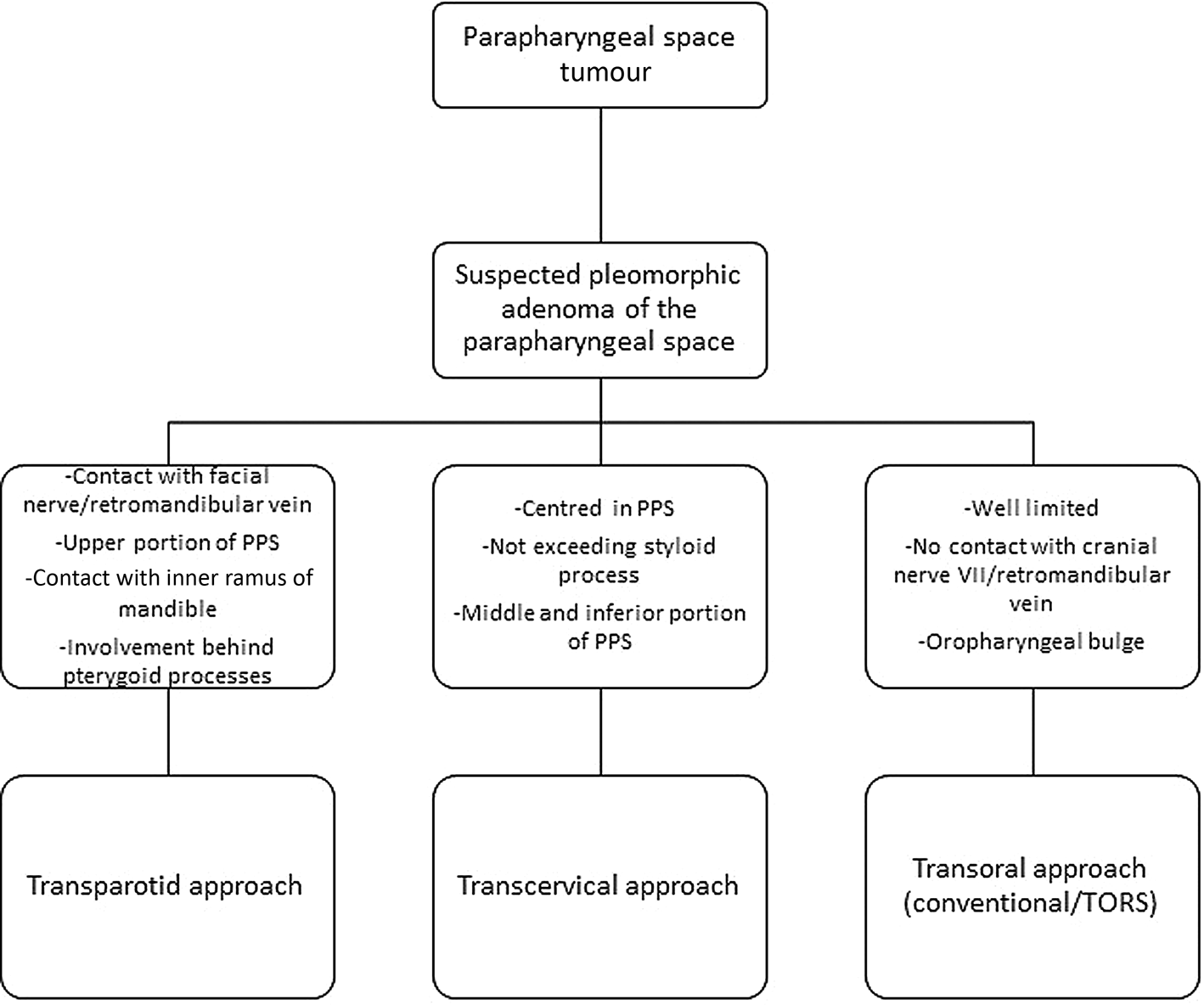
Our decision tree for management of pleomorphic adenoma of the parapharyngeal space was based on clinical examination and radiological findings (Figure 3). The transparotid-transcervical approach was preferred when close contact to the facial nerve was suspected. It should be preferred to the transcervical approach for salivary tumours. Transoral robotic surgery is a good option for selected, well-defined masses in the parapharyngeal space with oropharyngeal bulge clinically and radiologically. The classic transoral approach should be avoided if transoral robotic surgery is available because it is associated with significant risk of neurovascular injury with limited capability for intra-operative management. Indications for mandibulotomy must remain exceptional.
Proposal of decision-tree for management of pleomorphic adenoma of the parapharyngeal space (PPS). TORS = transoral robotic surgery.

Conclusion
Pleomorphic adenomas of the parapharyngeal space are rare tumours, often asymptomatic and largely underdiagnosed. Pre-operative assessment is paramount in order to choose the most suitable surgical approach. In selected patients, transparotid-transcervical, transoral robotic surgery and conventional transoral approaches can provide adequate tumour visualisation, a high rate of clear excisional margins and an acceptable morbidity. Indications for mandibulotomy for pleomorphic adenoma of the parapharyngeal space should be exceptional.
Competing interests
None declared