Introduction
Printing and dyeing chemical wastewater refers to the wastewater produced during the production and use of dyes, which contains a large amount of organic compounds [Reference An, Guo, Lee, Jeong, Zhao, Wang and Leiknes1]. These organic pollutants, such as methylene blue (MB), cause severe damage to the human body and to the environment [Reference Albadarin, Collins, Naushad, Shirazian, Walker and Mangwandi2]. How to remove these pollutants efficiently has attracted the attention of scientists [Reference Schwarzenbach, Escher, Fenner, Hofstetter, Johnson, Von Gunten and Wehrli3, Reference Martinez-Huitle and Ferro4, Reference Glassmeyer, Furlong, Kolpin, Batt, Benson, Boone and Mash5, Reference Zhang, Gu, Chen, Li and Jiang6, Reference Zhang, Yang, Liu, Sun and Xu7, Reference Lu, Xin, Zhang, Tang and Xu8, Reference Xie, Zhang, Tang, Anpo and Xu9]. For the existing water treatment technology, the conventional physical methods cannot effectively remove most of the organic pollutants [Reference Robinson, McMullan, Marchant and Nigam10, Reference Stackelberg, Furlong, Meyer, Zaugg, Henderson and Reissman11, Reference Giebner, Ostermann, Straskraba, Oetken, Oehlmann and Wagner12]. The organic pollutants removed are only the phase transformation, which is very likely to cause secondary pollution. The biological treatment methods commonly used in the existing sewage treatment processes can degrade and remove most of the organic pollutants [Reference Ahmed, Zhou, Ngo, Guo, Thomaidis and Xu13, Reference Yu, Han and He14, Reference Oller, Malato and Sánchez-Pérez15]. But the removal efficiency is unstable, and it depends on the activity of the sludge and the types of organic pollutants. Advanced oxidation technology has a good effect on the removal of organic pollutants [Reference Bolong, Ismail, Salim and Matsuura16], and can directly use solar energy to drive a series of chemical reactions [Reference Tang and An17, Reference Mamaghani, Haghighat and Lee18, Reference Yu, Jiang, He, Liu, Zhou, Ji and Chen19]. It has the advantages of low energy consumption, mild reaction conditions, and no secondary pollution. It is an effective way to solve the organic pollutions. The key to achieving this process is to find and design efficient photocatalytic material. The current photocatalytic material is mainly composed of inorganic semiconductors [Reference Song, Li and Wang20, Reference Kattel, Ramírez, Chen, Rodriguez and Liu21, Reference Wang, Peng, Li, Zhao and Fu22, Reference Yan, Xu, Chai, Wang, Wang and Ren23, Reference Li, Zhu, Wang, Min, Fang, Huang, Liu and Wu24, Reference Li, Xie, Jiang, Yu and Zhang25, Reference Ma, Wang, Xu and Wang26, Reference Yang, Wang, Xu, Qu, Ding and Chen27, Reference Chen, Yang, Luo, Wang and Yu28], which are limited in practical application due to their low efficiency of absorption light.
Metal–organic frameworks (MOFs) are a kind of very promising heterogeneous material [Reference Zhao, Song, Song and Zhang29, Reference Zhu, Liu, Jiang and Sun30, Reference Ye, Gagliardi, Cramer and Truhlar31, Reference Mueller, Schubert, Teich, Puetter, Schierle-Arndt and Pastre32, Reference Huang, Zhang, Song, Chen, Han and Wen33, Reference Ye, Li, Yu, Chen and Quan34], which as photocatalytic material have been studied for hydrogen evolution [Reference Lan, Zhu, Veroneau, Xu, Micheroni and Lin35], pollutants degradation [Reference Liu, Wang, Ma and Zhang36], and CO2 reduction [Reference Kung, Audu, Peters, Noh, Farha and Hupp37]. Actually, several silver MOFs have been proved to be efficient catalysts for many organic reactions, such as photocatalytic degradation of phenol, cycloaddition of azides and alkynes, oxygenation of sulfide, and partial oxidation of primary amines [Reference Huang, Wu, Tian, Wang, Zhou and Li38, Reference Ma and Zhu39]. The use of some Ag-MOFs as biocide material in water treatment and biomedical applications also has been evaluated [Reference Martín-Betancor, Aguado, Rodea-Palomares, Tamayo-Belda, Leganés, Rosal and Fernández-Piñas40, Reference Wyszogrodzka, Marszałek, Gil and Dorożyński41]. In addition to the above applications, photocatalytic degradation of organic compounds in printing and dyeing chemical wastewater has been widely recognized as one of the most valuable applications. Therefore, it is very necessary to develop and design synthetic high-efficiency photocatalytic materials to solve the above problems.
In this paper, a novel silver-based metal–organic framework (Ag-MOF), [{Ag(H2btc)}{Ag2(Hbtc)}]n (1) (H3btc = 1,3,5-trimesic acid) is synthesized. The photocatalytic degradation of MB mechanism and MB degradation pathway is analyzed by EPR and ESI-MS spectra. The results are helpful for in situ research of MOFs photocatalytic mechanism.
Results and discussion
Structure of 1
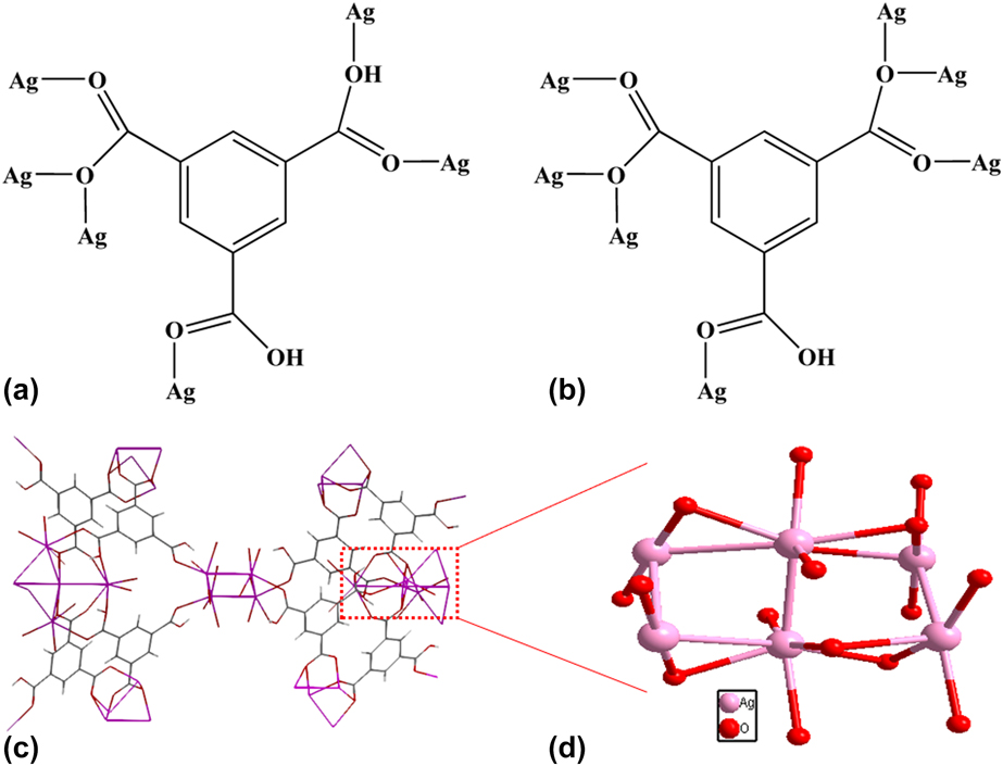
Here, a novel silver-based metal–organic framework (Ag-MOF), [{Ag(H2btc)}{Ag2(Hbtc)}]n (1) (H3btc = 1,3,5-trimesic acid) is synthesized by hydrothermal. To demonstrate the presence of Ag(I) inos in 1, the solids are characterized by X-ray photoelectron spectroscopy (XPS). The results show that both Ag3d5/2 and Ag3d3/2 peaks [Figs. 1(a) and 1(b)] can be divided into two separate peaks. Among them, the peaks at 367.4 and 373.4 eV are attributed to Ag+ in 1. An X-ray single-crystal diffraction analysis reveals that 1 crystallizes in orthorhombic system, Fddd space group, with a = 14.8812(7) Å, b = 25.8729(14) Å, c = 36.3742(18) Å, α = β = γ = 90°, and V = 14004.8(12) Å3. The coordination environment of Ag(I) ions in 1 is shown in Figs. 2(a) and 2(b). The empirical formula of 1 is C18H8Ag3O12, which consists of three Ag(I) ions, one H2btc− anion, and one Hbtc2− anion. There are Ag2(H2btc)2 (A) and Ag8(Hbtc)12/3 (B) whose two independent building units in 1. The coordination mode of H2btc− in A is shown in Fig. 2(a). There are three different coordination modes in three carboxyl groups of H2btc− anion. They include one of the carboxyl groups in H2btc− anion coordinated with one Ag(I) ion, one in H2btc− anion coordinated with two Ag(I) ions, and one in H2btc− anion coordinated with three Ag(I) ions. So, the H2btc− adopts a μ6-η1:η2:η3 bridging style to bridge six Ag(I) ions.
(a) XPS of core level region of Ag+ in 1; (b) XPS of 1.

(a) and (b) The coordination mode of 1,3,5-trimesic acid and Ag(I) in 1; (c) Three-dimensional structure is formed by Ag–Ag bonds; (d) The Ag–Ag bonds in 1.

However, there are only two coordination modes [Fig. 2(b)] for the carboxyl groups in B. Among them, two carboxyl functional groups (in which H+ has been removed) are coordinated with three Ag(I) ions, respectively; But the carboxyl that has H+ is only coordinated with one Ag(I) ion. Therefore, the Hbtc2− ligand in B connects to the A by a μ7-bridge to form a three-dimensional structure [Fig. 2(c)]. Since these Ag(I) ions are coordinated with the carboxyl anions of ligands, 1 is very stable within 405 °C (Fig. S1). The SEM image (Fig. S2) of 1 is consistent with its crystal structure. The surface area of 1 is determined using BET technique, and its specific surface area (Fig. S3) is 34.33 m2/g, which is very similar to P25. It is worth noting that there are Ag–Ag bonds [Fig. 2(d)] in 1. They are linked to each other in a three-dimensional structure. Moreover, these Ag–Ag bond distances are different, enriching the electronic transition and facilitating the absorption and utilization of light.
UV-vis diffuse reflectance spectrum and redox potential
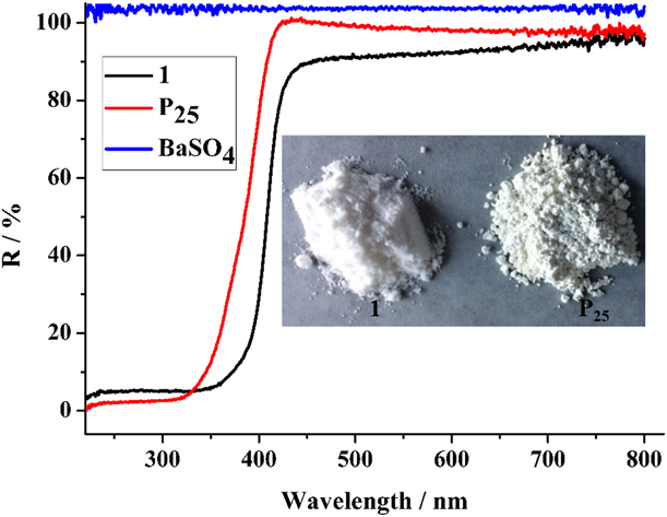
As can be seen from Fig. 3 that the reflection intensity of 1 is over 95%, and its absorbance is less than 5% in the visible range (421–800 nm). At 421 nm, the sample reflect the light very effectively, and it absorbs ultraviolet light in the wavelength range of 220–400 nm, which indicate that the absorption spectrum of 1 is dominated by visible light and ultraviolet light of wavelengths below 421 nm. However, UV-Vis DRS of P25 indicated that it was dominated by ultraviolet light of wavelengths below 400 nm. The cyclic voltammetry curve (Fig. S4) of 1 shows that there is a strong oxidation peak at 0.91 eV and a reductive peak at −1.73 eV. So, the bandgap (eV) of 1 is 2.64 eV. The HOMO = −5.35 eV and LUMO = −2.71 eV of 1 can be calculated by the following formula [Reference Bredas, Silbey, Boudreaux and Chance42]: E HOMO/eV = −4.44 − E onset (Ox); E LUMO/eV = −4.44 − E onset (Red).
UV-Vis diffuse reflectance spectrum of 1 and P25.

Photocatalytic performance
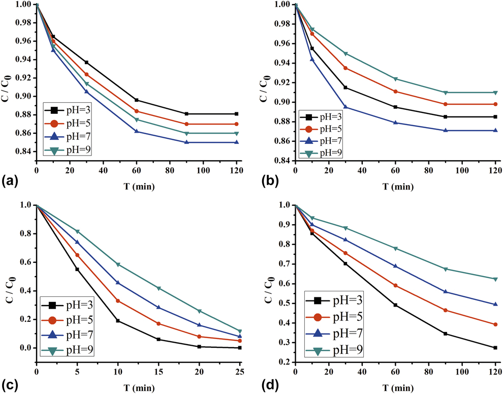
The main aim of this study was to assess the catalyst activity in MB oxidation reaction. The reaction conditions are as follows: MB initial concentration C 0 = 5 mg/L, pH = 3–9, and dosage: 50 mL; irradiated by UV light with a 175-W Hg lamp (λ > 350 nm, Toshiba SHLS-1002A), 250-mL round glass container with a circulating water jacket, and temperature (T) = 25 °C. The weight of 1 and P25 are 10 mg, respectively. The adsorption capacity and photocatalytic degradation MB performance of 1 and P25 are shown in Fig. 4. It can be seen from Figs. 4(a) and 4(b) that 1 and P25 adsorb MB for 90 min to achieve adsorption equilibrium. The adsorption capacity of MB is the largest at pH 7, reaching 0.00075 g/g and 0.0007 g/g, respectively. When the pH is less than 7, the adsorption capacity also decreases as the pH decreases. This shows that H+ or OH− is adsorbed on the surface of the catalyst under acidic or alkaline conditions, and they constitute a competitive adsorption with MB, resulting in a decrease in the amount of adsorption. It can be seen that MB can be adsorbed by 1 under the condition of pH = 3–9, indicating that 1 has the ability of photocatalytic degradation of MB.
The adsorbed amount of MB at pH 3–9 of 1 (a) and P25 (b); Photocatalytic degradation of MB at pH 3–9 of 1 (c) and P25 (d).

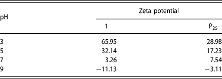
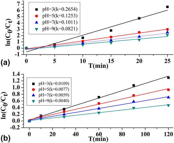
The P25 is a very promising heterogeneous catalyst, and it has good acid–base stability and visible light response, which has been studied for hydrogen evolution and pollutants degradation [Reference Shinde, Wadhai, Ponkshe, Kapoor and Thakur43, Reference Li, Shen, Ma, Chen and Xie44, Reference Xing, Zhang, Qiu, Tian, Anpo and Che45, Reference Tong, Yang, Xiao, Tian and Jiang46]. In order to verify the efficient photocatalytic degradation MB of 1, the reference experiments of P25 under the same conditions are performed. As seen from Figs. 4(c) and 4(d) that 1 and P25 both can be photocatalytic degradation MB. Among them, 1 can photocatalytically degrade 99.8% of MB in 20 min at pH = 3 [Fig. 4(c)]. However, under the same conditions, the rate constant of photocatalytic degradation MB by P25 is also very fastest, and about 72.9% of MB is photocatalytically degraded in 120 min at pH = 3 [Fig. 4(d)]. It can be seen that the photocatalytic decomposition of MB is more favorable under acidic conditions, mainly due to its specific surface charge. The zeta potential versus pH (Table I) of 1 and P25 shows that their zeta potentials at pH 3 are 65.95 mV and 28.98 mV, respectively. Because the photon quantum efficiency is determined by e−/h+ composite and the captured process [Reference Lan, Zhu, Veroneau, Xu, Micheroni and Lin35], an increase in the acidity of the photocatalyst surface will adsorb e− to the catalyst surface, prolonging the recombination time of e−/h+, thereby increasing quantum efficiency. So, the larger the surface charge of photocatalyst, the higher the photocatalytic quantum efficiency. In addition, there is a benzene ring in 1 (Fig. 2), which can adsorb MB through the Π–Π bond, resulting in the surface of 1 adsorbing more MB than P25 [Figs. 4(a) and 4(b)]. Due to these reasons, 1 is more efficient photocatalytic degradation of MB than P25. There is a good linear relationship between ln (C 0/C t) and time (t) of 1 and P25, indicating that the MB photocatalytic degradation reaction conforms to the first-order kinetic reaction (Fig. 5). The rate of 1 is 0.2654, which is 24 times higher than that of P25 at pH 3.
The zeta potential versus pH of 1 and P25.

The first-order kinetics of MB degradation in the presence of (a) 1 and (b) P25.

Photocatalytic mechanism
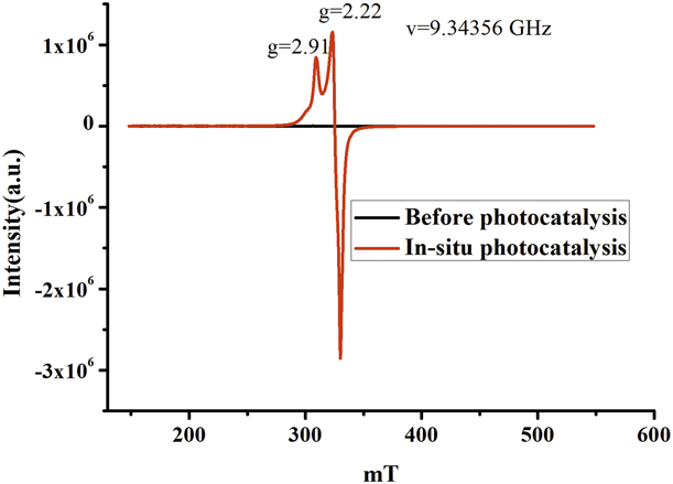
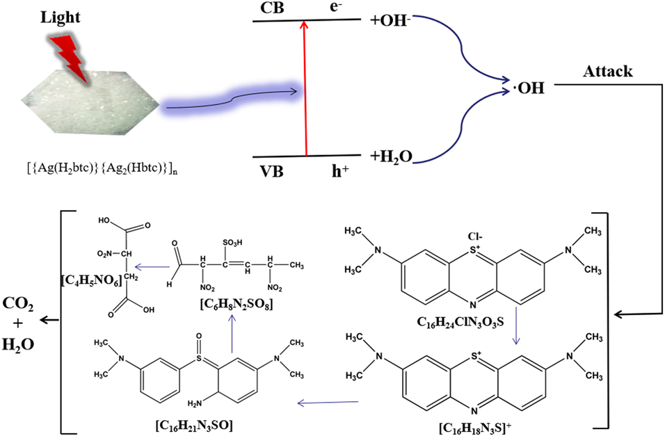
The in situ EPR (Fig. 6) indicates that there are single electrons in the photocatalytic process. These single electrons may be derived from Ag or Ag(II) ions because there are a large number of Ag–Ag bonds in 1, which are very prone to disproportionate when light is irradiated, and formed Ag and Ag(II) ions. The Ag(II) with super oxidizing ability can oxidize the organic compounds to CO2 and H2O. To further investigate the decomposition process of photocatalytic degradation MB by 1, the ESI-MS spectroscopy shows that four signals (Fig. S5) attributed to [C16H18N3S]+, [C16H22N3SO]+, [C6H9N2SO8]+, and [C4H6NO6]+ are observed. So, the degradation pathways of MB are shown in Fig. 7. This result is consistent with the DFT calculation that reported in the literature [Reference Li, Wei, Hu, Wu and Li47].
The in situ EPR spectrum of 1 sample under 175 W high-pressure mercury lamp radiation catalyzed the MB at 273 K.

The photocatalytic degradation pathway and mechanism of MB.

Recycling experiments of 1
To investigate the performance stability of this 1, the solid powders after the photocatalytic degradation MB of 1 are characterized by PXRD (Fig. S6). Results show that the peak and position of the test data are the same as the simulation of 1. This shows that the structure of 1 remains unchanged after photocatalysis. Therefore, recycling experiments is performed at pH 3, and the corresponding results (Fig. S7) show that the efficiency of 1 is nearly 100% for the first cycle and remained above 99% even after five cycles.
Conclusion
In summary, a metal–organic framework with Ag nodes and 1,3,5-trimesic acid linker has been constructed by hydrothermal, which can be employed as a highly effective catalyst for the degradation of MB. The mechanism of photocatalytic degradation MB shows that Ag(I) becomes Ag(II) during photocatalytic process, and the degradation pathways also show that MB is divided into multistep photocatalytic degradation. The present work not only provides some new insight into the photocatalytic mechanism of silver-based metal–organic frameworks but also confirms the photocatalytic degradation pathways of MB.
Experimental
Materials
All solvents and chemicals are commercial reagents and used without further purification. The titanium dioxide photocomplex (P25, 80% anatase, 20% rutile, BET area, approximately 50 m2/g) is kindly supplied by Degussa. The AgNO3 and 1,3,5-trimesic acid and MB are purchased from Energy Chemical Company (Shanghai, China).
[{Ag(H2btc)}{Ag2(Hbtc)}]n (1)
AgNO3 (0.0501 g, 2 mmol) and H3btc (0.042 g, 2 mmol) were mixed in 10 mL distilled water and then sealed the mixture in a 23-mL Teflon-lined stainless steel autoclave. The mixture is heated in an oven at 110 °C for 48 h and cooled to room temperature at a rate of 10 °C/h. Colorless massive crystals of 1 are obtained.: yield: 85% (based on H3btc). Anal. Calcd for C18H8Ag3O12: C, 29.20; H, 1.08; O, 25.95. Found: C, 29.23; H, 1.11; O, 25.98. IR(cm−1): 3117w, 3065w, 1681s, 1601s, 1560s, 1503s, 1434w, 1251s, 1199m, 934m, 745s, 670s, 509m, 406w.
X-ray crystallography
Colorless massive single crystal of 1 is mounted on the top of a glass fiber. The diffraction data for 1 are measured on a Bruker Smart APEX-II CCD diffractometer (Bruker, Karlsruhe, Germany) equipped with a graphite-monochromatized MoKa radiation (λ = 0.71073 Å) at 296(2) K. The anisotropic displacement parameters are applied to all nonhydrogen atoms in which full-matrix least-square refinements based on F2 are performed using SHELXL-2013. The structure is solved by direct methods using Olex2 program, Durham University, Durham, England [Reference Dolomanov, Bourhis, Gildea, Howard and Puschmann48] and refined with Olex2 program. [Reference Dolomanov, Bourhis, Gildea, Howard and Puschmann48] Anisotropic thermal parameters are assigned to all nonhydrogen atoms. The hydrogen atoms are placed at calculated positions and refined as riding atoms with isotropic displacement parameters. Crystallographic crystal data and structure processing parameters for 1 are summarized in Table SI. Selected bond lengths and bond angles for 1 are listed in Table SII. Hydrogen bonds for 1 are listed in Table SIII.
Photocatalytic experiments
Taking photocatalytic degradation of MB as an example, the photocatalytic performance of 1 is investigated and compared with P25. Reaction conditions are as follows: MB initial concentration C 0 = 5 mg/L, pH = 3–9, and dosage, 50 mL; a 175-W high-pressure mercury lamp is used as the light source, the distance between the light source and the solution to be measured is maintained at 15 cm, and a homemade cooling system is used to keep the solution temperature at room temperature; a 250-mL round glass container with a circulating water jacket, temperature (T) = 25 °C; the dosage of 1 and P25 is 10 mg. The concentration of MB is measured by UV-Vis 2550. The photocatalytic degradation performance is evaluated through the changes in the concentration of MB before and after the reaction. The degradation rate is calculated as follows:
 $$\eta = {{{C_0} - C} \over {{C_0}}} \times 100\% \quad ,$$
where η is the degradation rate (%) and C 0 and C are the quality of performance before and after degradation, respectively (mg/L).
Methodology section
Elemental analyses (carbon, hydrogen, and nitrogen) are performed with a Perkin-Elmer 240 elemental analyzer, PerkinElmer, Massachusetts. IR spectra are measured from KBr pellets on a Nicolet 5DX FT-IR spectrometer. The TGA is determined by Perkinelmer Pyris Diamond TG-DTA. The crystal structure is determined by a Bruker APEX-II CCD, Bruker, Karlsruhe, Germany. The XRD is performed using Rigaku’s D/max 2500 X-ray diffractometer (Rigaku, Kyoto, Japan) Cu Kα radiation (λ = 0.15604 nm), tube voltage is 40 kV, tube current is 150 mA, graphite monochromator, 2θ is 5 to 65°. The DRS-UV-Vis diffuse reflectance spectra are measured by UV-2700 from SHIMADZU, Kyoto, Japan, with BaSO4 as a reference. Scanning electron microscopy (HITACHI SU-8010, Hitachi, Kyoto, Japan) was used. The specific surface area was tested by AUTO CHEM II2920 from American Micro Instrument Company, Georgia. The photocatalytic degradation was studied using a 175-W high-pressure mercury lamp as a light source, and the irradiation intensity is approximately 30 mW/m2, as determined by a UV irradiation meter (Model UV-A; Photoelectric Instrument Factory of Beijing Normal University, Beijing, China). The concentration of MB in solution is measured with UV-Vis 2550 in 664 nm wavelength. The electrochemical cyclic voltammetry of 1 is conducted on a CHI 410B Electrochemical Workstation with Pt disk coated with the polymer film, Pt plate, and Ag/Ag+ electrode as working electrode, counter electrode, and reference electrode, respectively, in a 0.100 mol/L tetrabutylammonium hexafluorophosphate (Bu4NPF6) acetonitrile solution. The electrochemical potential is calibrated against Fc/Fc+.
Supplementary material
To view supplementary material for this article, please visit https://doi.org/10.1557/jmr.2018.507.
Author contributions
Xiu-Ju Yin and Long-Guan Zhu designed and conceived the experiments; Xiu-Ju Yin performed the experiments and analyzed the data; Xiu-Ju Yin wrote the paper with direction from Long-Guan Zhu; all authors participated in the analysis, interpretation, and review of the results and provided input in the writing process of the paper.
Acknowledgments
This work is supported by the project of education department of Guangxi province (No. YB2014331), the project of undergraduate teaching reform of higher education in Guangxi (No. 2018JGA254) and the subject of “12th Five-Year” Plan of Guangxi Education Science (No. 2015C408).