INTRODUCTION
Taenia multiceps inhabits the small intestine of wild and domestic canids while the larval stage develops in the central nervous system of sheep and other ungulates. Sheep, cattle or sometimes goats, deer, antelope and less commonly, human beings act as intermediate hosts of the parasites following ingestion of the eggs. The larvae hatch in the intestine through the blood stream towards different organs. The larvae, which reach the brain and spinal cord grow to the metacestode stage. Coenurus cerebralis will further mature in the brain and spinal cord. The clinical disease occurs in sheep and rarely in cattle (Soulsby, Reference Soulsby1982). The presence of cysts typically leads to neurological signs that cause the infected animals. As the location and depth of T. multiceps cysts are quite variable, there are no unique clinical signs. Ataxia, incoordination, paresis, head pressing, circling, blindness and coma may be observed in affected sheep. The disease may last several months and is commonly fatal (Abo-Shehada et al. Reference Abo-Shehada, Jebreen, Arab, Mukbel and Torgerson2002).
Turkey has a long tradition of rearing sheep and there is extensive local information on the management of sheep. Sheep are the main livestock of nomadic people in the east and southeast regions of Turkey. Although it is an offence to slaughter animals outside the abattoirs, illegal slaughtering of sheep is a common practice. Sheep are usually kept in large flocks, and mainly under free-range feeding. The prevalence of C. cerebralis in Turkey has been recorded in sheep, goat and cattle (Gicik et al. Reference Gicik, Kara and Arslan2007). The prevalence in sheep has been reported to be 15·5% in Kars, 1·3% in Istanbul and 16·3% in Konya (Akkaya and Vuruşaner, Reference Akkaya and Vuruşaner1998; Gicik et al. Reference Gicik, Kara and Arslan2007; Uslu and Guclu, Reference Uslu and Guclu2007). The prevalence of T. multiceps in dogs has been estimated to range between 0·32 and 12% in Turkey (Umur and Arslan, Reference Umur and Arslan1998).
Phenotypic differences in many instances reflect genotypic variability (McManus, Reference McManus2005). There have been very limited investigations about genetic variability within T. multiceps (Varcasia et al. Reference Varcasia, Lightowlers, Cattoli, Cansedda, Canu, Garippa and Scala2006; Al-Riyami et al. Reference Al-Riyami, Ioannidou, Koehler, Hussain, Al-Rawahi, Giadinis, Lafi, Papadopoulos and Jabbar2016). The presence of some clinical variation in coenurosis can indicate the existence of genetic intraspecific variability within the species (Varcasia et al. Reference Varcasia, Pipia, Dessi, Zidda, Tamponi, Pau, Scala and Boufana2016). Thus, for the aim of a molecular perspective, we described an investigation of genetic variability within Mardin isolates of T. multiceps in Turkey. DNA sequence variability was investigated within the cytochrome c oxydase subunit 1 (CO1) mitochondrial gene that it has been commonly used for the distinction of species and strains of other cestodes (McManus, Reference McManus2005).
MATERIAL AND METHODS
Sample collection
The C. cerebralis samples, were collected by a registered veterinary surgeon from sheep (n = 20) in Mardin province, which is situated in the southeast part of Turkey between the northern latitudes of 37° to 45° and eastern longitudes of 40° to 22°. Mardin has a hot-summer mediterranean climate with hot, dry summers and cold, wet and occasionally snowy winters. Temperatures in summer usually increase to 40 °C (104°F). Snowfall is quite common between the months of December and March, snowing for a week or two. Mardin has over 3000 h of sun per year. The highest recorded temperature is 42·5 °C (108·5°F). Average rainfall is about 641·4 mm (25 inches) per year (Turkish State of Meteorological Service). Cysts were obtained from the brain of individual sheep following either post mortem examination of clinically affected animals (by euthanizing of individual sheep) or following the routine slaughtering (by investigating the brain of all slaughtered sheep) in the abattoir during 2014. All cysts had protoscoleces according to morphological and microscopical examinations. Protoscoleces and/or membrane from individual cysts were fixed in 70% ethanol and then stored at −20 °C till used.
Genomic DNA isolation and polymerase chain reaction (PCR) amplification
Total genomic DNA was isolated from the protoscoleces and germinal membrane of the individual cysts with the use of a commercially available DNA extraction kit (MBI Fermentas, Vilnius, Lithuania) according to the manufacturer's instructions. A fragment (446 bp) of the CO1 gene was amplified with the use of the JB3 (5′-TTTTTTGGGCATCCTGAGGTTTAT-3′), and JB4.5 (5′-TAAAGAAAGAACATAATGAAAATG-3′) primer set (Bowles et al. Reference Bowles, Blair and McManus1992) under the following conditions: Pre-denaturating step at 95 °C for 5 min and 35 cycles (50 s 94 °C, 50 s 45 °C, 50 s 72 °C) followed by a final extension step (10 min 72 °C). Amplicons were electrophoretically separated on an ethidium bromide stained 1·4% agarose gel and visualized under UV. The CO1 bands were excised from the gel and purified with the use of QIAquick Gel Extraction Kit (Qiagen, Hilden, Germany). The sequences were automatically obtained by ABI PRISM Sequence Detection System, and their identity was determined by an NCBI BLAST search. Chromatograms’ quality was evaluated, the edges were trimmed and the ambiguities were corrected in FinchTV 1.4.0 (Geospiza Inc., Seattle Washington, USA) (http://www.geospiza.com).
Phylogenetic analyses
Multiple sequence alignment and phylogenetic tree construction of the obtained sequence of the mt-CO1 gene were performed with the use of CLC Main Workbench software (Knudsen et al. Reference Knudsen, Knudsen, Flensborg, Sandmann, Heltzen, Andersen, Dickenson, Bardram, Steffensen, Mansted, Lauritzen, Forsberg, Thanbichler, Bendtsen, Gorlitz, Rasmussen, Tordrup, Vaerum, Ravn, Hachenberg, Fisker, Dekker, Schultz, Hein and Sinding2007). Unidirectional DNA sequence analysis of the mt-CO1 gene of all samples was performed. Unreliable ends of the raw sequences were trimmed and then the phylogenetic tree was built using the neighbour-joining method (Saitou and Nei, Reference Saitou and Nei1987). Based on pairwise comparisons, sequence differences were calculated using the CLC Main Workbench software. The reliability of the inferred tree was evaluated by bootstrap analysis on 1000 replicates. Reference sequences of T. multiceps (JQ710587; FJ886783) were also included for phylogenetic analysis. Taenia saginata (JQ756979) and Echinococcus granulosus sensu stricto (EU006777) were used as an out group.
RESULTS
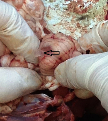
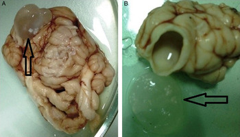
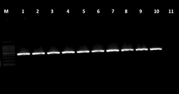
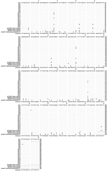
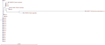
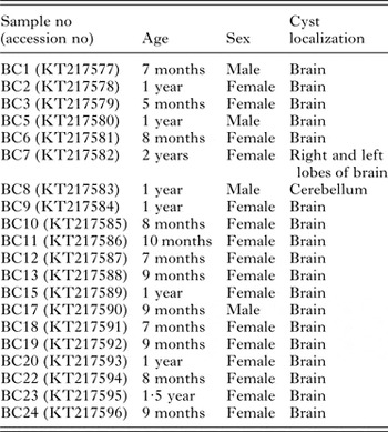
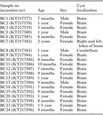
During this study 20 coenurus cysts of various sizes and locations from sheep were collected from slaughterhouses in Mardin province of Turkey. Among these, the most remarkable cerebellum cyst of sheep was showing the clinical signs. When skull was opened a massive nut size C. cerebralis cyst embedded into the cerebellum tissue was detected (Fig. 1). Distribution of cysts according to the age, sex and cyst localization has been shown in Table 1. We collected 19 cysts from brain however only one was obtained from cerebellum. Brain localized fluid-filled C. cerebralis cysts are shown in Fig. 2. All of those examined with the PCR method was also observed in the 446 bp band. Ten of them was shown in Fig. 3. Nucleotide sequences were deposited in GenBank under the accession nos: KT217577-596. According to the alignment results the most radical change was seen in two sites of the sequences. A/G and T/C nucleotide changing were observed in between BC10, BC11, BC22, T. multiceps reference sequences and the other samples in 150th and 252nd nucleotides, respectively (Fig. 4). Genetic tree view of alignment samples was done using neighbour-joining method of measurement with Kimura 80 (Fig. 5).
Coenurus cerebralis cyst in cerebellum.

(A) Coenurus cerebralis cyst localized in the brain tissue; (B) The cyst cavity formed in the brain tissue and the appearance of the protoscoleces in the cyst fluid.

mt-CO1 PCR results of Coenurus cerebralis cysts. M: Marker (100 bp), 1–9: randomly selected samples, 10: positive control, 11: negative control.

Sequence alignment of samples analysed in this study using the reference sequences (JQ710587 and FJ886783). Taenia saginata (JQ756979) and E. granulosus sensu stricto (EU006777) were used as an out group.

Genetic tree view of the aligned sequences.

Distribution of cysts according to the age, sex and cyst localization
According to the statistical evaluation of sequence data C + G rate was 121 and A + T rate was 249 in BC3 sample while these rates were 119 and 261 in BC10 and BC1 samples, respectively. First was 122 and the second was 258 in the other samples. Both the excess change rate of nucleotides and alignment results showed that BC3, BC10 and BC11 could be different variants.
DISCUSSION
Although coenurosis is common in almost every part of the world, it is more widespread in underdeveloped countries in sheep and goats (Scala et al. Reference Scala, Cancedda, Varcasia, Ligios, Garippa and Genchi2007). This study provides the first approach into the genetic characterization of T. multiceps from sheep in Turkey. A molecular-phylogenetic analysis of the coenurus cyst samples using mt-CO1 sequences was evaluated for the first time in Turkey. Mitochondrial genes are the most popular identification markers in population genetics and ecological studies of helminth parasites (Hebert and Gregory, Reference Hebert and Gregory2005). Some genetic markers like 28S rRNA, CO1, nad1, nad4, ITS rDNA and other nuclear protein-coding genes as rpbz, pepck and pold are used for the molecular characterization and phylogenetic studies in Taeniid tapeworms. However, multiplex-PCR and PCR-restriction fragment length polymorphism techniques were used for the identification of Taenia species and often CO1 and cytb genes that are conserved gene regions were preferred (Zhang et al. Reference Zhang, Hu, Jones, Allsopp, Beveridge, Schindler and Gasser2007). But, we used mt-CO1 sequence for genotyping of sheep coenurus isolates in the current study.
The life-cycle of the T. multiceps is very large extent rural, in which the dog-sheep cycle is the most important transmission dynamic. However, the farmer could often facilitate the contamination of the environment by opening the skull of infected sheep for interest or to confirm his own ‘diagnosis’, leaving the Coenurus cyst free to be eaten by dogs or, feeding them directly with it (Scala and Varcasia, Reference Scala and Varcasia2006). Similarly, every year hundreds of thousands sheep are slaughtered in festival of sacrifices without veterinary inspections and the infected brains may be given to stray dogs in Turkey. Therefore, the prevalance of parasite is higher in the country.
The differences in the clinical and pathology appearances in coenurosis suggest the presence of genetic intraspecific variability within the species, such as in other members of the genus Taenia and Echinococcus. For these reasons DNA sequence variability was investigated within the mt-CO1 gene in this study. Twenty samples were obtained from Mardin province of Turkey and examined through the sequencing of the mt-CO1 gene. A/G and T/C nucleotide changing were observed in three samples. Considering the analysed gene, it is possible to define three specific genetic variants in the current study.
Coenurosis affects sheep during their first year of age, mainly when small lambs of 3–4 months are left in the grass at the beginning of the spring season, when their immune system and rumen activity are not yet well developed (Hebert and Edwards, Reference Hebert and Edwards1984). Although 1 or 2-years-old sheep (15 and 21·7%, respectively) were more susceptible to the parasite (Gicik et al. Reference Gicik, Kara and Arslan2007) we detected the infection rate as 60% in under 1 year old lamb. This may be explained by the inadequate of an acquired immunity. It was reported that low amount of taenid eggs are capable to develop into mature metacestodes. Although 93% of Taenia hydatigena eggs cannot develop into a cyst, it can stimulate the immunity for the next exposure (Gemmell et al. Reference Gemmell, Lawson and Roberts1987). It can also be possible for the T. multiceps. Relatively low prevalence in older sheep may be due to the maternal immunity.
The coenurosis cyst can localize in any part of the brain, mostly in the cerebral hemispheres (Parihar, Reference Parihar1988). The great majority of the cysts (96·7%) was located in the cerebral hemispheres in another study (Gicik et al. Reference Gicik, Kara and Arslan2007). Scala et al. (Reference Scala, Cancedda, Varcasia, Ligios, Garippa and Genchi2007) reported that the brain cortex localization rate was 80·7 while 7·3% was for cerebellum. In the present study we detected only one cyst in the cebellum (5%). However we could not find any correlation between cyst localization and genotypic differences.
Concluding remarks
This is the first approach into the genotyping of C. cerebralis from sheep in Turkey. A molecular-phylogenetic analysis of coenurus cyst samples using mt-CO1 sequences was conducted and we detected three haplotypes for the first time. It is evident that intraspecific variations within T. multiceps may have biological or epidemiologic significance warrants for further studies. Thus the current study provides an insight for future studies of the genotyping of T. multiceps in possible hosts in Turkey.
ACKNOWLEDGEMENTS
The authors would like to thank all veterinarians and technicians for their kind help during sample collection.
FINANCIAL SUPPORT
This study has been supported by a grant from Firat University Scientific Research Projects Unit (Project No: VF.14.05, to S. S.).