I. INTRODUCTION
Diamond has a reputation for being radiation-hard. Reference Campbell, Choudhury, Mainwood, Newton and Davies1 Therefore, it is a suitable semiconductor to design detectors operating in high-radiation environments. Reference Berdermann2 Due to their fundamental properties, diamond color centers have been proposed as candidates for quantum information processing; therefore, they find application in other emerging areas of technology, such as magnetometry and cellular biomarking. Suitable centers are most efficiently created in synthetic diamond through electron irradiation or ion implantation followed by annealing. Reference Jelezko and Wrachtrup3,Reference Orwa, Greentree, Aharonovich, Alves, Van Donkelaar, Stacey and Prawer4
Ion implantation is a well-adapted technique for device fabrication since it allows one to introduce chosen impurities into a solid in a precise and well-controlled way. In the case of diamond, ion implantation offers the additional advantage that it provides a means to insert any possible dopant atom, independent of its size or solubility, into a tightly bound, thermodynamically metastable, crystal lattice. Reference Kalish5,Reference Prins6 However, ion implantation is a harsh, nonequilibrium method to insert atoms into a material. The interaction of high-energy ions with crystalline diamond may lead to defect formation and amorphization, and a successful technological application of this technique requires knowledge of the primary interaction processes. Radiation damage (RD) is, strictly speaking, a disturbance of the periodic order of the lattice atoms in a crystalline material. It, therefore, changes many properties of the material, which thereby can be taken as a measure for the RD level. RD in diamond has been studied for many years; however, significant progress in identifying the structure and properties of the simplest radiation defects, namely, the vacancy and the self-interstitial, has been made only recently.
The metastability of the sp 3 bonding of carbon in diamond yields, for fluences above a known critical value, graphitization of the material, Reference Uzan-Saguy, Cytermann, Brener, Richter, Shaanan and Kalish7 whilst the remaining surrounding material is restored to diamond during annealing, so that spatially well-defined structures can be created. This was used to demonstrate that square millimeter-sized slices of diamond can be lifted off intact from diamond crystals through a technique combining high-fluence ion implantation, annealing, and selective etching of a graphitized layer. Reference Yamada, Chayahara, Umezawa, Tsubouchi, Mokuno and Shikata8 The process of graphitization induced in diamond by ion implantation has been used to fabricate a single crystal diamond nanoelectromechanical switch, Reference Liao, Rong, Hishita, Imura, Koizumi and Koide9 three-dimensional buried conductive channels in single crystal diamond, Reference Olivero, Amato, Bellotti, Budnyk, Colombo, Jakšić, Manfredotti, Pastuović, Picollo, Skukan, Vannoni and Vittone10 components for diamond-based quantum coherent devices, Reference Greentree, Olivero, Draganski, Trajkov, Rabeau, Reichart, Gibson, Rubanov, Huntington, Jamieson and Prawer11 and 1D and 2D photonic crystals by means of the focused ion beam technique. Reference Zaitsev and Dobrinets12,Reference Riedrich-Moller, Kipfstuhl, Hepp, Neu, Pauly, Mucklich, Baur, Wandt, Wolff, Fischer, Gsell, Schreck and Becher13
Ion implantation is a method of creating a strongly nonuniform damage profile. It is known that an impinging ion first loses its energy by primarily interacting with electrons, until, at a low enough speed, the primary interactions become nuclear collisions that displace substrate atoms, producing vacancies and knock-on atoms. A primary recoil atom can generate a cascade of displaced particles. Defect production is, therefore, a cascade process in which the incident ion is simply the primary damage-producing particle. The total of all such events is commonly referred to as the collision cascade of the ion. High defect densities are generally associated with the vicinity of the ion track. Recent observations by using transmission electron microscopy highlight the presence of amorphous zones (pockets) in the cascade tracks, which is more consistent with heterogeneous amorphization models. Reference Hickey, Jones and Elliman14,Reference Fairchild, Rubanov, Lau, Robinson, Suarez-Martinez, Marks, Greentree, McCulloch and Prawer15 The model describes the damage as locally amorphized zones around each ion and the tracks of knock-out atoms. Reference Morehead and Crowder16–Reference Wendler18 Primary defects (vacancies and interstitial atoms) could escape the collision cascades into the surrounding crystalline material. As ion fluence increases, amorphous zones accumulate and overlap to form a continuous amorphous layer. Alternatively, the defect-induced model of amorphization has led to the postulate that the defects that form in the collision cascades, although not homogeneously distributed, consist primarily of vacancies and interstitial atoms. Reference Prins19 Point defects in diamond are immobile at room (ordinary) temperature; therefore, one can conclude that most of the implantation-induced damage is “frozen in.” In reality, the effects of dynamic annealing during implantation can be taken into account by shrinking amorphous zones and annihilation of Frenkel pairs. Reference Newton, Campbell, Twitchen, Baker and Anthony20 Implantation performed at liquid nitrogen temperature freezes the damage created during implantation. Reference Orwa, Santori, Fu, Gibson, Simpson, Aharonovich, Stacey, Cimmino, Balog, Markham, Twitchen, Greentree, Beausoleil and Prawer21
Naturally, a detailed analysis of RD in silicon very much facilitates the understanding of RD in diamond. Reference Watkins22,Reference Pelaz, Marqués and Barbolla23 Crookes was the first who started the study of RD in diamond as early as in 1904. Reference Crookes24 Nevertheless, the mechanisms of amorphization and graphitization of diamond due to RD need detailed analysis. Owing to its properties, diamond is used as a model material for investigating the nature of RD in covalent crystals. RD in diamond clearly manifests itself in optical Reference Amekura and Kishimoto25–Reference Collins27 and electrical properties, Reference Prawer and Kalish28,Reference Kalish, Reznik, Nugent and Prawer29 luminescence, Reference Steeds, Charles, Davis, Gilmore, Hayes, Pickard and Butler30,Reference Collins31 electron paramagnetic resonance, Reference Twitchen, Hunt, Newton, Baker, Anthony and Banholzer32 Raman scattering, Reference Amekura and Kishimoto25,Reference Orwa, Nugent, Jamieson and Prawer33 swelling, Reference Fairchild, Rubanov, Lau, Robinson, Suarez-Martinez, Marks, Greentree, McCulloch and Prawer15,Reference Amekura and Kishimoto25,Reference Kalish, Reznik, Nugent and Prawer29,Reference Prins, Derry and Sellschop34,Reference Bosia, Calusi, Giuntini, Lagomarsino, Giudice, Massi, Olivero, Picollo, Sciortino, Sordini, Vannoni and Vittone35 and mechanical properties Reference Bosia, Argiolas, Bazzan, Olivero, Picollo, Sordini, Vannoni and Vittone36 ; it is characterized by frozen-in defect structure Reference Newton, Campbell, Twitchen, Baker and Anthony20,Reference Orwa, Santori, Fu, Gibson, Simpson, Aharonovich, Stacey, Cimmino, Balog, Markham, Twitchen, Greentree, Beausoleil and Prawer21 and has a threshold indicator—graphitization. Reference Uzan-Saguy, Cytermann, Brener, Richter, Shaanan and Kalish7
Ion implantation allows one to produce any level of RD in thin layers, including very high levels. Reference Amekura and Kishimoto25 The local character of RD due to ion implantation opens up the possibility to study the defect structure of a material, especially by modern methods of electron microscopy and electron loss spectroscopy. Reference Hickey, Jones and Elliman14,Reference Fairchild, Rubanov, Lau, Robinson, Suarez-Martinez, Marks, Greentree, McCulloch and Prawer15 On the other hand, it hampers the study of RD by such methods as optical absorption, luminescence, Raman scattering, electron paramagnetic resonance, electric and magnetic measurements, measurements of mechanical stress and density, and, in general, by any method in which the contributions of layers with different RD levels are added up. Reference Bosia, Argiolas, Bazzan, Fairchild, Greentree, Lau, Olivero, Picollo, Rubanov and Prawer37 The main peculiarity of the present study is the fabrication of a layer with uniform RD by ion implantation in diamond. This layer can be investigated by the above-mentioned methods. One relevant consequence of the structural modifications due to ion implantation is a density variation in the damaged diamond, i.e., a constrained volume expansion leading to surface swelling in the irradiated region. The present study is a continuation of our paper Reference Khmelnitsky, Dravin, Tal, Latushko, Khomich, Khomich, Trushin, Alekseev and Terentiev38 and is aimed at determining the density of radiation-damaged diamond and mechanical stresses in it, assessing the role of defects and amorphized regions in radiation-damaged diamond, and studying the properties of a material with high level of RD.
II. EXPERIMENT
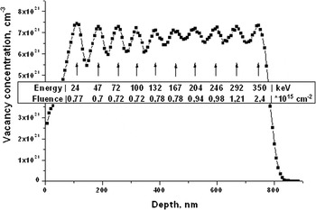
Experiments were carried out on polished plane-parallel disks with diameter 2L ∼ 3 mm and thickness h ∼ 120 μm cut out from (110) plates of natural single crystal diamond. For each implantation session, we prepared several (from 2 to 4) samples with different (low and high) concentrations of nitrogen (see Ref. Reference Khmelnitsky, Dravin, Tal, Latushko, Khomich, Khomich, Trushin, Alekseev and Terentiev38 for more detail). These samples were exposed to implantation with He+ ions with a set of energies (10 values ranging from the highest energy 350 keV to the lowest one 24 keV) and fluences that ensured uniform (±10%) RD in a 670-nm-thick layer (Fig. 1) through masks with circular apertures (with diameter slightly smaller than the diameter of the samples).
Profile of vacancies induced by He+ implantation with multiple energies and a total fluence of 1 × 1016 cm−2 as a result of Monte-Carlo simulation.

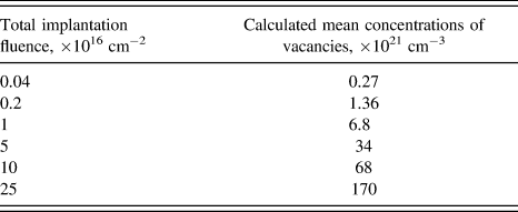
The implantation was carried out by a heavy ion accelerator of “High Voltage Engineering Europe.” To reduce the recombination rate of Frenkel pairs, the implantation was performed at liquid nitrogen temperature. Reference Orwa, Santori, Fu, Gibson, Simpson, Aharonovich, Stacey, Cimmino, Balog, Markham, Twitchen, Greentree, Beausoleil and Prawer21 To calculate the distribution of vacancies generated by multiple ion implantation, we used MRAC92 software system developed by one of us (V.A. Dravin, 1992). Reference Khmelnitsky, Dravin, Tal, Latushko, Khomich, Khomich, Trushin, Alekseev and Terentiev38 Like the well-known SRIM code, it is based on Monte-Carlo modeling, but has some advantages: (a) the scattering integral is calculated numerically and (b) some improvements have been made to the ion tracking algorithm. The inset in Fig. 1 shows the set of energies and fluences (for a total fluence of 1 × 1016 cm−2) at which He+ ions were implanted. One can see that, for the chosen values of energy, the deviation of vacancy concentration from its mean value is not greater than 10% throughout the subsurface layer depths from 90 to 760 nm. The implanted layer is covered on top with a thin (∼90 nm) layer with a low RD level. Table I shows the total implantation fluences and the calculated mean concentrations of radiation-induced vacancies. The ratio of the greatest total implantation fluence to the smallest one is 625. This range of implantation total fluences covers the whole relevant interval of RD levels from very low to complete graphitization of diamond.
Total implantation fluences of He+ and calculated mean concentrations of vacancies at a depth of from 90 to 760 nm.

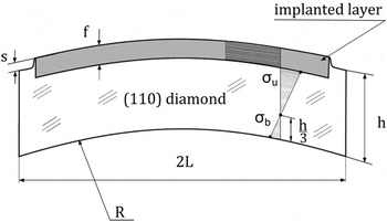
During the swelling, the implanted layer can freely expand only along the normal to the surface. Therefore, a step s appears on the boundary of the implantation region (Fig. 2). Expansion in the plane of the implanted layer induces mechanical compressive stress σf in this layer, which leads to the bending of the plate. We assume that mechanical stress in the layer arises only due to the swelling. The diameter of the implanted region is made slightly smaller than the diameter of the sample to allow for the same sample to measure the bending and the swelling.
Schematic cross section of an implanted sample with stress diagram. Here, 2L—a sample diameter, h—the thickness of a sample, f—the thickness of the implanted layer, R—the radius of curvature of a bent sample, and s—the height of a step on the boundary of the implanted area. The implanted layer experiences compressive stress σf; on the boundary of the implanted layer, the tensile stress is σu; the compressive stress σb attains its maximum at the back side of a sample.

The bending of the samples (R) and the surface swelling (s) were measured using a Zygo New View 5000 optical interferometer. Reference Khmelnitsky, Dravin, Tal, Latushko, Khomich, Khomich, Trushin, Alekseev and Terentiev38 The Raman light scattering in implanted samples was studied in confocal geometry by an HR800 optical spectrometer of Horiba Jobin Yvon (Horiba, France). An argon laser with a wave length of 488 nm was used as an excitation source. Transmission spectra over wavelengths of 200–900 nm were obtained on a Specord M400 (Carl Zeiss, Jena) spectrophotometer. To determine the graphitization threshold, we have annealed individual samples with total implantation fluences of (5 ÷ 10) × 1016 cm−2 in a vacuum graphite furnace at a temperature of 1450 °C. Reference Khomich, Khmelnitskiy, Dravin, Gippius, Zavedeev and Vlasov39
III. RESULTS AND DISCUSSION
The problem of bending of an elastic plate with a thin strained surface layer was solved by Stoney in 1909 for isotropic materials.
Reference Stoney40
In crystalline plates, one should take into account the anisotropy of mechanical properties of the material.
Reference Janssen, Abdalla, Van Keulen, Pujada and Van Venrooy41
The bending of a diamond plate is determined by the mechanical compressive stress in the implanted layer and the biaxial moduli of elasticity, which are equal to M
100 = 1170 GPa and M
110 = 1320 GPa, respectively, in two mutually perpendicular directions <100> and <110> in the {110} plane of the diamond plate.
Reference Khmelnitsky, Dravin, Tal, Latushko, Khomich, Khomich, Trushin, Alekseev and Terentiev38
As a result, the bending of the plate has the shape of an ellipsoid defined by two different radii of curvatures R
100 and R
110 in the directions <100> and <110>, respectively. The data on the plate thickness h and the thickness of the implanted layer (f = 670 nm) with regard to swelling s allow one to determine the mechanical compressive stress σf in the implanted layer by the Stoney formula 6R(f + s)σf = Mh
2.
Reference Khmelnitsky, Dravin, Tal, Latushko, Khomich, Khomich, Trushin, Alekseev and Terentiev38
We assume that mechanical stress in the implanted layer is uniform and isotropic. The nonuniformity of stress in the plane of the plate is determined by the nonuniformity of the implantation fluence, which does not exceed 1%, while the in-depth homogeneity is provided by the energy and fluence distributions (Fig. 1). The disordering of material due to RD should guarantee the isotropy of stress in the plane of the implanted layer. Therefore, we assume that the mechanical stress in the implanted layer is uniform and isotropic. This assumption is confirmed experimentally by two facts: (a) the swelling s is the same over the whole perimeter of the implantation region and (b) the bending of the plate has the shape of an ellipsoid, where the ratio of R
100 to R
110 corresponds to the ratio of the biaxial moduli of elasticity in these directions M
100 and M
110 within the measurement accuracy; i.e., this ratio is determined only by the anisotropy of elastic properties of the diamond plate. Our case fully corresponds to the applicability conditions of the Stoney formula.
Reference Zhang and Zhao42
For example, we assumed that the following geometrical relations are satisfied:
 $R \gg h \gg f$
and
 $2L \gg h$
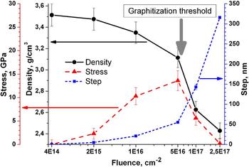
. In Refs. Reference Amekura and Kishimoto25, Reference Prins, Derry and Sellschop34, and Reference Khomich, Khmelnitskiy, Dravin, Gippius, Zavedeev and Vlasov39, it was shown that the effect of an implanted impurity on the swelling of diamond can be neglected. Figure 3 shows the measured values of swelling s of the as-implanted material and the calculated mechanical compressive stress σf in the implanted layer as a function of the total implantation fluence.
Measured swelling and calculated mechanical stresses and density of diamonds (with low concentration of nitrogen) implanted with different total fluences.

An important result of the measurements and calculations is the nonmonotonic dependence of σf on the RD level. At its maximum, σf can reach a value of 14 GPa. This value corresponds to the region of thermodynamic stability of diamond in the phase diagram of carbon Reference Bundy43 ; however, it is much less than the calculated values of 40 (Ref. Reference Bosia, Argiolas, Bazzan, Fairchild, Greentree, Lau, Olivero, Picollo, Rubanov and Prawer37) and 170 GPa. Reference Fairchild, Rubanov, Lau, Robinson, Suarez-Martinez, Marks, Greentree, McCulloch and Prawer15 We observed no micro- or macrocracking at the edges of the implanted region. The nonmonotonic dependence of σf on the total implantation fluence allows one to divide the range of RD for low-nitrogen diamonds at least into two regions, the boundary corresponding to a total fluence of about 5 × 1016 cm−2. In some samples with such implantation total fluence, the implanted layer was restored to diamond after annealing at a temperature of 1450 °С, while in some other samples, the implanted layer was graphitized. The difference is mainly determined by the concentration of nitrogen in diamond. Generally, in all our experiments, the higher the concentration of nitrogen in diamonds, the higher the RD level in diamond, conditions being equal. Reference Khmelnitsky, Dravin, Tal, Latushko, Khomich, Khomich, Trushin, Alekseev and Terentiev38 Therefore, the annealing of samples with a nitrogen concentration of above 1 × 1018 cm−3 and total implantation fluence of 5 × 1016 cm−2 leads to the graphitization of the implanted layer. Thus, the total implantation fluence in our experiments, 5 × 1016 cm−2, is close to the critical fluence for graphitization. According to Monte-Carlo modeling, this fluence corresponds to the calculated concentration of vacancies 3.4 × 1022 cm−3 (see Table I), which is about 2–2.5 times less than the values obtained for the graphitization threshold under monoenergetic implantation. Reference Hickey, Jones and Elliman14,Reference Fairchild, Rubanov, Lau, Robinson, Suarez-Martinez, Marks, Greentree, McCulloch and Prawer15,Reference Bosia, Argiolas, Bazzan, Olivero, Picollo, Sordini, Vannoni and Vittone36,Reference Khomich, Khmelnitskiy, Dravin, Gippius, Zavedeev and Vlasov39 This means that one should be careful when describing the RD level in diamond by the calculated concentration of vacancies.
In the range of total fluences (1–5) × 1016 cm−2, one can observe a saturation of s and σf as a function of total fluence. This range can be called as a range of amorphization fluences. The saturation of RD occurs at high radiation doses, where knocking out already knocked on atoms Reference Prins44 and disordering already disordered regions do not change the RD level of the material. Reference Amekura and Kishimoto25,Reference Bosia, Argiolas, Bazzan, Fairchild, Greentree, Lau, Olivero, Picollo, Rubanov and Prawer37 Such behavior is characteristic for Si Reference Pelaz, Marqués and Barbolla23 and other semiconductors Reference Wendler18 and is attributed to the amorphization of the implanted layer. A characteristic feature of diamond is its graphitization at RD levels corresponding to complete amorphization in other materials. This fact leaves open the question of whether complete amorphization without graphitization is possible under RD in diamond. Perhaps, to this end, one should increase the temperature of diamond under irradiation, as in Ref. Reference Braunstein and Kalish45.
A comparison of the parameter s, which describes the deformation of the as-implanted diamond, with mechanical stress σf in diamond allows one to estimate the elastic properties of the implanted material and their variation under increasing RD level. An increase in the total implantation fluence from 1 to 5 × 1016 cm−2 leads to a 2.5 times increase in s, while σf increases only by a factor of 1.2–1.4. This fact points to the degradation of the mechanical properties of diamond with increasing RD level, which can be attributed to a 2–3 decrease in Young's modulus of the implanted material under the assumption that plastic deformation plays an insignificant role at fluences of up to 5 × 1016 cm−2. Reference Khmelnitsky, Dravin, Tal, Latushko, Khomich, Khomich, Trushin, Alekseev and Terentiev38 As the total fluence increases from 5 × 1016 cm−2 to 2.5 × 1017 cm−2, mechanical stress in the implanted layer decreases by a factor of 100 (Fig. 3). This fact can be given with two explanations: (a) the modulus of elasticity of the material implanted at a total fluence of 2.5 × 1017 cm−2 is hundreds of times less than that of diamond and (b) above the graphitization threshold, the material becomes ductile. The first assumption seems to be improbable, because even the modulus of elasticity of dense graphite is only 30 times less than that of diamond (Ref. Reference Pierson46, p. 131).
We will focus on the density of the radiation-damaged layer of the implanted material in the absence of stresses. The relative variation of the density can be obtained from the expression that takes into account the swelling and the in-plane deformation of the implanted layer Δρ/ρ = s/(f + s) + σf(1 − νf1)/E f + σf(1 − νf2)/E f, where E f is the Young's modulus of the implanted diamond, νf1 and νf2 are the Poisson's ratios of the implanted diamond in the [110] and [100] directions, respectively. We assume that νf is equal to the Poisson's ratio of diamond νs. The main contribution to the density is made by expansion, which manifests itself in the swelling s. However, one should also take into consideration the compression of the material under stresses. Reference Khmelnitsky, Dravin, Tal, Latushko, Khomich, Khomich, Trushin, Alekseev and Terentiev38 The compression is significant only for large σf in the range of total fluences of (1–5) × 1016 cm−2, i.e., in the range of amorphization fluences. One should also take into account that Young's modulus of implanted diamond decreases. The uncertainty of the Young's modulus and Poisson's ratio of the implanted material contributes to the errors of density data in Fig. 3. Figure 3 illustrates a decrease in the density of the material as the total implantation fluence increases. In Refs. Reference Fairchild, Rubanov, Lau, Robinson, Suarez-Martinez, Marks, Greentree, McCulloch and Prawer15 and Reference Bosia, Argiolas, Bazzan, Fairchild, Greentree, Lau, Olivero, Picollo, Rubanov and Prawer37, it was established that the graphitization threshold corresponds to the density of 2.95 g/cm3 of the implanted material, and our data agree with this result (Fig. 3). Diamond implanted at a total fluence of 2.5 × 1017 cm−2 has a density of 2.4 g/cm3, which is slightly greater than the theoretical density of graphite of 2.26 g/cm3 (Ref. Reference Pierson46, p. 51). Such a strongly damaged material is virtually free of mechanical stress.
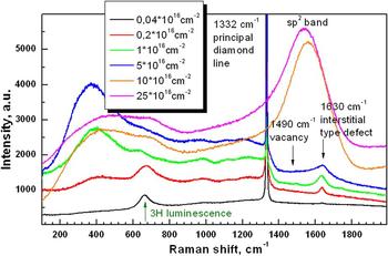
Figure 4 shows the Raman spectra of the as-implanted samples. The line νLO ≈ 1332 cm−1 corresponds to undamaged diamond, which is located directly under the implanted layer. Reference Khmelnitsky, Dravin, Tal, Latushko, Khomich, Khomich, Trushin, Alekseev and Terentiev38 For small total implantation fluences (0.4 and 2 × 1015 cm−2), there is also the photoluminescence line of the 3H center, which is emitted by the material under the implanted layer. All the remaining rich pattern of Raman scattering refers to implanted diamond. This is the subject of separate consideration. Right now, it is important that the components above the diamond line νLO ≈ 1332 cm−1 are associated with defects in crystalline diamond, while those below this line, with amorphous phase. Reference Prawer, Rosenblum, Orwa and Adler47 When the total fluence is just doubled from 5 to 10 × 1016 cm−2, the spectrum is drastically changed. Due to the sharply increased optical absorption, the emission of the line νLO ≈ 1332 cm−1 from the material under the implanted layer is not detected through the layers implanted at total fluences above the critical fluence of graphitization, 1 and 2.5 × 1017 cm−2. The emission of this line is not detected in the implanted layers. It is still more remarkable that spectral bands related to defects in the diamond phase are missing above the diamond line νLO ≈ 1332 cm−1. Instead, the G-band is strongly pronounced, which is related to the sp 2 phase and has a maximum at about 1580 cm−1. This is indicative of the fact that there is nearly no crystalline diamond phase in the material implanted at fluences above the graphitization threshold, while the sp 2 phase is formed immediately under implantation at fluences above the graphitization threshold.
Raman spectra of implanted diamonds as a function of total fluence.

Such a sharp change in the microstructure of the material and its properties above the graphitization threshold needs explanation. It may be associated with a decrease in the displacement energy in the disordered material. This is probably due to a strong decrease in the thermal conductivity of the disordered material. Reference Khomich, Khmelnitskii, Hu, Khomich, Popovich, Vlasov, Dravin, Chen, Karkin and Ralchenko48 Nanodomains of material, where fast ions are slowed down, are strongly heated. Reference Saada, Adler and Kalish49 The heating increases if the region is surrounded by a poorly heat-conducting amorphous material. The temperature of the heated region may reach the melting point of the material (for carbon, this point is higher than 4000 K Reference Bundy43 ). This situation corresponds to a transition to the thermal spike regime. Reference Wendler18,Reference Pelaz, Marqués and Barbolla23 Upon cooling of the melt, nanodomains of sp 2 graphite-like material are formed.
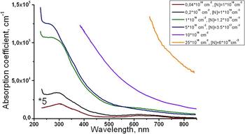
RD in diamond manifests itself in optical absorption in the range of wavelengths above 220 nm. The optical absorption spectra of the as-implanted diamonds are shown in Fig. 5. In samples with total implantation fluences of 0.4 and 2 × 1015 cm−2, optical absorption is mainly determined by vacancies, which yield two broad absorption bands with maxima at about 300 and 620 nm. Reference Khomich, Khmelnitskiy, Dravin, Gippius, Zavedeev and Vlasov39 In samples with total implantation fluences of 1 × 1016 cm−2, optical absorption is contributed from a component (or components) responsible for continuous absorption, which decreases from UV to IR domain, while the contribution of isolated vacancies is halved. We associate the continuous component of optical absorption with the amorphized material. In samples with total implantation fluences of 5 × 1016 cm−2, the continuous absorption component slightly increases, while absorption due to vacancies decreases by another factor of 5. As a result, the total absorption remains almost unchanged, but the absorption spectrum is changed so that one can even observe the clarification of the material in the range of wavelengths above 550 nm. This is a manifestation of the saturation of the properties of the material as RD increases in the range of amorphization fluences. The reduced role of defects in the diamond phase also manifests itself in the Raman scattering in the spectral region above the diamond line νLO ≈ 1332 cm−1 (Fig. 4). Above the graphitization threshold, optical absorption sharply increases. This is attributed to the rapid growth of the sp 2 phase in the implanted layer.
Optical absorption spectra of diamonds implanted at different total fluences (for a total fluence of 4 × 1014 cm−2, the absorption coefficient is increased by a factor of 5 for clarity).

Why does the concentration of vacancies (and, generally, point defects) decrease as the RD level increases in the range of amorphization fluences? We can point out several reasons. The first is that the total amount of nondiamond (amorphous, or, generally, disordered) regions increases with the RD level, while the fraction of crystalline diamond with single vacancies decreases. The second is that, as the vacancy concentration increases, the probability of formation of complex defects, multivacancy complexes, Reference Hounsome, Jones, Martineau, Fisher, Shaw, Briddon and Öberg50 and V–C = C–V defects Reference Hyde-Volpe, Slepetz and Kertesz51 rapidly increases. The third reason is that the vacancies can diffuse short distances under local heating of nanoscopic regions of the material. Reference Saada, Adler and Kalish49 The local heating of the region of crystalline diamond material with defects increases when this region is surrounded by poorly thermal conducting regions. Reference Khomich, Khmelnitskii, Hu, Khomich, Popovich, Vlasov, Dravin, Chen, Karkin and Ralchenko48 This stimulates the recombination of vacancies and reactions in which they take part. The concentrations of vacancies and interstitials in type IaA diamonds increase with the concentration of the A-form of nitrogen. Reference Kiflawi, Collins, Iakoubovskii and Fisher52 This effect is attributed to the trapping of vacancies and interstitials in the strain field of nitrogen. The trapping produces “strained” vacancies and partly immobilizes interstitials, thus reducing the vacancy–interstitial recombination during irradiation. However, even in the range of amorphization fluences, when there are practically no vacancies, the RD level in crystals rises with higher nitrogen concentrations, conditions being equal.
A sharp decrease in mechanical stress in the implanted layer as the RD level increases above the graphitization threshold suggests a hypothesis that graphitization of diamond is determined by the internal pressure in the material. Reference Bosia, Argiolas, Bazzan, Fairchild, Greentree, Lau, Olivero, Picollo, Rubanov and Prawer37 The idea is that, while the internal pressure in the implanted layer during annealing is a few GPa or more, the material is annealed under the conditions of thermodynamic stability of diamond Reference Bundy43 and, hence, restores its diamond sp 3 structure. If the internal pressure in the implanted layer drops below the diamond/graphite boundary in the phase diagram, the material disordered under RD is transformed into a graphite-like sp 2 structure. To solve this problem, we carried out experiments on the annealing of diamonds implanted above the graphitization threshold under local and static pressure.
To create a local pressure, we fabricated diamond Berkovich indenters (in the form of trihedral pyramids). Such an indenter is pressed under a load of 100 g to the implanted surface of diamond during annealing in a furnace. According to calculations, the pressure at the indenter tip is at least 20 GPa, which corresponds to the conditions of thermodynamic stability of diamond. We carried out experiments on crystals implanted at total fluences of 1 and 2.5 × 1017 cm−2. In the experiments under local pressure, we also used diamonds implanted by He+ ions with the energy of 350 keV and at the fluence of 3 × 1016 cm−2, which is above the graphitization threshold. Annealing under local pressure was carried out at a temperature of 1450 °С for an hour.
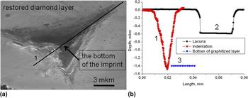
After annealing, we found an impression in the place where pressure was exerted [Fig. 6(a)]. The implanted layer was graphitized everywhere except for the impression area, where no graphitized layer was found—this place was optically transparent. However, the restoration of diamond did not occur either. Figure 6(b) shows that the bottom point of the impression is at a depth of 1.48 μm. For comparison, consider the profile of a small unimplanted region (lacuna) on the same sample. In this case, the swelling of the graphitized material is 0.58 μm. The difference 1.48 μm − 0.58 μm = 0.9 μm corresponds to the lower boundary of the implanted layer, or even to a deeper level (see Fig. 1). That is, the material was squeezed out from under the indenter. It did not stick to the indenter, since Fig. 6(a) clearly shows a thin diamond layer restored (although slightly cracked) under annealing, which covers the graphitized material on the top side. The implanted material was squeezed out from under the indenter, which points to the ductility of the material. One can hardly see any bulge of squeezed-out material around the impression neither in the SEM image [Fig. 6(a)], nor in the topogram (is not shown), nor in the profile along the symmetry axis of the impression [Fig. 6(b)]. This unexpected fact is indicative of large plastic yielding of the implanted material. Assuming that the thickness of the implanted layer of diamond ranges from 0.8 (as in Fig. 1) to 0.9 μm [as in Fig. 6(b)] and taking the value 0.58 μm of swelling, we can find that the density of the graphitized layer is 2.04–2.14 g/cm3. This value is slightly less than the density of crystalline graphite [2.26 g/cm3 (Ref. Reference Pierson46, p. 51)] and corresponds to the data of Refs. Reference Fairchild, Rubanov, Lau, Robinson, Suarez-Martinez, Marks, Greentree, McCulloch and Prawer15, Reference Bosia, Argiolas, Bazzan, Fairchild, Greentree, Lau, Olivero, Picollo, Rubanov and Prawer37, and Reference Khomich, Khmelnitskiy, Dravin, Gippius, Zavedeev and Vlasov39.
The result of annealing of diamond implanted with the total fluence of 2.5 × 1017 cm−2 under local pressure with Berkovich indenters: (a) SEM image, (b) surface profile along the line (1) [shown in panel (a)], surface profile across the nonimplanted region (2) and depth position of the bottom of graphitized layer (3).

Diamonds implanted at fluences above the graphitization threshold were annealed under hydrostatic pressure corresponding to the region of thermodynamic stability of diamond. The samples were annealed under a static uniaxial compression of 5.5 GPa at a temperature of 1550 °С in a CsCl environment for an hour using the universal high-pressure units (model URS-01/V, TISNCM). Annealing under static pressure yields a result similar to that obtained in the case of annealing under local pressure. Diamond implanted at fluences above the graphitization threshold is not restored under pressure annealing in the region of thermodynamic stability of diamond. Instead, it is transformed into a graphitized material. Hence, the hypothesis that the graphitization of diamond is determined by internal pressure in the material was not confirmed in our experiments, due to complexity of the structure transformations in heavily damaged diamond. One must take into account the possibility of forming the mixture of diamond, graphite, and other carbon allotropes. The material of the implanted layer is squeezed out to tens and hundreds of micrometers almost everywhere along the edges of the plates, and the thin diamond layer that covered the implanted layer on its top side is cracked at these places and is broken under pressure. This again provides evidence of high ductility of the material implanted at fluences above the graphitization threshold. In our opinion, it is responsible for a sharp decrease in the mechanical stress in the layers implanted at total fluences of 1 and 2.5 × 1017 cm−2 (Fig. 3).
IV. CONCLUSIONS
At low doses of radiation, the RD level in diamond and its properties are determined by point radiation defects—vacancies and interstitial atoms. In this case, the swelling and the associated mechanical stress are small. In the range of amorphization fluences, one can observe strong swelling, saturation of RD with increasing radiation fluence, the reduced role of point defects (vacancies), and amorphization of the material, which is accompanied by a change in its mechanical properties. The mechanical stress in the material may reach 14 GPa. Thus, the swelling of the material is determined by the formation of amorphization regions. In this case, in the range of fluences below the graphitization threshold, the annealing of the radiation-damaged diamond leads to almost complete restoration of the diamond lattice, just as in Si. Reference Pelaz, Marqués and Barbolla23 The RD level in diamond increases as the concentration of nitrogen increases.
Above the graphitization threshold, the diamond phase completely disappears in the material as the RD level increases. On the other hand, the fraction of sp 2 material sharply increases. Such a material is very ductile. It cannot be restored to diamond even by annealing under high pressure.
ACKNOWLEDGMENT
The study was supported in part by the Russian Foundation for Basic Research (Project No. 14-02-00597 and 14-03-00936).