Coarctation of the aorta accounts for roughly 5% of those born with congenital heart disease. Reference Reller, Strickland, Riehle-Colarusso, Mahle and Correa1 Surgical repair of this cardiovascular lesion was first reported in 1945 and if unrepaired, the average age of survival was 35 years. Coarctation of the aorta is not a focal lesion and complications reported in unrepaired patients include hypertension, stroke, aortic dissection, early-onset coronary artery disease, and heart failure. Reference Warnes, Williams and Bashore2 The first association of coarctation of the aorta and ruptured intracranial aneurysm was reported as early as 1871 by Eppinger. Reference Eppinger3 Studies over the last few decades have demonstrated an estimated 10% risk of intracranial aneurysm formation in adults with a history of coarctation of the aorta as well as an increased risk of subarachnoid haemorrhage. Reference Connolly, Huston, Brown, Warnes, Ammash and Tajik4–Reference Pickard, Gauvreau and Gurvitz8 While these are important findings, there may have been some era effect and referral bias for these previously published studies. The prevalence of intracranial aneurysms in the general population is estimated at 3.2% by a recent meta-analysis. Reference Vlak, Algra, Brandenburg and Rinkel9 Intracranial aneurysms are clinically significant when they spontaneously rupture and cause intracranial haemorrhage. Ruptured intracranial aneurysms cause the majority of subarachnoid haemorrhages, which are responsible for 25% of all cerebrovascular deaths. Reference Wardlaw and White10 Intracranial aneurysms in patients with coarctation of the aorta do not have one clear aetiology, however, suggested possibilities include long-standing hypertension, inherent vascular anomalies, and shear wall stress. Reference Singh, Marzo and Staicu11 The high prevalence of intracranial aneurysms in patients with coarctation of the aorta compared to the general population has led to a recommendation in the 2018 American Heart Association/American College of Cardiology Adult Congenital guidelines to be aware that coarctation alone may not be sufficient for the development of intracranial aneurysms and other factors may play a role in development and progression of aneurysms, including age and hypertension. The 2018 guidelines state that it is reasonable to screen patients with a history of coarctation of the aorta with a head computed tomography angiography (CT angiography) or magnetic resonance angiography (MR angiography) for intracranial aneurysm. Reference Thompson, Brown and Amin-Hanjani12 This recommendation is Class IIb based on limited populations evaluated and non-randomised trials (level of evidence B). Reference Stout, Daniels and Aboulhosn13 Of note, in prior studies finding a 10% rate of intracranial aneurysms, the patients’ mean age at diagnosis coarctation was 17 years and the mean age at which the MR angiography was performed was 41 years. Reference Connolly, Huston, Brown, Warnes, Ammash and Tajik4 A recent prospective study by Donti et.al was performed on a younger population and demonstrated that this screening may be overly aggressive and unnecessary. In their population of 80 young adults (mean age 16 years), all of whom underwent coarctation of the aorta repair during early childhood, no intracranial aneurysms were identified by MR angiography screening. In this study, the age at which these patients were screened was younger than the age at which other studies have screened their patients. Reference Donti, Spinardi and Brighenti14 Taking this study into account, the question remains whether or not patients with coarctation of the aorta warrant costly screening for intracranial aneurysms. If warranted, further information is needed to determine the age at which it would be reasonable to obtain a screening brain CT angiography, MRI, or MR angiography. There may also be a subset of patients who are at increased risk based on other comorbidities and thus should be screened (i.e., hypertension, smoking, family history of intracranial aneurysm, and genetic syndromes). Reference Thompson, Brown and Amin-Hanjani12,Reference Vlak, Rinkel, Greebe and Algra15 We sought to describe the prevalence of intracranial aneurysms in patients with coarctation of the aorta who underwent brain imaging at our centre.
Materials and methods
A retrospective cohort study was performed at the Utah Adult Congenital Heart Disease programme. Patients were included in the study if they had undergone a brain CT angiography, MRI, or MR angiography between January, 2000 and February, 2018, were aged 13 years and older at the time of their brain imaging, and were seen by the Utah Adult Congenital Heart Disease programme during this study period. Patients were excluded if they were younger than 13 years at the time of their brain imaging, or if they did not have adequate brain imaging performed. The MRI scanners at the University of Utah are Siemens 3 Tesla and at the Primary Children’s Hospital are GE 3 Tesla architect. MR angiography sequencing at both institutions was performed with the 3D acquisition of 0.5 mm slices. MRI at the Primary Children’s Hospital obtained 1.6 mm slices and when a vascular concern was noted, star-weighted angiography was performed for enhanced assessment of vasculature. MRI at the University of Utah obtained 5 mm thick, no gap slices and when a vascular concern is noted, MR angiography may be added to the study. CT angiography was performed with a GE Revolution scanner and sequences included 0.625 mm slices. We collected demographic information and risk factors for intracranial aneurysms as well as details regarding their history of coarctation of the aorta, age of intervention, type of intervention, and recurrent coarctation of the aorta. We also collected data regarding the age at which they underwent brain imaging, the type of brain imaging, and the indication for brain imaging. Study data were collected and managed using REDCap electronic data capture tools hosted at the University of Utah. We used descriptive statistics including the percentage, median, and range for these data points to characterise this cohort.
Results
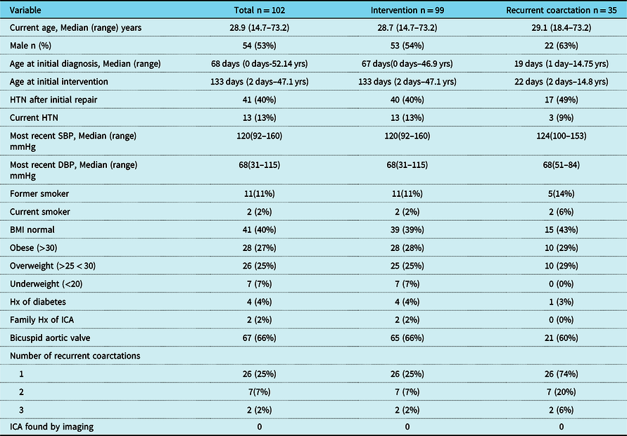
There was a total of 102 patients with coarctation of the aorta who underwent adequate brain CT angiography, brain MRI, or brain MR angiography from 2000 to 2018. Patient demographics and characteristics are summarised in Table 1. The baseline demographics included 53% men and a median age of 28.8 years (range 14.7–73.3 years). The most common additional cardiac diagnoses included bicuspid aortic valve in 65.7% and ventricular septal defect in 27.5% of patients. Additional cardiac and intracranial aneurysm risk factors that were collected included hypertension following coarctation of the aorta repair (40.2%), ongoing hypertension with or without antihypertensive therapy (12.7%), history of tobacco use (10.8%), current tobacco use (4%), history of diabetes (3.9%), family history of early coronary artery disease (7.8%), and known family history of intracranial aneurysm (2%). There was one patient with Moyamoya and no patients with genetic syndromes or connective tissue disorders (other than bicuspid aortic valve) included in the study. The median age of diagnosis of coarctation of the aorta was 68 days (range 0 days–52 years) with the initial intervention being surgical repair in 90.2% of patients. Of note, there were three patients who have not undergone coarctation of the aorta intervention, suggesting a less severe coarctation of the aorta. The median age for repair was 4.4 months (range 2 days–47 years). Overall, 34% of patients had recurrent coarctation of the aorta requiring re-intervention; 25% of patients underwent a single re-intervention, 7% had 2 re-interventions, and 2% had 3 re-interventions.
Baseline demographics and risk factors for cardiovascular disease and intracranial aneurysms

Abbreviations: BMI = body mass index; DBP = diastolic blood pressure; HTN = hypertension; Hx = history; ICA = intracranial aneurysm; SBP = systolic blood pressure; Yrs = years.
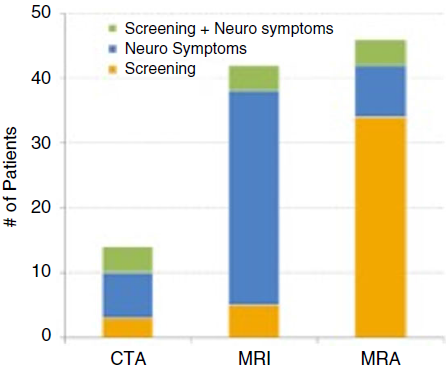
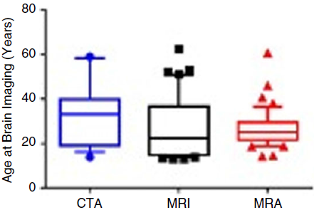
All patients included in this study underwent adequate brain imaging for intracranial aneurysm screening, however, not all brain imaging was performed for the specific indication of intracranial aneurysm screening. The indications and type of imaging study are summarised in Fig 1. Of our patients, 13.7% underwent brain CT angiography, 42.1% underwent brain MRI, and 46.1% underwent brain MR angiography. Upon review of indications for these imaging studies, 42% had the testing ordered for screening purposes alone and 34% of these studies were a brain MR angiography. Another 48% underwent imaging for neurologic symptoms alone (ranging from headaches to stroke symptoms) and 33% of these studies were a brain MRI. 12% of studies performed for a combination of screening as well as neurologic symptoms. These studies were equally divided amongst the imaging modalities at 4% each. The age at which patients underwent each type of imaging is summarised in Fig 2. CT angiography was performed at a median age of 33.3 years (range 13.9–59.1 years), MRI was performed at a median age of 22.4 years (range 13–62.5 years), and MR angiography was performed at a median age of 25 years (14.2–60.6 years). In our cohort of patients, there were no intracranial aneurysms diagnosed by brain CT angiography, MRI, or MR angiography.
Distribution of patients undergoing computed tomography angiography (CTA), MRI, and magnetic resonance angiography (MRA) and the indications for each of these studies (screening for intracranial aneurysm alone, neurologic symptoms alone, or combination of neurologic for peer-review symptoms, and screening for intracranial aneurysm).

Box and whisker plot demonstrating the median and interquartile ranges for the age at which each patient underwent computed tomography angiography (CTA), MRI, and magnetic resonance angiography (MRA).

Discussion
In this cohort of 102 patients with coarctation of the aorta, there were no intracranial aneurysms diagnosed by brain CT angiography, MRI, or MR angiography. Although the historical prevalence of intracranial aneurysms in patients with coarctation of the aorta has been reported at 10%, we did not find a similar prevalence in our patient population. We included imaging studies performed for indications other than screening purposes. The inclusion of patients with neurologic symptoms could have increased the likelihood of identifying an intracranial aneurysm, but despite including patients that underwent brain imaging for neurologic symptoms, there were no intracranial aneurysms identified in our cohort. Our experience suggests that not all patients with coarctation of the aorta are at increased risk for developing intracranial aneurysms and further research is warranted to identify patients who may be at increased risk for intracranial aneurysm development.
Our population may differ from those in prior studies due to a younger age at diagnosis and repair, a more recent era of surgical intervention, and different risk factors. Given our findings, we feel that in the current era, screening brain imaging may be warranted only for select patients who are older or have additional risk factors. As we did not identify any patients with intracranial aneurysms, we cannot make a recommendation for an appropriate age to start screening. The median age at which both MRI and MR angiography were performed in our patients was under 30 years and the median age for CT angiography was 33 years. Based on these findings, delaying screening until at least 30 years of age may be considered. There may be select patients with additional risk factors for whom screening at a younger age may be considered (i.e., genetic syndromes). Delaying screening may avoid unnecessary medical expenditure or radiation exposure when CT angiography is indicated over MR angiography.
Limitations of this study include the relatively small number of patients in a single-centre cohort. This population may have selection bias with only 2% of our patients being current smokers. According to the Center for Disease Control and Prevention in 2015, the rate of nationwide tobacco use was 16.8% and in Utah was 9.1%. 16 In addition, there is a potential selection bias, and in that only a patient who presented for physician visits and imaging studies were included. We may be excluding patients lost to follow-up or who died, possibly of intracranial aneurysms. Although current recommendations are for CT angiography and MR angiography, we also included patients who underwent brain MRI. In patients who have neurologic symptoms such as headaches, a brain MRI is frequently ordered for the dual purpose of evaluating these symptoms as well as for aneurysm screening. There are limitations to including brain MRI as it is not as sensitive for the detection of small aneurysms <4 mm in size, with one small study finding it to have a sensitivity of 79%. Reference Caliskan, Pekcevik and Kaya17 Aneurysms that are <7 mm in size are felt to be a low risk of rupture and do not typically warrant intervention. Reference Villablanca, Duckwiler and Jahan18 Although there is a chance that small aneurysms are missed by conventional MRI, these studies are utilised often in this patient population and many patients in this cohort underwent this imaging modality. In a future study with a larger cohort, it would be ideal to only include those who have undergone the preferred modalities of CT angiography and MR angiography.
This study differs significantly from prior studies evaluating intracranial aneurysms in the coarctation of the aorta population, finding no increased risk for intracranial aneurysms. This study calls into question the utility of the current recommendation to broadly consider the screening of all patients with a history of coarctation of the aorta for intracranial aneurysms and the 2018 American Heart Association/American College of Cardiology Adult Congenital guidelines recognise this topic as an area of interest for future research. Reference Stout, Daniels and Aboulhosn13 In the current era of congenital heart disease treatment and follow-up, there may be a combination of factors leading to this lower risk including young age at repair, relatively low rate of ongoing hypertension, and low rate of tobacco use. Additional studies consisting of a larger patient population are needed to determine the risk factors for intracranial aneurysms in patients with coarctation of the aorta in the current era.
Acknowledgements
None.
Financial support
This manuscript utilised resources made available by the University of Utah Center for Clinical and Translational Sciences in Salt Lake City, Utah with grant support (8UL1TR000105 (formerly UL1RR025764) NCATS/NIH).
Conflicts of interest
None.