INTRODUCTION
Cervical dystonia (CD) is the most common form of idiopathic focal dystonia (Albanese et al., Reference Albanese, Bhatia, Bressman, Delong, Fahn, Fung and Teller2013). It usually begins in adulthood, and it can last for life. CD is the third most common movement disorder following essential tremor and Parkinson’s disease. Its prevalence ranges from 15 to 30 per 10,000 people (Nutt, Muenter, Melton, Aronson, & Kurland, Reference Nutt, Muenter, Melton, Aronson and Kurland1988), with some estimates suggesting an increase of 732 per 100,000 people for individuals aged 50 years and older (Müller et al., Reference Müller, Kiechl, Wenning, Seppi, Willeit, Gasperi and Poewe2002). Cervical dystonia is characterized by patterned involuntary contractions (Albanese et al., Reference Albanese, Bhatia, Bressman, Delong, Fahn, Fung and Teller2013) involving neck muscles leading to head and neck twist (torticollis [TC]) or bending forward (anterocollis), backward (retrocollis), or sideways (laterocollis [LC]).
The most common dystonic vectors are rotational TC and LC (Chan et al., 1991), where spasms of the sternocleidomastoid, trapezius, and other neck muscles, usually more prominent on one side than the other, cause turning or tilting of the head (Wilkins & Rengachary, Reference Wilkins and Rengachary1996). Rotational TC is the most common form of neck dystonia and is characterized by a partial rotation or torsion of the head occurring along the longitudinal axis and involving the contralateral sternocleidomastoid and ipsilateral splenius muscles (Brashear, Reference Brashear2004). Since there are a large number of muscles implicated in head rotation, the degree of displacement depends on the number of muscles misperforming and the strengths of the spasms.
In LC, the head is pulled sideways and downward toward the shoulder and is usually the result of abnormal muscle activity in the sternocleidomastoid, splenius, and/or levator scapulae in the side of the body the head is being pulled toward (Wilkins & Rengachary, Reference Wilkins and Rengachary1996). Thus, in TC, the affected muscles seem to be ipsilateral as well as contralateral, whereas in LC, the affected muscles are mainly ipsilateral. The pathophysiology of cervical dystonia is unknown and it is also uncertain if several mechanisms might play role in the different forms.
Motor symptoms in dystonia are associated with deficient cortical inhibition, as indexed by excessive motor evoked potential facilitation, shorter cortical silent period, and increased corticospinal motor output (Allam, Frank, Pereira, &Tomaz, Reference Allam, Frank, Pereira and Tomaz2007; Quartarone & Hallett, Reference Quartarone and Hallett2013). The lack of inhibition would lead to alterations in the topography and response properties of motor as well as somatosensory brain areas (Hallett, Reference Hallett2011).
Despite the “motor” definition of dystonia, there is increasing evidence that non-motor, sensory features are also present (Fabbrini et al., Reference Fabbrini, Berardelli, Moretti, Pasquini, Bloise, Colosimo and Berardelli2010; Kuyper, Parra, Aerts, Okun, & Kluger, Reference Kuyper, Parra, Aerts, Okun and Kluger2011). Tinazzi, Fiorio, Fiaschi, Rothwell, and Bhatia (Reference Tinazzi, Fiorio, Fiaschi, Rothwell and Bhatia2009), in a recent behavioral study, found compromised sensory functions in patients with different types of primary dystonia (focal dystonia and in genetically characterized DYT1 dystonia). In focal dystonia, the authors observed disorders of temporal and spatial tactile discrimination, integration of sensory visual and tactile stimuli, proprioceptive afferent processing, and movement representation (Tinazzi et al., Reference Tinazzi, Fiorio, Fiaschi, Rothwell and Bhatia2009). Of interest, these symptoms affected both dystonic and non-dystonic body parts. These findings would reflect the presence of diffuse neurophysiological abnormalities in dystonia (Lange, Seer, Dengler, Dressler, & Kopp, Reference Lange, Seer, Dengler, Dressler and Kopp2016) and support the hypothesis that the movement disorder might be the result of a localized disorder superimposed upon widespread sensorimotor dysfunction (Tinazzi et al., Reference Tinazzi, Fiorio, Fiaschi, Rothwell and Bhatia2009).
Few earlier investigations provide evidence of non-motor symptoms in patients with CD, in the domain of visuospatial processing. Duane (Reference Duane1991) assessed attentional visual search (on a letter cancellation task) and auditory verbal learning in 108 Torticollis patients and found attentional impairment, during visual search, in nearly 50% of the patients. The authors interpreted their results as due to impairment of an attentional mechanism mediated by a circuit including the basal ganglia and the frontal lobes. Hinse et al. (Reference Hinse, Leplow, Humbert, Lamparter, Junge and Emskötter1996) observed lower performance on visuospatial tasks requiring mental manipulation of personal and extrapersonal space (body-scheme, route-walking, and road-map evaluations) in dystonic patients than in age-matched healthy controls. Consistent with these findings, Leplow and Stubinger (Reference Leplow and Stubinger1994) found marked deficits of orientation in extrapersonal space in patients with spasmodic TC, when they followed a specific path drawn on a map (route-walking test). In addition, patients made atypical displacement errors to the right when requested to align a rod with the apparent subjective vertical (subjective vertical task).
These results were in line with previous findings showing that Parkinsonian patients with left-sided symptoms did not exhibit the expected displacement of the visual vertical to the left when the body was bent to the right (Proctor, Riklan, Cooper, & Teuber, Reference Proctor, Riklan, Cooper and Teuber1964; Starkstein, Leiguarda, Gershanik, & Berthier, Reference Starkstein, Leiguarda, Gershanik and Berthier1987). In addition, they are consistent with data in normal subjects (Schneider & Bartley, Reference Schneider and Bartley1962, Reference Schneider, Feifel, Ott, Schumacher, Lücking and Deuschl1994) showing atypical displacement errors of the subjective vertical when the neck muscle tone was altered experimentally (the bias was in the direction of the altered muscle). Leplow and Stubinger’s (Reference Leplow and Stubinger1994) results were largely independent of the clinical characteristics of the disease. The authors attributed the pattern of results to a subtle attentional deficit underlying complex measures of visuospatial functions, due to a discrete dysfunction of the striatal-frontal circuits, at least in a subgroup of patients.
The above-reported earlier studies suggest the presence of a deficit of spatial attention in CD. However, they used complex tasks and do not provide information on the presence of a specific directional bias (i.e., toward or opposite to the affected side) and/or evidence of a relationship with specific forms of CD. Some more recent preliminary evidence, in patients with a different form of focal dystonia (i.e., affecting the upper and/or lower limb) showed biased visuospatial attention toward the side of the dystonic muscles using a simple line bisection task (Ricci, Salatino, Siebner, Mazzeo, Nobili, Reference Ricci, Salatino, Siebner, Mazzeo and Nobili2014; Ricci, Mazzeo, Celentano, Nobili, & Salatino. Reference Ricci, Mazzeo, Celentano, Nobili and Salatino2015). This finding was attributed to hyperactivity of posterior parietal cortex (PPC) contralateral to the dystonic limb. Indeed the PPC plays a crucial role in line bisection performance together with the cerebellum (Fink et al., Reference Fink, Marshall, Shah, Weiss, Halligan, Grosse-Ruyken and Freund2000; Ricci et al., Reference Ricci, Salatino, Li, Funk, Logan, Mu and George2012; Salatino, Poncini, George, & Ricci, Reference Salatino, Poncini, George and Ricci2014; Thiebaut de Schotten et al., Reference Thiebaut de Schotten, Dell’Acqua, Forkel, Simmons, Vergani, Murphy and Catani2011). Consistent with the above hypothesis, inhibitory rTMS over PPC, contralateral to the dystonic hand, improved the attentional bias (Ricci et al., Reference Ricci, Salatino, Siebner, Mazzeo and Nobili2014, Reference Ricci, Mazzeo, Celentano, Nobili and Salatino2015).
In the present study, we further investigated whether non-motor symptoms in patients with CD might also involve higher level visuospatial processes. In particular, we aimed to assess the presence of a directional bias in the deployment of visuospatial attention and its possible relationship with two different types of the disease, that is, LC versus TC. Specifically, we asked whether LC and/or TC patients might also manifest differences in the deployment of visuospatial attention. To this end, we used the line bisection task, since it provides one of the most frequently used and reliable (Learmonth, Gallagher, Gibson, Thut, & Harvey , Reference Learmonth, Gallagher, Gibson, Thut and Harvey2015; Pierce, Jewell, & Mennemeier, Reference Pierce, Jewell and Mennemeier2003) measures of visuospatial attention.
This task is commonly used in patients with unilateral spatial neglect, a deficit of contralesional spatial attention (Ricci, Calhoun, & Chatterjee, Reference Ricci, Calhoun and Chatterjee2000; Ricci & Chatterjee, Reference Ricci and Chatterjee2001; Savazzi, Posteraro, Veronesi, & Mancini, Reference Savazzi, Posteraro, Veronesi and Mancini2007; Schenkenberg, Bradford, & Ajax, Reference Schenkenberg, Bradford and Ajax1980) as well as in patients with other brain diseases (Laudate, Neargarder, & Cronin-Golomb, Reference Laudate, Neargarder and Cronin-Golomb2013; Lee, Harris, Atkinson, & Fowler, Reference Lee, Harris, Atkinson and Fowler2001; Ricci et al., Reference Ricci, Salatino, Siebner, Mazzeo and Nobili2014, Reference Ricci, Mazzeo, Celentano, Nobili and Salatino2015) and in healthy individuals (Chieffi et al., Reference Chieffi, Lavarone, Iaccarino, La Marra, Messina, De Luca and Monda2014; Thiebaut de Schotten et al., Reference Thiebaut de Schotten, Dell’Acqua, Forkel, Simmons, Vergani, Murphy and Catani2011). Healthy young adults typically mis-bisect horizontal lines erring to the left of veridical center, a phenomenon called “pseudoneglect” (Bowers & Heilman, Reference Bowers and Heilman1980; Jewell & McCourt, Reference Jewell and McCourt2000). Pseudoneglect is reduced or even reversed (i.e., rightward bisection bias) in older adults (Benwell, Thut, Grant, & Harvey, Reference Benwell, Thut, Grant and Harvey2014; Learmonth, Benwell, Thut, & Harvey, Reference Learmonth, Benwell, Thut and Harvey2017).
Performance on line length judgements seem to reflect asymmetry of visuospatial attention due to right hemisphere dominance for visuospatial attention in the healthy young brain (Jewell and McCourt, Reference Jewell and McCourt2000; Thiebaut de Schotten et al., Reference Thiebaut de Schotten, Dell’Acqua, Forkel, Simmons, Vergani, Murphy and Catani2011) and reduced hemispheric lateralization in elderly (Benwell et al., Reference Benwell, Thut, Grant and Harvey2014; Learmonth et al., Reference Learmonth, Benwell, Thut and Harvey2017). Moreover, the direction of the attentional bias can also be affected by hand (Marzoli, Prete, & Tommasi, Reference Marzoli, Prete and Tommasi2014) and ocular dominance (Roth, Lora, & Heilman, Reference Roth, Lora and Heilman2002), or other physiological (Fukatsu, Fujii, Kimura, Saso, & Kogure, Reference Fukatsu, Fujii, Kimura, Saso and Kogure1990; Salatino et al., Reference Salatino, Poncini, George and Ricci2014; Thiebaut de Schotten et al., Reference Thiebaut de Schotten, Dell’Acqua, Forkel, Simmons, Vergani, Murphy and Catani2011) and/or pathological conditions (see for example, Finney et al., Reference Finney, Williamson, Brandon Burtis, Drago, Mizuno, Jeong and Heilman2015; Ishihara et al., Reference Ishihara, Revol, Jacquin-Courtois, Mayet, Rode, Boisson and Rossetti2013; Rao, Arasappa, Reddy, Venkatasubramanian, & Reddy, Reference Rao, Arasappa, Reddy, Venkatasubramanian and Reddy2015; Ricci & Chatterjee, Reference Ricci and Chatterjee2001; Ricci et al., Reference Ricci, Salatino, Siebner, Mazzeo and Nobili2014; Savazzi et al., Reference Savazzi, Posteraro, Veronesi and Mancini2007).
Thus, the observation of a specific directional bias in association with a specific form of the disease, for example toward the affected side as shown in focal limb dystonia (Ricci et al., Reference Ricci, Salatino, Siebner, Mazzeo and Nobili2014, Reference Ricci, Mazzeo, Celentano, Nobili and Salatino2015), or opposite to it, as reported in Parkinson’s disease (Proctor et al., Reference Proctor, Riklan, Cooper and Teuber1964; Starkstein et al., Reference Starkstein, Leiguarda, Gershanik and Berthier1987), might provide important insights into the mechanisms underlying differences in patients with CD, and then be crucial to the design of effective rehabilitation treatments.
METHODS
Participants
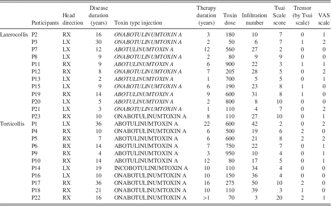
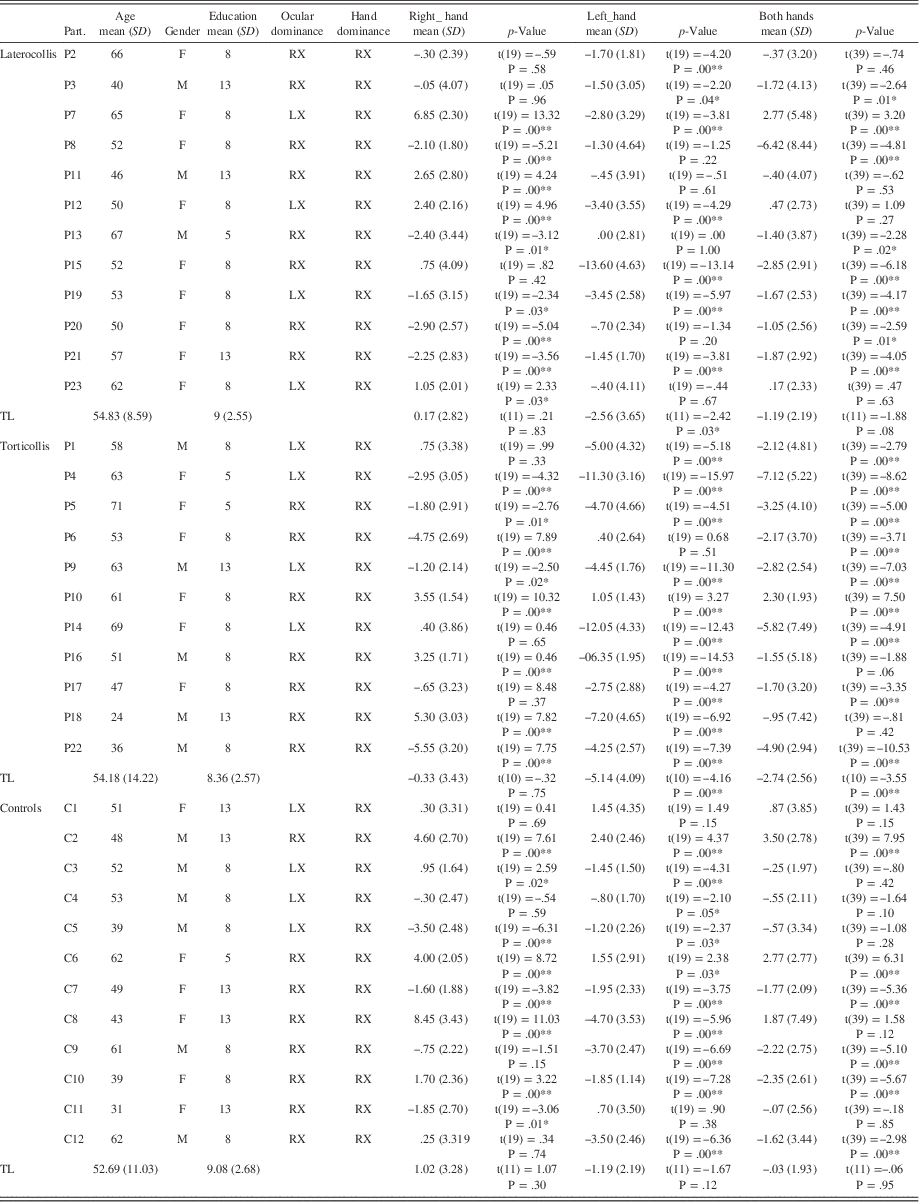
The participants’ demographic and clinical data are reported in Tables 1 and 2. Twenty-three participants (15 females and 8 males) with idiopathic cervical Dystonia were recruited from the Movement Disorders Centre of the University of Messina. Twelve healthy controls (7 females and 5 males; mean age 52.69±11.03 years) constituted the control group. They were recruited from the community through word-of-mouth. Patients were matched to controls for age as well as hand and ocular dominance. Moreover, they were matched for years of education and gender. All participants were right-handers according to Edinburgh Handeness Inventory (Oldfield, Reference Oldfield1971). Twenty-eight of them were right-eye dominant and the remaining 17 were left-eye dominant, according to the ocular dominance test (Yang, Blake, & McDonald, Reference Yang, Blake and McDonald2010).
Clinical Data of Dystonic Patients

RX=right; LX=left.
Demographic Data and Bisection Performances for the Three Groups of Participants

Note. Mean bisection errors (mm) and relative SDs are reported for each participant. Results of one-sample t-tests are also reported. An asterisk indicates significant p-values<.05. A double asterisk indicates significant p-values<.001.
RX=right; LX=left; TL=total.
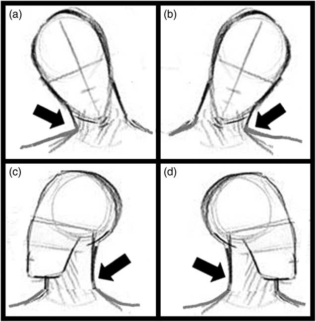
Only patients exhibiting a prevalence of unilateral symptoms were included in the study. They were subdivided into two subgroups according to the head posture and direction (see Table 1 and Figure 1): 12 of them were classified as LC (mean age, 54.83±8.59 years) and 11 as TC (54.18±14.22). Five of 12 LC patients (42%) and 8 of 11 TC patients (73%) showed an aberrant head posture to the right side. Furthermore, 8 of 12 patients (67%) in the LC group and 7 of 11 patients (64%) in the TC group were right-eye dominant.
Sketch of the two forms of CD. (a) LC with right head inclination. (b) LC with left head inclination. (c) TC with right head rotation. (d) TC with left head rotation. The arrows indicate the side of the main affected muscles: in LC, ipsilateral sternocleidomastoid muscles, whereas in TC, contralateral sternocleidomastoid muscles.

All patients underwent extensive neurological examination, laboratory and neuroimaging (i.e., computed tomography or magnetic resonance imaging) investigations to rule out acquired causes of dystonia. None of the enrolled patients have never been treated with drugs blocking the dopamine receptor. All drugs affecting the central nervous system were discontinued at least 1 week before the beginning of the study. All patients were receiving botulinum toxin therapy and were examined at least 3 months after the last injection and just before the periodic injection of botulinum toxin. It is worth noting that since many of these patients have had botulin treatment for many years (see Table 1), the abnormal posture of their head was very subtle (even though they were tested at least 3 months after the last botulin injection). The local ethical committee approved the research protocol and all participants signed an informed consent before examination.
Stimuli and Procedure
Before undergoing the cognitive tasks, the patients were assessed on the TSUI (Tsui, Eisen, Stoessl, Calne, & Calne, Reference Tsui, Eisen, Stoessl, Calne and Calne1986) and Visual Analogue Scale (VAS) scales (Caraceni et al., Reference Caraceni, Mendoza, Mencaglia, Barbatella, Edwards, Forjaz and Cleeland1996) to evaluate the severity and duration of the cervical movements as well as tremor the first one, pain perception the second.
Participants’ hand dominance and ocular dominance were first assessed. Then, they were asked to perform the Line Bisection Task. The Edinburgh Inventory by Oldfield (Reference Oldfield1971) was used to assess hand dominance. The Hole-in-the card test (Yang et al., Reference Yang, Blake and McDonald2010) was performed by participants for assessment of ocular dominance. A red cross (3×3 cm) was presented approximately 5 m in front of the participant. The participant held a sheet of paper with both hands, at arm’s length, and moved the card until the cross was seen through a hole in the center of the card (1.5 cm in diameter) with both eyes open. Then the participant was instructed to close one or the other eye, alternatively, and report whether the cross remained in his/her line of view. The eye that allowed the observer to maintain the view of the cross was identified as the preferred sighting eye (Yang et al., Reference Yang, Blake and McDonald2010).
Line Bisection Task (Schenkenberg et al., Reference Schenkenberg, Bradford and Ajax1980) was performed. Participants were asked to mark, with a pencil, the middle of a series of 200-mm-long and 1-mm-thick black horizontal lines. Each line was centered on an A4 white sheet of paper and oriented along its major axis. Stimuli were centered on the participants sagittal midplane and presented on a table at a distance of approximately 50 cm. Twenty lines were bisected using the right hand (condition A) and 20 lines using the left hand (condition B). Half of the participants started with the right hand, and the other half with the left hand. The order of the starting hand was balanced across subjects.
Statistical Analysis
On the Line Bisection Task, the deviation of the subjective midpoint from the true center of the line was measured to the nearest millimeter. The rightward bisection errors were expressed in millimeters, using positive values (i.e., they were preceded by+), and the leftward errors using negative values (i.e., they were preceded by –). This measure constituted the dependent variable. The rightward and leftward bisection errors were analyzed using repeated measures analysis of variance (ANOVA). In the ANOVA, Group (TC, LC, controls) was the between-subjects factor, while Hand (right hand, RH; left hand, LH) and repetition (across all 20 trials) were the within-subjects factors.
For ANOVA, age, gender, ocular dominance, and education were included as covariates in the model. Greenhouse-Geisser degrees of freedom (df) correction was used to account for potential assumptions violation in the model. Greenhouse-Geisser method was the most conservative choice for our method available within the SPSS software (Bagiella, Sloan, & Heitjan, Reference Bagiella, Sloan and Heitjan2000). When necessary, Bonferroni correction was applied on post hoc tests to obtain a global significance threshold of 0.05.
In addition, one sample t tests were performed to test whether individual biases were significantly different from 0. At the time of the study, patients were receiving different botulin toxin types; moreover, the disease duration and the toxin therapy duration was quite variable. Thus, we tested whether those variables, together with CD severity and pain perception might influence the bisection bias. To this end, we estimated Pearson correlation coefficients between bisection bias and disease duration, as well as toxin therapy duration and type of injection, TSUI and Pain scales. These analyses were performed both on global measures obtained by pooling the two hand conditions together, as well as for each separate hand condition.
RESULTS
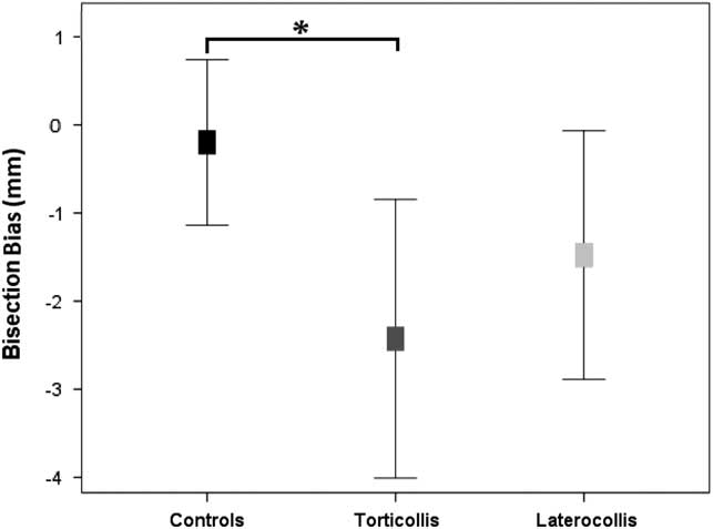
The participants’ demographic and clinical data are reported in Tables 1 and 2. The mean bisection bias for the three groups and the relative differences are presented in Table 2. The ANOVA showed that there was a significant effect of the Group factor [F(2,126.414); p=.024; partial η2=0.209]. Post hoc analyses revealed that, overall, TC patients (corrected p-values p=.05, see Figure 2) showed a greater leftward deviation (TC mean=−2.74 mm; SD=2.56) than the control group (control mean=−0.33; SD=1.93). Moreover, there was a significant effect of the Hand [F(1,180.97)=15.397; p<.001, partial η2=0.325]. On average participants showed a greater leftward deviation when they bisected with the left (mean=−2.87; SD=3.70) than with the right hand (mean=0.30; SD=3.13). No interactions resulted to be significant.
Mean bisection error for each group. Bars represent standard error. Asterisks indicate significant differences between groups at 0.05 threshold.

One sample t test showed a leftward deviation significantly different from 0 both for the mean bisection bias of the two hands [(t(10)=−3.550; p=.005)] and for the left hand condition [t(10)=−4.169; p=.0019] in TC patients. Also, LC patients showed a significant leftward bias when bisecting with their left hand [t(11)=−2.426; p=.034 ], while controls did not show any significant bias. For single case analyses, one-sample t tests showed that the bisection bias of LC patients was significantly different from 0 in 9 of 12 patients (75%) when they used their right hand (5/9 patients [55.6%] showed a leftward deviation), and in 7 of 12 patients (58.3%) when they used their left hand (7/7 participants [100%] showed a leftward deviation). In TC patients, a significant bisection bias was found in 8 of 11 patients (72.7%) when bisecting with their right hand (of them 5/8 participants [ 62.5%] showed a leftward deviation), and in 10/11 patients (90.9%) when bisecting with their left hand (9/10 participants [ 90%] showed a leftward deviation). Lastly, the bisection bias was found to be significant in 8/12 healthy controls (66.7%) when performing with their right hand (5/8 participants [62.5%] showed a leftward deviation) and in 10/12 participants (83.3%), when performing with their left hand (8/10 participants [80%] showed a leftward deviation).
To analyze whether a specific directional bias was associated with a specific form of disease and/or head direction, within each group and for each hand, we analyzed the percent of patients that showed a specific directional bias. In the TC group, 8 of 11 (72.7%) patients had their head slightly turned to the right while 3/11 (27.3%) had their head slightly turned to the left. During right hand line bisection, 5 of 8 patients with right head turn (62.5%) showed a significant leftward deviation, while 2 of the 3 patients with left head turn (P1 and P14) did not show any significant bias and the third one (P16) manifested a rightward bias. In the LC group, 5 of 12 (41.7 %) patients had their head slightly tilted to the right while 7/12 (58.3%) had their head slightly inclined to the left. For the LC patients, 3 of 5 patients (60%) with right head tilt showed a significant rightward deviation (one did not have any bias), while 4 of 7 patients (57%) with left head tilt showed a leftward deviation. In both groups, performances with the left hand did not seem to discriminate between different patients.
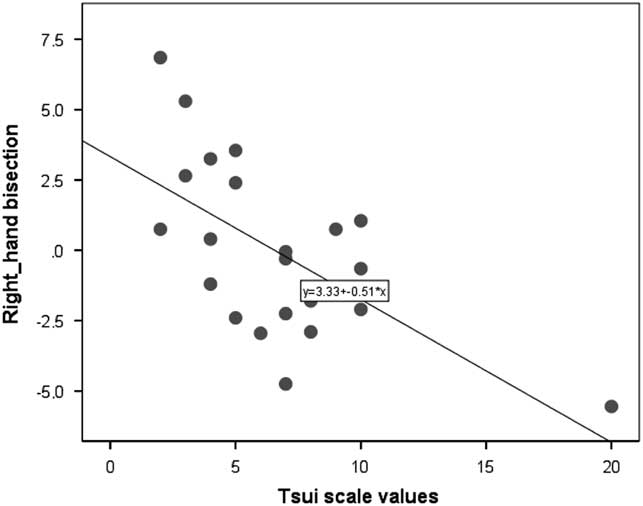
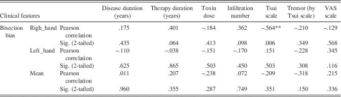
In agreement with the above findings, CD patients showed CD patients showed a significant correlation between bisection bias for the right hand condition and the Tsui Scale score (see Figure 3). No other significant correlation was observed between the bisection bias and clinical features (see Table 3).
Correlation between mean bisection error during right hand execution and Tsui Scale scores. An increase of the leftward bisection bias is linked to increased severity of dystonic symptoms.

Correlations between Bisection Performances (Separately for Right Hand, Left Hand and Their Mean) and Clinical Data (Disease Duration, Therapy Duration, Infiltration Number, Tsui Scale Severity, Tsui Scale Tremor, VAS)

Note. A double asterisk indicates significant p-values<.001.
DISCUSSION
With the present study, we provide evidence of consistent leftward attentional bias in patients with asymmetric symptoms of idiopathic cervical dystonia, and in particular in TC patients.
On the other hand, control participants did not show any significant bias, in agreement with the evidence that pseudoneglect is reduced in older individuals (Benwell et al., Reference Benwell, Thut, Grant and Harvey2014; Learmonth et al., Reference Learmonth, Benwell, Thut and Harvey2017). In accordance with the literature (Marzoli et al., Reference Marzoli, Prete and Tommasi2014), the hand used to perform the task affected the overall performance. Indeed, participants deviated more to the left of true center when using their left hand than when using their right hand. Of interest, the leftward bisection deviation, indexing asymmetrical distribution of visuospatial attention toward the left, was significant in TC patients (except for the right hand condition) and in LT patients for the left hand condition. However, only TC patients performed significantly different from age-matched healthy controls.
On the basis of preliminary findings in different types of focal dystonia (Ricci et al., Reference Ricci, Salatino, Siebner, Mazzeo and Nobili2014, Reference Ricci, Mazzeo, Celentano, Nobili and Salatino2015), we expected to observe biased attention toward the side of the dystonic muscles. Interestingly, here we observed, in TC patients, hyper-attention toward the side contralateral to the head turn and, likely, in the direction of the contralateral sternocleidomastoid muscle (causing the rotation of the head to the opposite side). It is worth noting that the majority of TC patients had subtle right-side head turns and overall the group showed a leftward bias.
In agreement with the above hypothesis, two of the three patients with left head turn did not show any bias, and the third one showed an opposite, rightward bias. A possibility might also be that the head posture might have affected these patients’ performance. In other words, the attentional bias could simply be due to the effect of misaligning the head from other egocentric coordinate systems and its preferential orientation toward one side of the egocentric space. However, if this were the case one would expect to observe an attentional bias in the same direction of the head orientation (and, therefore, opposite to what we observed).
Indeed, Schindler & Kerkhoff (Reference Schindler and Kerkhoff1997) found reduced rightward bisection bias (i.e., increased leftward deviation) in patients with left neglect, when they performed the task with their head rotated to the left, while they were not affected by right-side head rotation. Importantly, head rotation did not affect line bisection performance in healthy controls (Schindler, & Kerkhoff, Reference Schindler and Kerkhoff1997). Thus, given the above evidence it seems unlikely that the observed behavior might be explained by the (slightly) deviated head posture. However, testing the patients before and after treatment, in future studies, might offer a better insight into the mechanisms underlying the attentional bias.
As suggested by preliminary evidence in focal limb dystonia (Ricci et al., Reference Ricci, Salatino, Siebner, Mazzeo and Nobili2014), we propose that the leftward attentional bias might be an index of pathological hyperactivity of (right) attentional circuits contralateral to the main dystonic muscles. Picazio, Ponzo, and Koch (Reference Picazio, Ponzo and Koch2015) propose the idea that the right parietal lobe and the left cerebellar hemisphere work together for directing attention toward the left hemi-space. Abnormal hyper-activity of this neural network might explain our findings. Although idiopathic cervical dystonia has long been considered to be related to dysfunction of the basal ganglia, recent evidence also suggests the involvement of PPC (de Vries et al., Reference De Vries, De Jong, Bohning, Hinson, George and Leenders2012; Premi et al., Reference Premi, Diano, Gazzina, Cauda, Gualeni, Tinazzi and Borroni2016; Ricci et al., Reference Ricci, Salatino, Siebner, Mazzeo and Nobili2014) and cerebellum (Filip, Lungu, Shaw, Kasparek, & Bares, Reference Filip, Lungu, Shaw, Kasparek and Bares2013; Kuoppamaki, Giunti, Quinn, Wood, & Bhatia, Reference Kuoppamaki, Giunti, Quinn, Wood and Bhatia2003; Perruchoud, Murray, Lefebvre, & Ionta, Reference Perruchoud, Murray, Lefebvre and Ionta2014; Prudente, Hess, & Jinnah, Reference Prudente, Hess and Jinnah2014) in its pathophysiology.
Abnormalities in circuits involving the cerebellum have been observed in clinically unaffected carriers of the DYT1 dystonia mutation, during learning of visuo-motor sequences (performed with the right hand) with a compensatory increased activation in the left ventral prefrontal cortex (Quartarone et al., Reference Quartarone, Rizzo, Terranova, Milardi, Bruschetta, Ghilardi and Girlanda2014), and decreased activation in the posterior medial cerebellum (Ghilardi et al., Reference Ghilardi, Carbon, Silvestri, Dhawan, Tagliati, Bressman and Eidelberg2003). In line with the above hypothesis, non-invasive brain stimulation over the PPC (Ricci et al., Reference Ricci, Salatino, Siebner, Mazzeo and Nobili2014) or cerebellum (see Cho and Hallett, Reference Cho and Hallett2016 for a review) can improve dystonic symptoms.
The present findings are consistent with preliminary evidence (Chillemi et al., Reference Chillemi, Calamuneri, Morgante, Terranova, Rizzo, Girlanda and Quartarone2017) supporting the idea that, while sensory abnormality might be mainly present in LC patients, higher level cognitive impairment might specifically affect TC patients. In details, in our previous study (Chillemi et al., Reference Chillemi, Calamuneri, Morgante, Terranova, Rizzo, Girlanda and Quartarone2017), we observed that patients with TC were less accurate than patients with LC in judging the temporal duration of visual stimuli. Relevant to this finding, the cerebellum seem to play a critical role in temporal attention processing (Bares et al., Reference Bares, Lungu, Liu, Waechter, Gomez and Ashe2007, Reference Bares, Lungu, Liu, Waechter, Gomez and Ashe2011; Bares, Lungu, Husárová, & Gescheidt, Reference Bares, Lungu, Husárová and Gescheidt2010; Husarova et al., Reference Husarova, Lungu, Marecek, Mikl, Gescheidt, Krupa and Bares2011). In line with these findings, we suppose that the cerebellum might be mainly involved in TC rather than in LC. Similarly, the current finding of a stronger attentional bias in TC than in LC might suggest an aberrant enrollment of the posterior parietal and cerebellum circuit in the regulation of cortical activity specific for TC. Of course, these interpretations are largely speculative. In the next future, we plan to further investigate the above hypotheses by using neuroimaging and non-invasive brain stimulation techniques.
Lastly, it is worth noting that, for the CD group, the only significant correlation we observed between experimental task outcomes and clinical features, was a negative correlation between right hand bisection performance and the Tsui Scale score. Of interest, this result seems to reflect a relationship between the magnitude of the leftward bias and CD severity, strengthening, therefore, the hypothesis that a specific cognitive impairment involving attention might play a critical role in cervical dystonia.
Nevertheless, we recognize that our study present a series of limitations and methodological weaknesses. The heterogeneity as well as the small sample size of the two groups of patients limited the conclusions that can be drawn from the present findings. In addition, the small sample size did not allow to stratify the patients according to the side (i.e., right or left) of the aberrant head posture. Future studies using more extensive clinical examination in larger groups of patients will be necessary to clarify the differences of attentional performance that we observed within subtypes of cervical dystonia. The information coming from these studies might offer a rationale for targeting specific sites with non-invasive brain stimulation in prospective rehabilitative interventions in CD patients.
Acknowledgments
Drs. Ricci and Quartarone have equally contributed to the present work. The authors have no conflicts of interest or financial disclosures to make. The authors have no professional or financial affiliations that might be perceived as having biased the presentation.