

INTRODUCTION
Cnidarians represent the simplest and perhaps the most basic structural organization within the Eumetazoa (Collins et al., Reference Collins, Cartwright, McFadden and Schierwater2005). Therefore, the investigation of their organization is essential for understanding the earliest steps of metazoan evolution. The soft tissue (coenosarc) of the cnidarians can be sketched as a double-layered sac with a single opening—the mouth, leading to a gastric cavity and surrounded by tentacles. Their body wall is composed of two epithelial layers (the inner gastrodermis and the outer epidermis) separated by the mesoglea (Thomas & Edwards, Reference Thomas, Edwards, Harrison and Westfall1991).
Numerous hydroid species form colonies. From the outside, the coenosarc is covered by the rigid skeleton, the chitinous perisarc (Naumov, Reference Naumov1969; Bouillon et al., Reference Bouillon, Medel, Pagès, Gili, Boero and Gravili2004). The perisarc is shaped at the endings of the branched shoots and does not change its form later on. The endings of the branched colony are occupied either by hydrants or by specific elements—the growing tips—responsible for the morphogenesis of new structures (Hale, Reference Hale1960; Kosevich, Reference Kosevich1990). The system of coenosarc interconnects the zooids, and thus serves for the distribution of nutrients and for the physiological integration of the colony.
Two main groups of colonial hydroids are recognized—athecates and thecates (subclass Anthomedusae and subclass Leptomedusae respectively (Bouillon & Boero, Reference Bouillon and Boero2000)).
Athecates are characterized by the location of hydranths at the endings of branches (stems), pronounced enlargement of hydranths, the elongation of colony shoots by intercalate growth at the base of the hydranth and an absence of skeleton housing the hydranths (the perisarc covers stolons and shoots, but ends up at the base of the hydrants).
The group of thecates is distinguished by the presence of a rigid outer skeleton all over the soft tissues including the hydranths—the hydrothecae. Therefore, the hydranths in the thecate species are fully formed prior to their emergence and functioning; afterwards they do not grow in size. The growth of the thecate colony completely depends on the functioning of the growing tips located at the endings of shoots and stolons. The growing tips have a regular periodic growth and form the soft tissues of the colony and the outer skeleton. The new perisarc is shaped at the apexes of the tips (Naumov, Reference Naumov1969; Bouillon & Boero, Reference Bouillon and Boero2000; Kossevitch et al., Reference Kossevitch, Herrmann and Berking2001; Marfenin & Kosevich, Reference Marfenin and Kosevich2004).
According to a simplified schema of the cnidarian organization, the coenosarc is nothing but a branched, double-layered tube. Nevertheless, some investigations carried out on athecates have already revealed that the anatomy of colonial hydroids can be more complicated. The investigations have shown for instance, the presence of special chordal tissue in the dactylozooids and hydrorhiza of Paracoryne huvei, Picard 1957 (Bouillon, Reference Bouillon1974, Reference Bouillon1975) and in the stems and the hydrant base of Tubularia sp. (Tardent, Reference Tardent1980). This tissue, presumably with a supportive function, is formed by highly vacuolated gastrodermal cells which lost their epithelial organization (Thomas & Edwards, Reference Thomas, Edwards, Harrison and Westfall1991).
The anatomy of thecate hydroids is much less studied. It is difficult to keep thecates in the laboratory, and also their rigid perisarc in most cases is too thick and almost impenetrable to various substances used in histology, electron microscopy and immunocytochemistry.
On the other hand, the thecate hydroids in particular are characterized by diverse colonies with large, complex shoots (Marfenin & Kosevich, Reference Marfenin and Kosevich2004). It can thus be expected that they also have a more complicated inner organization. Moreover, when looking through the translucent perisarc of some thecate hydroids, it is evident that the soft tissues are not just a double-layered tube, but are split into several tubes forming a three-dimensional structure (Figure 1). The present investigation summarizes observations made on the soft tissue in three representatives of thecate hydroids.
The base of the stem of Diphasia fallax. h, hydrotheca; s, stem of the shoot. Scale: 400 µm.

MATERIALS AND METHODS
For the examination we chose three thecate species belonging to the family Sertulariidae: Hydrallmania falcata (L., 1758), Sertularia mirabilis (Verrill, 1873) and Diphasia fallax (Johnston, 1847). The colonies of these species grow to large dimensions, reaching up to 60 cm in height (H. falcata (Naumov, Reference Naumov1969)). They are highly integrated colonies with complex large, monosiphonic, pinnate shoots.
The specimens were collected at a depth of 7–10 m in the neighbourhood of the N.A. Pertsov White Sea Biological Station of the Moscow State University, Kandalaksha Bay, White Sea, Russia (66° 34′N 33° 08′E).
For light microscopy, TEM and SEM examination the specimens were fixed in 2.5% glutaraldehyde in phosphate buffer (0.83 Osmol) at pH 7.3–7.4 (Millonig, Reference Millonig1964) according to the following protocol:
(1) Fixation for 1 hour at 4°C.
(2) 3 washes for 10 minutes in the same buffer.
(3) Post-fixation in 1% OsO4 in the buffer for 1 hour at room temperature.
(4) 3 washes for 10 minutes in the buffer.
(5) Dehydration in 30%, 50% and 70% ethanol (2 washes for 15 minutes each).
Several months later, specimens for light microscopy and TEM were embedded in the standard mixture of Araldite (Serva) and Epon (Fluka) (Mironov et al., Reference Mironov, Komissarchik and Mironov1994). Blocks were sectioned on ultramicrotomes (Sorvall Ultra Microtome Dupont MT5000 and УМТП-3). Semi-thin sections ranging in thickness from 1–2 µm were stained with a mixture of toluidine blue and methylene blue (Mironov et al., Reference Mironov, Komissarchik and Mironov1994) and viewed with an Axioplan 2 imaging microscope (Zeiss). Ultra-thin sections were stained with uranyl acetate and lead citrate and examined with a JEOL JEM 100B electron transmission microscope. Specimens for SEM were dehydrated in ethanol series and acetone, critical point dried, mounted on stubs, sputter coated with platinum and palladium, and viewed with CAM SCAN, SCAN JSM 63 LA and Hitachi S-405A electron scanning microscopes.
RESULTS
Hydrallmania falcata
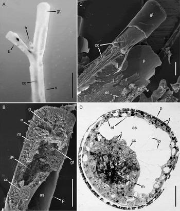
The growing tip (Figure 2A, B & C) of the shoot stem is about 460–550 µm in diameter. The perisarc thickness on the sides of the tip is about 15 µm. The coenosarc occupies the whole lumen of the perisarc tube. The epidermis of the tip consists of narrow and high, columnar, closely packed cells. Highly vacuolated cells constitute the gastrodermis. A thin mesoglea separates two layers.
The upper part of Hydrallmania falcata shoot. (A) An appearance of the shoot; (B) a longitudinal section (SEM); (C) the shoot with dissected perisarc (SEM); (D) a cross-section below the growing tip. as, axial space; b, branch; cc, coenosarc canal; e, epidermis; el, epidermal lining; g, gastrodermis; gc, gastric cavity; gt, growing tip; h, hydrotheca; lp, lamellipodia; m, mesoglea; n, nematocyst; p, perisarc; s, stem of the shoot; t, tanning cell. Scale: A, 1 mm; B, C, 300 µm; D, 100 µm.

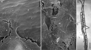
The upper part of the stem (Figure 2A, B, C & D) (just below the growing tip) is about 500 µm in diameter, the perisarc is 20–25 µm thick. The coenosarc canal is much narrower than the lumen of the perisarc tube. The wall of the coenosarc tube is simple and consists of two epithelial layers—epidermis and gastrodermis, separated by a thin mesoglea (Figure 2D). The gastric cavity is not visible at the level the cross-section was made (Figure 2D), but is apparent at the SEM longitudinal sections (Figure 2B). The coenosarc tube is eccentric, thus a part of the perisarc tube is not occupied by soft tissue. The surface of the epidermis of the coenosarc tube forms deep longitudinal folds (Figure 2C). Where the coenosarc gets in contact with the perisarc, some epidermal cells lose the connection with the mesoglea and hence their typical form. They become flattened, look like fibroblasts, form slender and extensive filopodia (Figure 3A), and migrate along the inner surface of the perisarc. More proximally to the growing tip, these fibroblast-like cells cover the entire inner surface of the perisarc and thus constitute a thin layer lining the skeleton tube (Figures 2D & 3B). The epidermal cells on the outside of the coenosarc tube and the epidermal lining form slender projections (Figures 2D & 3C); further SEM examination of the other species (see Sertularia mirabilis) has revealed that these projections are flattened and resemble lamellipodia. The lamellipodia get in contact with each other repeatedly and thus partly occupy the axial stem space (the space within the perisarc tube). Among the cells of the epidermal lining and the coenosarc canal there are numerous tanning cells (Figure 2D), which take part in the sclerotization of the perisarc (Knight, Reference Knight1970; Letunov & Stepanjants, Reference Letunov, Stepanjants, Fedyakov and Lukanin1986; Kossevitch et al., Reference Kossevitch, Herrmann and Berking2001).
Epidermis reorganization in the Hydrallmania falcata shoots. (A) An epidermal lining formation just proximally the growing tip (SEM); (B) the epidermal lining surface (SEM); (C) attaching lamellipodia in the axial stem space. e, epidermis; el, epidermal lining; lP, lamellipodia; p, perisarc. Scale: A, 3 µm; B, 10 µm; C, 500 nm.

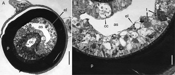
The middle part of the stem (Figure 4A) is approximately 500 µm in diameter. The perisarc here can reach 100 µm in thickness and it has a laminated structure. The coenosarc canal partly occupies the inner space within the perisarc tube; it consists of an epithelial epi- and gastrodermis, separated by a thin mesoglea. The gastric cavity lumen is apparent in the centre of the canal.
The middle part of the stem of Hydrallmania falcata shoot. (A) A cross-section; (B) the epidermal lining (a cross-section). as, axial space; cc, coenosarc canal; e, epidermis; el, epidermal lining; p, perisarc; t, tanning cell. Scale: A, 100 µm; B 20 µm.

The epidermal lining covers the entire inner surface of the perisarc (Figure 4B). The lining is rather thick and occupies a large portion of the axial stem space. The epidermal cells of the lining are highly vacuolated, and some tanning cells are visible here.
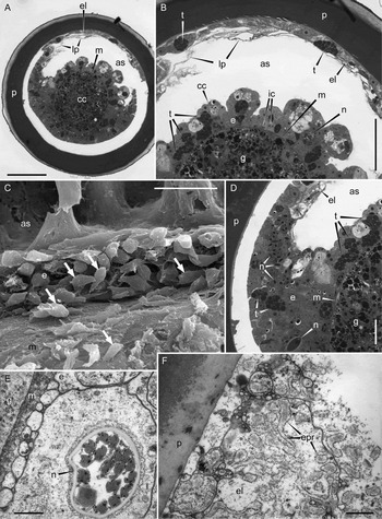
The stem base (Figure 5A) is about 400 µm in diameter and the perisarc thickness here is approximately 50 µm. The coenosarc canal is eccentric. The epidermal surface of the canal bears deep longitudinal folds. Inside the folds, nematocysts, numerous tanning cells, and interstitial cells are visible (Figure 5B). SEM examinations have revealed that inside these folds there are many cells which migrate along the mesoglea surface. These cells are streamlined (one end is wide, the other end is narrow) and they possess filopodia on the surface (Figure 5C). The gastrodermis of the canal is separated from the epidermis by a thin mesoglea (Figure 5A, B, D & E). A gastric cavity is not visible.
The stem base of the Hydrallmania falcata shoot. (A) A cross-section (the space within the coenosarc canal and the perisarc inner surface most probably is an artefact); (B) the epidermal lining and the fragment of the coenosarc canal (a cross-section); (C) the dissected epidermis of the coenosarc canal, ‘running’ cells are shown by arrows (SEM); (D) an area, where the epidermis of the coenosarc canal turns to the epidermal lining (the space within the coenosarc canal and the inner perisarc surface most probably is an artefact); (E) a nematocyte in immediate proximity to the mesoglea (TEM); (F) the epidermal lining (TEM). as, axial space; cc, coenosarc canal; e, epidermis; el, epidermal lining; epr, granular endoplasmic reticulum; g, gastrodermis; ic, interstitial cells; lp, lamellipodia; m, mesoglea; n, nematocyst; p, perisarc; t, tanning cell. Scale: A, 100 µm; B, 50 µm; C, 30 µm; D, 20 µm; E, F, 500 nm.

Epidermal cells line the entire inner surface of the perisarc (Figure 5A, B). The cells of epidermal lining form some lamellipodia. The lining includes some tanning cells. The lining cells bear numerous projections, filled with granular endoplasmic reticulum (Figure 5F). In other words, the cells within the lining move and show a high synthetic activity. Where the epidermis of the coenosarc canal turns into the lining of the perisarc, numerous differentiating nematocysts are apparent (Figure 5D, E).
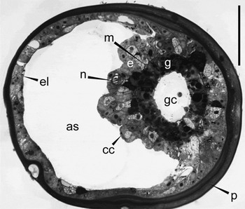
The branches (Figure 6)—both the first-order and second-order ones—are characterized by the same inner organization. The coenosarc canal consists of epithelial epi- and gastrodermis, separated by thin, wavy mesoglea. The gastric cavity lumen is present. The canal joins the inner perisarc surface on one side. The epidermis of the canal includes nematocysts, and the surface of the epidermis forms longitudinal folds. Epidermal cells spread over the inner surface of perisarc, forming the lining.
A cross-section of a first-order branch of Hydrallmania falcata. as, axial space; cc, coenosarc canal; e, epidermis; el, epidermal lining; g, gastrodermis; gc, gastric cavity; m, mesoglea; n, nematocyst; p, perisarc. Scale: 50 µm.

Sertularia mirabilis
The growing tip (Figure 7A, B) is cylindrical, 0.5–1.0 mm in diameter. The perisarc is 2–3 µm thick at the tip apex and is about 7 µm at the tip sides. The coenosarc completely occupies the inner space within the perisarc. The epidermis is epithelial, consisting of densely packed cells. The gastrodermis is highly vacuolated, 2–3 times thicker, than the epidermis. Both layers are filled with tanning cells. At the apex of the tip, a gastric cavity lumen is visible.
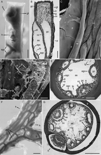
The stem of Sertularia mirabilis shoot. (A) an appearance of the distal part of the stem; (B) a longitudinal section of the distal part of the shoot; (C) an area where the coenosarc canal is adjacent to the inner perisarc surface, just more proximally from the growing tip (SEM); (D) a dissected middle part of the stem (SEM); (E) a cross-section of middle part of the stem; (F) an appearance of the stem base; (G) a cross-section of the stem base (the space within the epidermal lining and the inner perisarc surface and within the epidermis surrounding the canals and the mesoglea most probably is an artefact). agc, additional gastrodermal canal; as, axial space; b, branch; cc, coenosarc canal; e, epidermis; el, epidermal lining; g, gastrodermis; gc, gastric cavity; gt, growing tip; h, hydrotheca; lp, lamellipodia; m, mesoglea; p, perisarc; s, stem of the shoot. Scale: A, B, F, G, 250 µm; C, 30 µm; D, E, 100 µm.

In the upper part of the stem (Figure 7A, B) (just below the growing tip) the coenosarc canal becomes 2–3 times narrower and is eccentric, adnate to the perisarc wall. The organization of the epi- and gastrodermis is epithelial. The epidermis surface produces deep longitudinal folds (Figure 7C). Some epidermal cells spread over the inner perisarc surface, forming the lining (identically as in H. falcata).
A short distance from the growing tip the main coenosarc canal starts branching and several anastomosing additional canals appear (Figure 7A, B). These canals are formed by gastrodermis and surrounded with a common epidermis. The gastric cavity is evident in the centre of the canals. The canals run along the inner surface of the skeleton tube. Several lamellipodia, formed by the epidermal cells, occur in the axial stem space.
The middle part of the stem (Figure 7D, E) is about 0.6–1.0 mm in diameter. The perisarc is greatly thickened, laminated, up to 100 µm thick. Numerous gastrodermal canals run along the inner surface of the perisarc tube. The gastrodermis of the canals is epithelial. These canals anastomose and form a kind of irregular net. The canals are enclosed in a common epidermis. In close proximity to the mesoglea that covers the gastrodermal canals, the epidermis is epithelial. Between the canals the epidermal cells lose the connection to the mesoglea and take a form of the cells of the perisarc lining. It is easy to distinguish the main canal from the others. This canal continues from the growing tip just along one side of the perisarc tube and produces the additional canals. The epidermal surface of the main canal forms longitudinal folds (Figure 7D). The gastrodermal cells, constituting the wall of the main canal, are higher and larger in comparison to the cells composing additional canals. The epidermal cells form numerous lamellipodia. The lamellipodia repeatedly attach to each other and loosely occupy the axial stem space (Figure 7D, E).
The stem base (Figure 7F, G) is about 1 mm in diameter. The perisarc is 45–65 µm thick. Mostly the coenosarc is characterized by the same organization as in the middle part of the stem. But the network composed of the anastomosing gastrodermal canals is denser (additional canals are present), and the epidermal cells produce much more interconnecting lamellipodia, which occupy the axial stem space (Figure 7G).
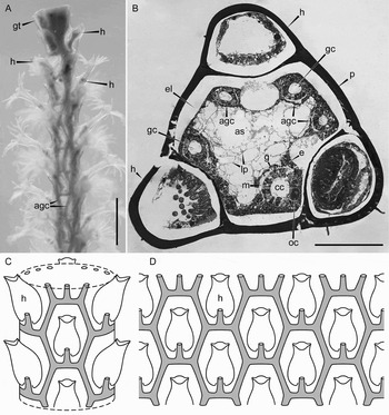
The branches (Figure 8A, B) are about 550–650 µm in diameter. The perisarc thickness is about 35 µm. Where the stem produces the branch, the main coenosarc canal also divides. The main canal produces additional canals, which run along the inner surface of the perisarc, anastomosing and forming a regular hexagonal network (Figure 8C, D). The network is formed by the curved longitudinal coenosarc canals, which merge in pairs forming bridge-like connections. Every connection is situated right below a hydrotheca and it branches a short canal in the direction of a hydranth. The canals are composed of epithelial gastrodermis and surrounded by epidermis. Between the canals the epidermis lines the perisarc. The epidermal cells produce numerous lamellipodia, which attach to each other and partly occupy the axial branch space (Figure 8B). Sometimes it is possible to distinguish the main canal, which grows from the branch growing tip, from the other canals of the network by its bigger diameter.
The branch of Sertularia mirabilis shoot. (A) An appearance of the shoot; (B) a cross-section (the space within the epidermal lining and the inner perisarc surface most probably is an artefact); (C) the scheme of gastrodermal canals, constituting a regular hexagonal network, located over the inner perisarc surface (the gastrodermal canals are shown by grey colour, hydrants inside hydrothecae are not shown); (D) scanning image of the regular hexagonal network, formed by gastrodermal canals (the gastrodermal canals are shown by grey colour, hydrants inside hydrothecae are not shown). agc, additional gastrodermal canal; as, axial space; cc, coenosarc canal; e, epidermis; el, epidermal lining; g, gastrodermis; gc, gastric cavity; gt, growing tip; h, hydrotheca; lp, lamellipodia; m, mesoglea; oс, oocyte; p, perisarc. Scale: A, 1 mm; B, 250 µm.

Diphasia fallax
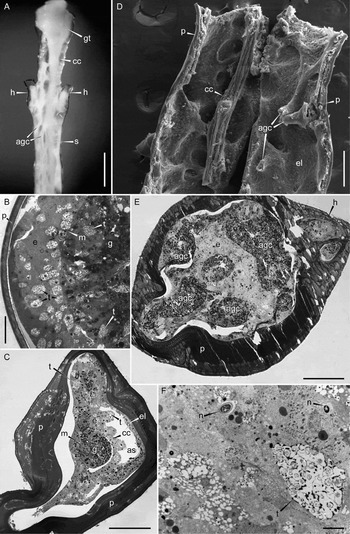
The growing tip (Figure 9A, B) is about 480 µm in diameter. The coenosarc occupies all the inner space within the perisarc tube, its organization is epithelial. The cell boundaries are difficult to distinguish. The gastric cavity is not visible. Both epithelia are full of numerous tanning cells. Some nematocysts are visible in the epidermis.
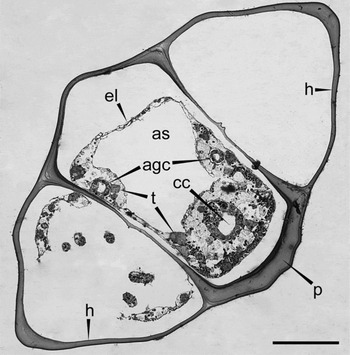
The stem of Diphasia fallax shoot. (A) An appearance of the distal part of the stem; (B) a cross-section of the growing tip; (C) a cross-section of the upper part of the stem (the space within the epidermal lining and the inner perisarc surface most probably is an artefact); (D) a dissected middle part of the stem (SEM); (E) a cross-section of the stem base; (F) an epidermal tissue of the stem base (TEM). agc, additional gastrodermal canal; as, axial space; cc, coenosarc canal; e, epidermis; el, epidermal lining; g, gastrodermis; gt, growing tip; h, hydrotheca; m, mesoglea; n, nematocyst; p, perisarc; s, stem; t, tanning cell. Scale: A, 500 µm; B, 40 µm; C, E, 100 µm; D, 300 µm; F, 5 µm.

The upper part of the stem (Figure 9A, C) (just below the growing tip) is about 380 µm in diameter. The laminated perisarc is up to 100 µm thick. The coenosarc canal is much narrower than the inner diameter of the perisarc tube. The gastric cavity is not visible. The canal is eccentric. The epidermal surface of the canal forms deep folds. The gastrodermis and the epidermis are separated by a thin mesoglea. The epidermal lining covers the inner perisarc surface. The epidermis of the canal and the lining include numerous differentiating nematocysts and some tanning cells. Only a small axial space is not occupied by the coenosarc.
In the middle part of the stem (Figure 9A, D), almost just below the growing tip, the coenosarc canal starts branching. The anastomosing additional canals run along the inner perisarc surface. The canals are formed by gastrodermis and enclosed in a common epidermis. The main coenosarc canal is thicker than additional ones, and the epidermal surface of the canal forms numerous folds.
The stem base (Figures 1 & 9E) is about 470 µm in diameter. The laminated perisarc is up to 100 µm. The coenosarc occupies all the inner space within the perisarc tube and represents a system of branching gastrodermal canals, enclosed in a common epidermal tissue. At this level it is not possible to distinguish the main coenosarc canal from the additional canals. The gastrodermal canals are separated from the epidermal tissue by a thin mesoglea. The gastric cavity is not visible inside the canals. The gastrodermal cell boundaries are difficult to distinguish. The epidermal tissue looks like not epithelial (Figure 9E, F): the epidermal cells lose their typical structure and fill all the axial space between the gastrodermal canals. The epidermal tissue consists of various cells (including tanning cells and nematocytes), which are located randomly, but closely packed. Some epidermal cells include electronic-dense vesicles.
The branches (Figure 10) are about 40 µm wide. The perisarc here is up to 40 µm thick. The coenosarc represents several gastrodermal canals, which run along the inner perisarc surface. The epidermis surrounds the canals and lines the inner perisarc surface. The gastrodermis is separated from the epidermis by a thin mesoglea. Some tanning cells are evident in the epidermis. The large axial space is not occupied by the coenosarc.
A cross-section of the branch of Diphasia fallax shoot (the space within the epidermal lining and the inner perisarc surface most probably is an artefact). agc, additional gastrodermal canal; as, axial space; cc, coenosarc canal; el, epidermal lining; h, hydrotheca; p, perisarc; t, tanning cell. Scale: 100 µm.

DISCUSSION
The soft tissue organization of the shoots in sertulariid species Hydrallmania falcata, Sertularia mirabilis and Diphasia fallax was investigated. The coenosarc was examined in the most distal parts of the shoots (the youngest parts) up to the basal shoot parts (the oldest ones). In this way we could trace the pattern of the soft tissue reorganization during the growth of the colony. The coenosarc of the examined sertulariid species takes a form of simple double-layer epithelial tube only in the most distal parts of a colony—in the growing tips of stolons and shoots. Proximally to the tip the coenosarc is reorganized significantly.
The coenosarc canal occupies the entire lumen of the perisarc tube only in the growing tips (Figures 2A, 2B, 7A, 7B, 9A & 9B). Below the tip the coenosarc canal becomes much narrower and adnate to the inner side of the perisarc tube (Figures 2B & 7B). At the same time, the large space within the perisarc tube (axial space) is free of soft tissue. The axial space has no connection to the gastric cavity of the coenosarc canal and is obviously filled with some sort of liquid, most probably seawater. Beneath the growing tip, an epidermal cell lines the inner surface of the perisarc (Figure 2D, 2C & 7B). The lining occurs everywhere in the stem and the branches of the examined species. The lining is formed by epidermal cells of the coenosarc canal which have lost the connection to mesoglea, became flattened, and have spread over the inner surface of a perisarc tube (Figures 3A & 7C).
Moreover, in two of three examined species (S. mirabilis and D. fallax), the coenosarc canal starts branching almost just below the growing tip (Figures 7A, 9A & 9D). The additional canals are formed by gastrodermis and enclosed in common epidermis (Figure 7E, G). Casual observations of additional coenosarc canals formation were already made in the papers by Nutting (Reference Nutting1904) and Allman (Reference Allman1871). Nutting just mentioned in passing about the system of canals in some sertulariids, but did not describe its organization. Allman briefly described the canals of Antennularia antennina (Linnaeus, 1758) (Plumulariidae) (p. 126), but according to his drawings, the additional canals start directly from the growing tip. This is in contrast to the present results and the rules of the growth and morphogenesis in hydroids which state that the growing tip is a ‘blind end’ of a single coenosarc canal; several growing tips cannot fuse forming one large tip (Davis, Reference Davis, Lenhoff and Davis1971; Beloussov & Dorfman, Reference Beloussov and Dorfman1974; Kosevich, Reference Kosevich2006). In the investigated sertulariid species, the main coenosarc canal begins producing the additional anastomosing gastrodermal canals a short distance below the growing tip.
Most likely, the epidermal lining and the additional gastrodermal canals formation is explained by the large size of the shoots of the examined sertulariid species (Naumov, Reference Naumov1969; Cornelius, Reference Cornelius1979). Probably, the main coenosarc canal cannot occupy the whole lumen of the perisarc tube because of physical and hydrodynamic restrictions. The hydroplasma movement contributes to the whole colony integration (Huxley & de Beer, Reference Huxley and de Beer1923; Saint-Hilaire, Reference Saint-Hilaire1930; Hale, Reference Hale1960; Karlsen & Marfenin, Reference Karlsen and Marfenin1984). But if the diameter of the coenosarc tube is too large, the fluid inside the gastric cavity (hydroplasma) could not be mixed properly. Mixing of hydroplasma is necessary for effective uptake of the food particles by gastrodermal cells. In part the hydroplasma mixing depends on the effect of the gastrodermal cilia (Allman, Reference Allman1871; Josephson & Mackie, Reference Josephson and Mackie1965), and it becomes better firstly when the diameter of coenosarc canal is comparable with the length of the digestive cell cilia and secondly when a ratio of the gastrodermal surface to the hydroplasma volume becomes bigger. In other words, the hydroplasma mixing becomes more effective when the wide coenosarc canal is transformed into the system of branched tubes of smaller diameter.
The system of coenosarc canals within the stems of S. mirabilis and the stems and branches of D. fallax is irregular (Figures 7F, 9A & 9D). In most cases, the main canal is distinguishable due to the larger size of its gastrodermal cells (Figure 7E, G). The additional canals have a thinner gastrodermal layer. Moreover, the main canal continues from the growing tip just along one side of the perisarc tube and its epidermal surface forms longitudinal folds (Figures 7D & 9D). In general, it looks like the branching of the canal becomes more pronounced with the age of the shoot part. But the spatial organization of the system of additional canals remains irregular.
In contradistinction to the stems and branches of D. fallax and the stems of S. mirabilis, in the branches of S. mirabilis the gastrodermal canals form a regular hexagonal network (Figure 8A, C & D). Obviously, the regularity of the network owes its existence to the regular arrangement of the hydrothecae. In the branches of S. mirabilis, the hydrothecae are arranged very regularly: in four to eight longitudinal rows with the hydrothecae of one circle alternating with gaps in the flanking circles (Nutting, Reference Nutting1904; Naumov, Reference Naumov1969; Pyataeva & Kosevich, Reference Pyataeva and Kosevich2008).
The formation of the epidermal lining can be explained as an accommodation of the hydroid for prolonged perisarc secretion. As is well known, epitheliomuscular cells take part in the perisarc secretion and tanning cells participate in perisarc sclerotization (Knight, Reference Knight1970; Letunov & Stepanjants, Reference Letunov, Stepanjants, Fedyakov and Lukanin1986; Kossevitch et al., Reference Kossevitch, Herrmann and Berking2001). Thus, the perisarc continues to become thicker throughout the hydroid's life only when it is in contact with soft tissues. This must be the reason why the perisarc is thicker and laminated in the older colony parts (Figure 7E). The lining secures the permanent contact of the soft tissues to the inner perisarc surface when the coenosarc canal occupies the axial space only partly. The cells of the lining show high synthetic activity (their cytoplasm is full of granular endoplasmic reticulum (Figure 5F)) and many tanning cells are evident among them (Figures 2D, 4B & 5B). For such species as sertulariids, a thick skeleton is essential for supporting large, complex colony shoots.
The cells within the epidermal lining and the epidermis of the coenosarc canal are mobile (Figures 3B & 5C), as is well known for most hydrozoan cells (Koizumi & Bode, Reference Koizumi and Bode1986; Kossevitch, Reference Kossevitch1999). Just below the growing tip, the axial stem space becomes partly occupied by tiny protrusions of the epidermal cells. A scanning microscopy study revealed that mainly these protrusions are flattened and similar to lamellipodia (Figures 2D, 3C, 7D, 7G & 8B). Perhaps these protrusions contribute the best colony soft tissues' physiological integration. In D. fallax the epidermal cells even fill all the space within the perisarc tube between the gastrodermal canals (Figure 9E). The newly formed structure consists of shapeless and randomly tightly located cells of various types (Figure 9F) and can be defined as ‘mixed parenchyma’ (Beklemishev, Reference Beklemishev1969). In contrast to the chordal tissue of gastrodermis origin in Paracoryne huvei (Bouillon, Reference Bouillon1974, Reference Bouillon1975) and Tubularia sp. (Tardent, Reference Tardent1980) the ‘mixed parenchyma’ of epidermis origin in D. fallax includes no highly vacuolated cells and hence it is unlikely supportive. Most probably the ‘mixed parenchyma’ is a source of epitheliomuscular cells and tanning cells for prolonged perisarc secretion like epidermal lining and contributes the best colony coenosarc physiological integration as well.
In general the described soft tissue organization of H. falcata, S. mirabilis and D. fallax is a striking example of structural complexity that does not contravene the limits of the ground plan of the phylum Cnidaria. Their tissues are still organized as two-layered, branched tube. But the enlargement of the colony structures leads to the formation of a divaricated system of canals inside the outer skeleton tube that assumes the protective and supportive function. This allows to overcome the trade-offs between growth of colony-size and the functional efficiency of a circulatory system (see above). The epidermal lining on the inner surface of skeleton tube or the ‘mixed parenchyma’ of epidermis origin solves the problem of skeleton strengthening by extention of the perisarc secretion and sclerotization that is not possible without permanent contact between the living cells and the outer skeleton.
ACKNOWLEDGEMENTS
We thank two anonymous referees for helpful reviewing of the manuscript and Rohan Bates for improving its English. Support was provided by the Russian Foundation for Basic Research (grants No. 05-04-48662-a and 07-04-00736-a).