Dieting is a global emerging trend in recent years as more people strive to adhere to food restriction plans in order to achieve a desired slim body or body weight and form. Dieting is a first choice for weight management in obese people to prevent the onset of serious co-morbidities such as diabetes and hypertension. A recent trend among youth is the maintenance of a perfect and healthy figure through dieting. Unfortunately, this strategy may have unforeseen repercussions that may result in the onset of various disorders including malnutrition, weakened immune system and reproductive potential such as hormonal and menstrual disorder, sub-fertility and even infertility(Reference Habib, Ali and Nazir1). In addition, the intermittent fasting (complete food deprivation for a day or two days) is very common among young girls and women in several countries that may generate potential risk on their reproductive health(Reference Mao, Liu and Zhang2).
Mammalian females are born with a fixed number of germ cells housed in functional units known as follicles. Each follicle contains an oocyte as well as specialised somatic granulosa cells required for the growth, development, maturation and survival of follicular oocytes. Pituitary gonadotropins such as follicle-stimulating hormone (FSH) and luteinising hormone (LH) surge at the time of puberty that trigger follicular growth and development. The selective recruitment occurs in the ovary that enables only dominant follicles to progress further, while others are eliminated from the cohort of the ovary through the process called follicular atresia(Reference Holesh, Bass and Lord3).
The massive depletion of a majority of germ cells (> 99 %) from the cohort of fetal ovary prior to birth has been reported in most of the mammalian species including human females(Reference Hułas-Stasiak and Gawron4–Reference Yadav, Tiwari and Gupta8). Players and pathways that trigger massive depletion of germ cells via follicular atresia that may lead to premature ovarian failure and thereby infertility remains poorly understood. Therefore, players and pathways that drive rapid germ cell/oogonia/oocyte depletion have gained global attention in the recent past.
Growing evidences suggest that a variety of apoptotic and non-apoptotic cell death pathways are involved during germ cell depletion from the cohort of the ovary(Reference Edinger and Thompson9–Reference Yadav, Pandey and Premkumar13). The apoptosis, autophagy, necroptosis and necrosis pathways have been reported during germ cell depletion in mammalian ovaries(Reference Yadav, Tiwari and Gupta8,Reference Tiwari, Prasad and Tripathi11,Reference Hutt14,Reference Tilly15) . Although in order to maintain the highest-quality oocytes capable of developing into the appropriate female gametes, elimination of poor-quality germ cells may be part of the selection process(Reference Mork, Tang and Batchvarov16), and the rapid depletion of germ cells and oocytes may exhaust ovarian reserve and trigger premature ovarian failure that may cause infertility(Reference Edinger and Thompson9–Reference Tiwari, Prasad and Tripathi11).
Apoptosis and autophagy have been observed in oocytes at all stages of oogenesis and in the ovum after ovulation under in vitro culture conditions(Reference Tiwari, Prasad and Tripathi11,Reference Hutt14,Reference Tilly15,Reference Chaube, Prasad and Tiwari17,Reference Lobascio, Klinger and Scaldaferri18) . Some pathways work together or in parallel to destroy germ cells from the ovary under various inducing factors, pathological conditions and ageing(Reference Tiwari, Prasad and Tripathi11,Reference Hutt14,Reference Aitken, Findlay and Hutt19) . Emerging evidences suggest that along with the apoptosis, autophagy plays an essential role during germ cell depletion from the ovary in mammals(Reference Galluzzi, Kepp and Trojel-Hansen20,Reference Schwartz, Seeley and Zeltser21) .
Autophagy is a regulated cell death pathway mediated by autophagy-related genes (ATG) that regulate autophagosome formation(Reference Liu, Tang and Zhou22). The autophagosomes then fuse with liposomes to degrade the internal components. Studies suggest that impaired autophagy or excessive autophagy may lead to cell death(Reference Schwartz, Seeley and Zeltser21) so called autophagy-mediated cell death(Reference Kroemer, Galluzzi and Vandenabeele23). The involvement of impaired autophagy has recently been proposed in follicular somatic as well as germ cells in mammalian ovary(Reference Pandey, Yadav and Premkumar12,Reference Yadav, Pandey and Premkumar13,Reference Pandey, Yadav and Premkumar24) . Emerging evidences suggest that both apoptosis and impaired autophagy could be actively involved in germ cell depletion from mammalian ovary and may be interconnected during developmental-regulated cell death(Reference Yadav, Tiwari and Gupta8,Reference Pandey, Yadav and Premkumar12,Reference Pandey, Yadav and Premkumar24,Reference Pandey, Yadav and Premkumar24) .
Mammalian ovary is a dynamic organ that performs several functions including folliculogenesis, oogenesis, steroidogenesis and generation of competent oocytes required for successful fertilization and early embryonic development(Reference Pandey, Yadav and Premkumar12,Reference Yadav, Pandey and Premkumar13,Reference Pandey, Yadav and Premkumar24–Reference Prasad, Tiwari and Koch26) . Stress affects the ovarian functions and oocyte quality in several mammalian species including human(Reference Pandey, Yadav and Premkumar24,Reference Prasad, Tiwari and Koch26) . Although the negative impact of dietary stress on various reproductive health issues has already been documented(Reference Khaled, Hundley and Tsofliou27–Reference Sun, Shen and Liu29), the molecular mechanism by which dietary stress induce cell death in the ovary remains poorly understood. Therefore, the present study was designed to investigate whether dietary stress generated due to complete food deprivation (fasting) or partial food deprivation (half diet food restriction for one or two estrous cycle) could induce autophagy and apoptosis signalling in ovary of female rats, Rattus norvegicus.
Material and methods
Chemicals and experimental animals
Chemicals used in the present study were purchased from Sigma-Aldrich unless otherwise specified. Adult female rats of 1 year old (body weight 220 (sem 10) g) of Charles-foster strain were separated from the existing colony of departmental animal facility and subjected to the determination of their estrous cycle following standard procedure. In brief, a small amount of fluid collected from the vagina using normal saline was used to prepare vaginal smear on the cleaned slide and then observed under a microscope. A vaginal cytology with abundance of non-nucleated cornified epithelial cells was considered as estrus stage and 24 female rats having almost 5 d of estrous cycle with estrus stage were subjected to the determination of half diet under a 12 h light/dark cycle with food and water ad libitum in the departmental animal facility. All procedures were approved by the Institutional Animal Ethical Committee (Wide letter no. BHU/DoZ/IAEC/2018–2019/015) and performed in accordance with the Institutional guidelines.
Determination of half diet for experimental female rats
The half diet for experimental female rats was determined by analysing the daily food intake following the methods published earlier(Reference Claassen30,Reference Seki, Yamaguchi and Marumo31) . For the purpose, 1 year aged 24 adult female rats in the estrus stage were subjected to routine diet observation for 1 week and the daily food intake was monitored for all individual rats under controlled light (12 h of light/dark cycle), temperature (24 ± 2℃), food and water ad libitum. The daily food intake was determined as 5–6 g and expressed per 100 g body weight for experimental female rats. Accordingly, the 50 % diet (half diet) was calculated from the total food intake of each female rat every after 24 h based on the total body weight.
Generation of dietary stress
Studies suggest that aged ovaries exhibit increased sensitivity to stress and dysregulated cell death and survival pathways(Reference Sasaki, Hamatani and Kamijo32,Reference Yan, Zhao and Li33) . Therefore, dietary stress was induced in 1-year-old adult female rats in the present study that roughly corresponds to advance reproductive ageing and considered the equivalent of perimenopausal stage in humans. These reproductive-aged female rats represent a biologically relevant model to study the decline in ovarian function, altered hormonal milieu and increased susceptibility to cellular stress responses such as autophagy and apoptosis. A short-term dietary stress was induced in experimental female rats either by subjecting them to complete food deprivation for 48 h or partial food deprivation for 5 (one estrous cycle) and 10 d (two estrous cycle) in order to find out short-term dietary stress response of ovary at the cellular and molecular levels during reproductive ageing. In brief, after determination of half diet, twenty-four experimental female rats were randomly divided into four groups and each group has six experimental rats (n 6/group). The first group of experimental female rats was maintained as control and provided food and water ad libitum. The second group was completely deprived of food for 48 h and then immediately subjected to sample collection and further analysis of various parameters. The third group of experimental rats was supplied with the 50 % of normal diet (half diet) for 5 d, while the fourth group was supplied with 50 % of the normal diet (half diet) for 10 d. At the end of the experimental period, control and experimental groups of rats were subjected to ovary isolation and blood serum collection.
Blood serum collection and ovary isolation
The blood serum samples were collected from all twenty-four experimental female rats from caudal vein after mild ether anaesthesia. Immediately thereafter, animals were sacrificed by cervical dislocation, and their ovaries were isolated in ice-cold PBS. After 15 min at room temperature, blood samples were centrifuged for 15 min at 2400 g in order to separate and collect the serum. The blood serum samples were stored at −20℃ and used for the analysis of FSH, LH and estradiol-17β using commercially available ELISA kits purchased from G-Biosciences, MO, USA.
Analysis of serum levels of follicle-stimulating hormone, luteinising hormone and estradiol-17β using ELISA
The serum levels of FSH, LH and estradiol-17β were analysed using commercially available ELISA kits (Rat FSH ELISA Kit Cat No. ITLK01315; Rat LH ELISA Kit Cat No. ITLK02367; Rat Estradiol ELISA Kit Cat No. ITLK08714; G-Bioscience) following the company manual protocol. In brief, kits as well as serum samples were brought to the laboratory temperature (22 ± 2℃). Subsequently, a 50 µl solution containing either a working standard or a serum sample was introduced into each well and then 50 µl of biotinylated conjugate was added. After gentle shaking in order to mix the sample loaded in each well, the plate was sealed and incubated at a temperature of 37°C for 1 h. At the end of the incubation period, the plate was washed three times with wash buffer 200 µl. The wash buffer residual in the plate was eliminated by inverting the plate and pressing it against a clean blotting towel. The 100 µl Streptavidin-HRP conjugate was added in each well and further incubated for 1 h at a temperature of 37°C. At the end of the incubation period, the plate was washed five times with wash buffer and then 90 µl of substrate solution was added to each well and the plate was placed in dark room at 37°C for 20 min. Thereafter, 50 µl of stop solution was added in each well that resulted in the development of colours. The optical density of the microplate was measured at 450 nm using an ELISA reader (Thermo Fisher Scientific). The concentration of hormones in the sample was determined by constructing a standard curve, which involved plotting the optical density values against the known concentrations of the hormone in the samples. Each sample was run in duplicate and minimum six serum samples obtained from each group of experimental animals were subjected to analyse FSH, LH and estradiol-17β levels.
Histology of the ovary
For histology, one ovary from all twenty-four experimental female rats was fixed in 4 % paraformaldehyde in PBS for 24 h and then washed in PBS then underwent a series of dehydrations in various grades of ethanol. The tissue was then cleared using the xylene (Qualigen) three times each lasting 30 min and then dipped in a mixture of prewarmed paraffin wax and xylene (Merck, Darmstadt, Germany) and held in a hot air oven for 45 min. The procedure was repeated and then the tissue was submerged in molten wax for 30 min three times to completely permeate it with wax. Additionally, using steel moulds and cassettes the paraffin blocks were prepared. The semiautomatic Leica ultramicrotome was used to sectioning the paraffin block containing the tissue into thin sections, each measuring 6 µm in thickness. The microsections floating in hot water bath were then transferred to Poly-L-Lysin (Sigma) coated slides where they were allowed to dry for the next day before being stored at 4°C until further analysis.
The slides were removed and kept in a hot air oven for 30 min at 55°C to allow the wax to soften. The slides were subsequently immersed in xylene three times in order to remove the wax. Thereafter, slides with tissue sections were exposed to serial rehydration by being dipped twice for 5 min each in a graduated ethanol solution starting at 100 % ethanol and ending with 30 % ethanol and then distilled water. The sections were then stained with hematoxylin for 20 min, rinsed with running water and exposed to acid alcohol before being maintained in distilled water for 15 min to allow for colour differentiation. Thereafter, slides were dehydrated by submerging two times for 5 min each in ethanol that was gradually increased in strength ranging from 70 % ethanol to absolute ethanol. The cytoplasmic stain, eosin, was used to counterstained the slides after which they were washed twice for 5 minutes each in absolute ethanol and xylene. The slides were then used for imaging after being mounted with cover slips using DPX (Merck) mounting medium. Minimum of six sections from each experimental group (n 6) were selected, photographed and analysed for cellular changes in the histoarchitecture of the ovary using a Brightfield Trinocular Research Microscope (Model: BX63, Olympus Tokyo) outfitted with cellSens Imaging software at the Central Discovery Center, Banaras Hindu University and a representative photograph from each experimental group is shown in the result section.
Analysis of autophagy and proapoptotic proteins expression using western blotting
Another ovary from each experimental female rat of all groups was made free of fat and other tissues using forceps. Thereafter, ovary weight was taken quickly and then lysed for an overnight period at 4℃ in RIPA buffer (ThermoFischer) mixed with a protease inhibitor cocktail (Sigma) and phosphatase inhibitor cocktail (Sigma). The total protein was analysed using Bradford assay in various lysates. The total protein content was equated and then subjected to various autophagy marker proteins analysis using Western Blotting.
In brief, equated protein samples were denatured by boiling at 95°C for 5 minutes in 2× lamellae buffer (Sigma;) in a 1:1 v/v ratio. Samples were loaded onto 8 % or 12 % SDS-PAGE (Bio-Rad) until it was completely resolved. The resolved proteins were then electroblotted on a Polyvinylidene Fluoride (PVDF) membrane (Millipore, MA, USA) with a pore size of 0·45 µm for 3 h at 350 mA. Thereafter, blotted membrane was exposed to 3 % Bovine-serum albumin in Tris-buffered saline-tween 20 (TBS-T) for overnight at 4°C to block the non-specific binding sites. The blocked membranes were then incubated either with the p-mTOR antibody (rabbit monoclonal, Cat No. 5536T, Cell Signaling Technology), mTOR (rabbit monoclonal mTOR antibody, Cat No. 2983S; Cell Signaling Technology), p-ULK-1 antibody (rabbit polyclonal, Cat No. ITA0629, G-Bioscience), ULK-1 antibody (rabbit monoclonal, Cat No. 8054S, Cell Signaling Technology), p-Beclin-1 antibody (rabbit polyclonal, Cat No. PA5-104576, Invitrogen), Beclin-1 antibody (rabbit monoclonal, Cat No. 3495T, Cell Signaling Technology) or LC-3 antibody (rabbit monoclonal, Cat No. L8918, Sigma) at 1:1500 dilution.
The blocked membranes were also exposed to Lamp-1 antibody (mouse monoclonal, Cat No. sc-20011, Santa Cruz Biotechnology), Bax antibody (mouse monoclonal, Cat No. sc-7480, Santa Cruz Biotechnology), Bcl-2 antibody (mouse monoclonal, Cat No. sc-7382, Santa Cruz Biotechnology), cytochrome-c antibody (mouse monoclonal, Cat No. sc-13156, Santa Cruz Biotechnology), β-Actin antibody (mouse monoclonal, Cat No. sc-47778, Santa Cruz Biotechnology) at 1:300 dilution in 1 % bovine-serum albumin (SRL) in TBS-T for overnight at 4°C.
The membrane was then washed three times in TBS-T buffer for 5 minutes each while being continuously shaken at room temperature. The membrane was then incubated either with goat anti-mouse HRP secondary antibody (Cat No. ab6789, Abcam) or with goat anti-rabbit HRP secondary antibody (Cat No. ab6721, Abcam) at 1:10 000 dilution in 1 % bovine-serum albumin for 90 min at room temperature while being continuously shaken. The membrane was then washed three more times for 5 minutes each in TBS-T buffer before receiving a single wash in Tris-buffered saline (TBS). By employing a femtolucent chemiluminescence detection kit (G-Biosciences), specific autophagy and apoptotic protein markers band were developed and visualised using the high-resolution Charge Coupled Device (CCD) camera on the Azure 600 Western Blot Imager (Azure Biosystems) and analyzed by AzureSpot Pro image analysis software (Azure Biosystems). The images and the intensity of the protein of interest were normalised using the intensity of housekeeping proteins such as β-actin.
Statistical analyses
The data are the mean +standard error of mean (sem) of six replicates. Data are analysed by ANOVA followed by post hoc Tukey analysis using SPSS software version 16·0 (IBM). The *P < 0·05 was considered statistically significant.
Results
Dietary stress reduced body weight and ovary weight
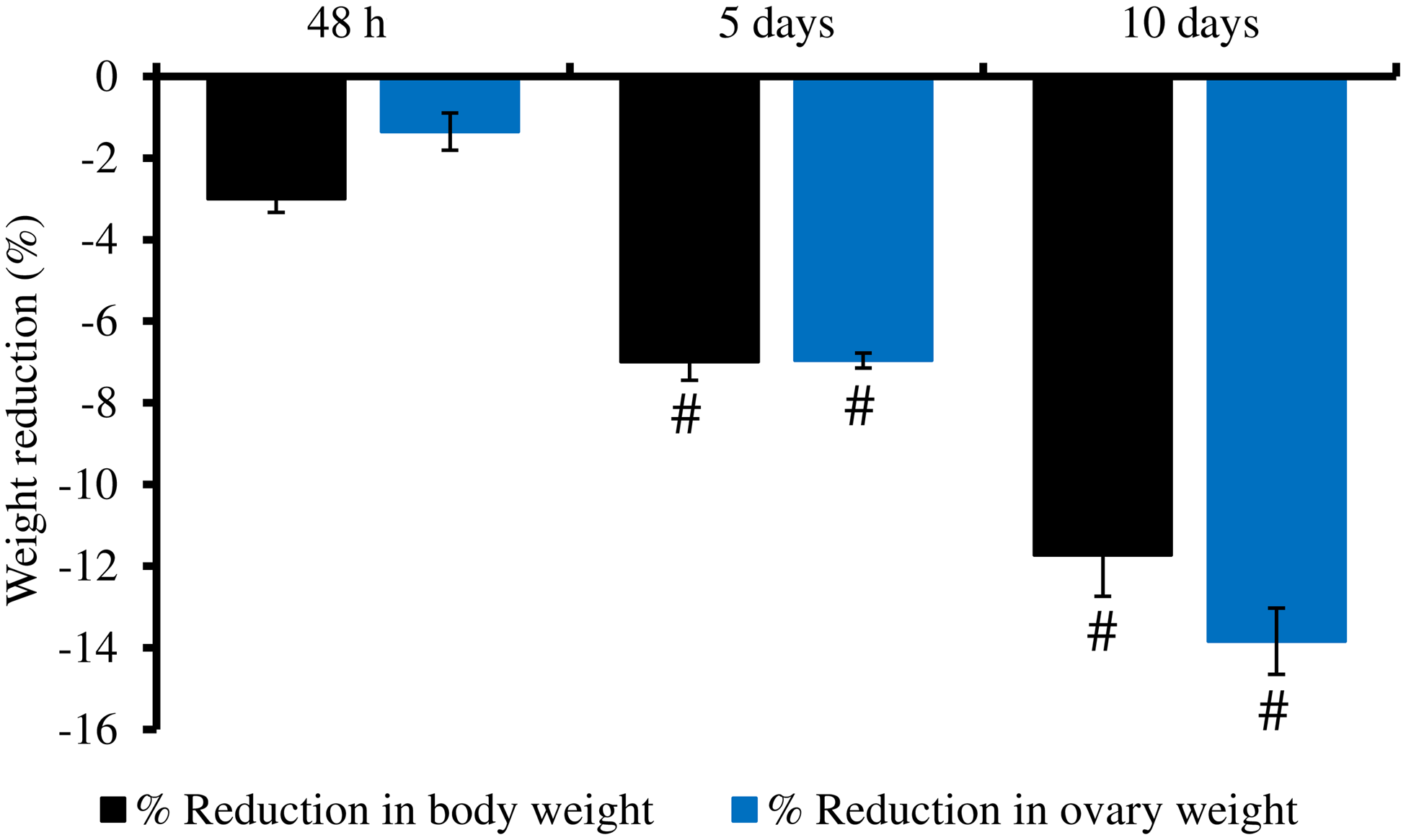
It is generally understood that the dietary stress or partial food deprivation may lead to reduced body weight and a lower ovary weight in mammals. This notion is further confirmed by the data of the present study that the complete food deprivation for 48 h reduced body weight by 3 %. The dietary stress caused due to 5 d partial food deprivation (half diet) reduced body weight by 7 %. The body weight loss was further increased by almost 12 %, if the experimental female rats were subjected to 10 d partial food deprivation as compared with the control group. Although complete food deprivation for 48 h did not reduce the ovary weight, a significant decrease in the weight of the ovary was observed if the experimental female rats were subjected to partial food deprivations for more than 5 d and 10 d (Figure 1).
Impact of dietary stress generated due to food deprivation or restriction on the body weight and ovary weight of the female rats. Data are mean (sem) of six replicates and analysed by one-way ANOVA followed by a post hoc Tukey test. ‘#’ denotes a significant decrease (P < 0·05) as compared with control.

Dietary stress reduced serum level of reproductive hormones
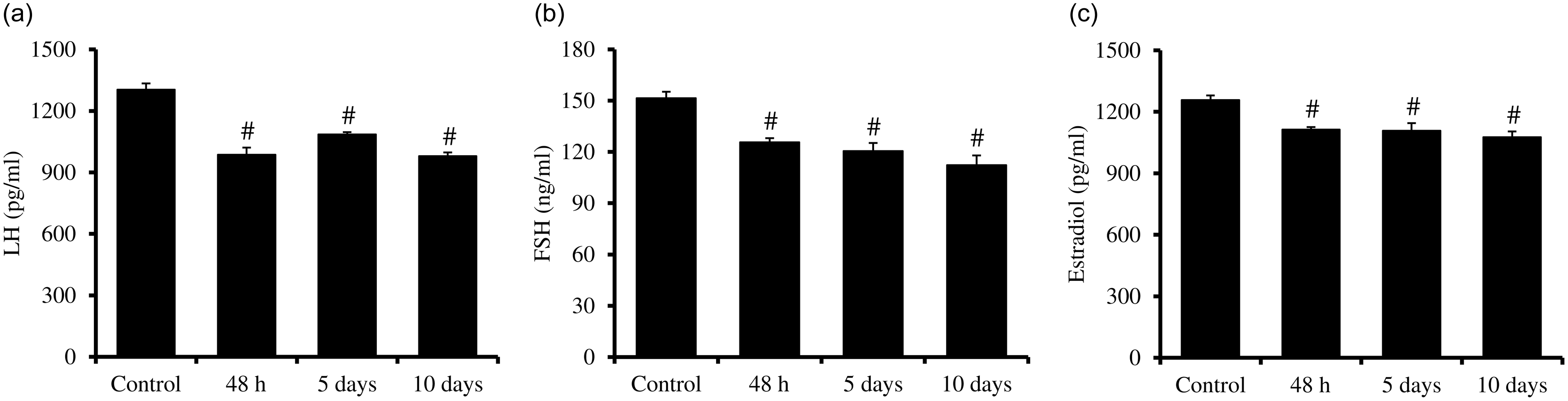
The impact of food deprivation on the serum levels of gonadotropic and steroid hormones is shown in Figure 2. A significant decrease in the levels of FSH and LH was observed if the female rats were completely deprived of food for 48 h. The decreased serum levels of both the gonadotrophin were maintained even after 5 or 10 d of partial food deprivation (Figure 2(a) and (b)). Similarly, food deprivation also declined the serum level of estradiol-17 β in female rats that were subjected to complete food deprivation for 48 h and partial food (half diet) food deprivation for 5 and 10 d (Figure 2(c)).
Impact of dietary stress generated due to food deprivation or restriction on the serum level of (a) LH, (b) FSH and (c) estradiol-17β in female rats. Data are mean (sem) of six replicates and analysed by one-way ANOVA followed by a post hoc Tukey test. ‘#’ denotes a significant decrease (P < 0·05) as compared with control.

Dietary stress affects histoarchitecture of the ovary
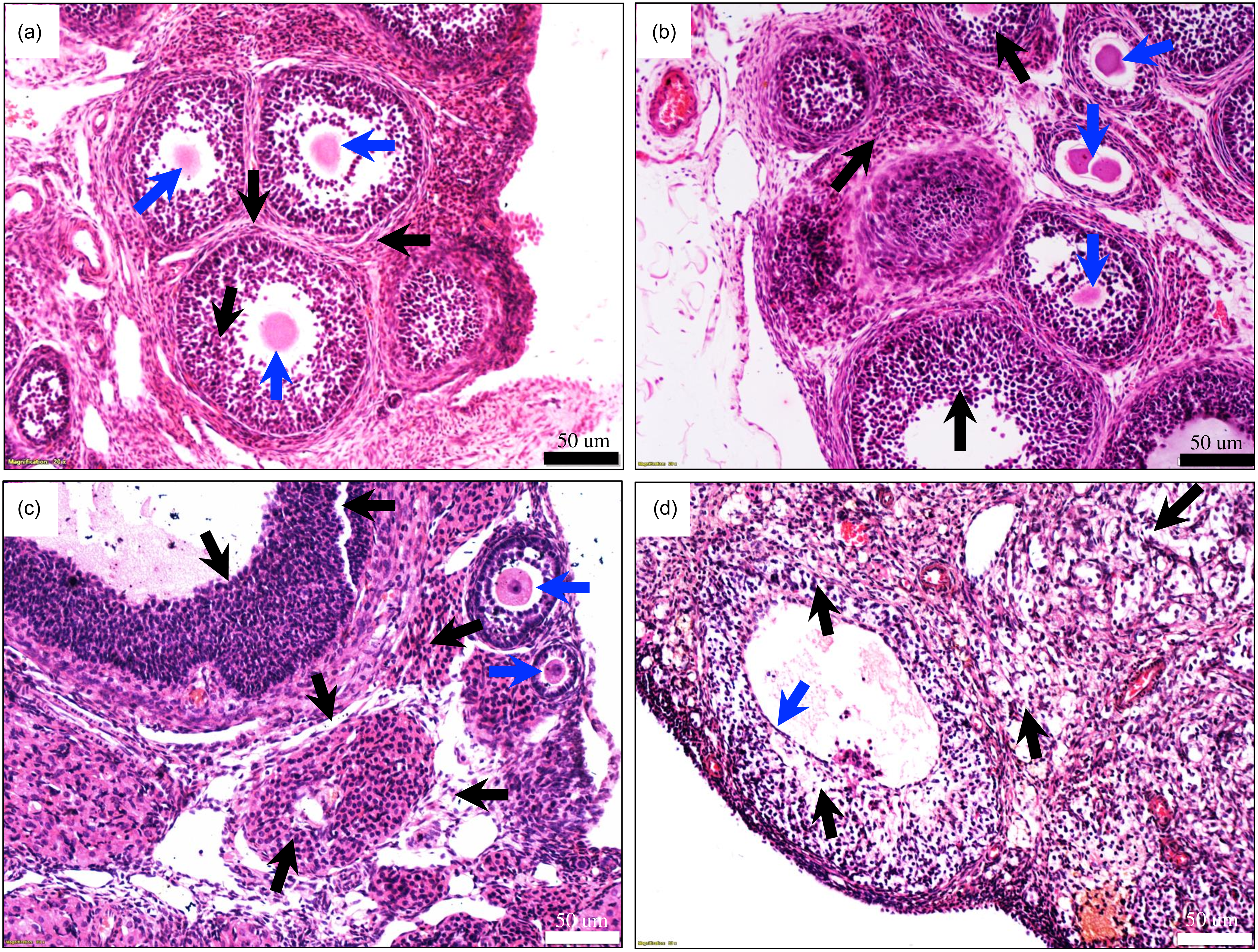
As shown in Figure 3(a), a normal histoarchitecture that possesses primordial germ cells, primary follicles and secondary follicles is seen with normal morphology of epithelial cell lining (black arrows). The cumulus oocyte complex encircling with several layers of granulosa cells with outer layer of theca cells is evident (black arrows). The oocyte with germinal vesicle and nucleolus is visible (blue arrows) in the control ovary (Figure 3(a)). Complete food deprivation for 48 h did not significantly alter the histoarchitecture of the epithelial cells, theca cells as well as granulosa cells with the follicles (black arrows) of the ovary (Figure 3(b)) and was almost similar to control (Figure 3(a)). However, increased numbers of fragmented, irregular shaped oocytes were evident (blue arrows) in the antral follicles (Figure 3(b)).
Representative photographs showing the impact of dietary stress generated due to food deprivation or restriction on the histoarchitecture of ovary. (a) Control group, (b) 48 h group, (c) 5 d group and (d) 10 d group. The 50 µm scale shows the 20× magnification image of the phase contrast microscope.

The supplementation of half diet for 5 d significantly altered the histoarchitecture of the ovary as evident by the reduction in the number of primary follicles (Figure 3(c)). The detachment of epithelial lining of the wall of the follicles and derailment of follicular somatic cells such as theca externa, theca interna, mural granulosa as well as cumulus granulosa cells are observed (black arrows), while oocytes are showing normal morphology (black arrows) with germinal vesicle in the centre (Figure 3(c)). However, the severity of histoarchitectural changes with the reduction in the number of primary follicles and formation of inner lining of steroidogenic cells within the follicle are evident in the histoarchitecture of the ovary, if the female rats were subjected to 10 d on half diet (blue arrow; Figure 3(d)). The disruption of epithelial cells, dissociation and damage of various cells including theca as well as granulosa cells are seen (black arrows) in the ovary (Figure 3(d)).
Dietary stress inhibits p-mTOR and induces autophagy signalling in the ovary
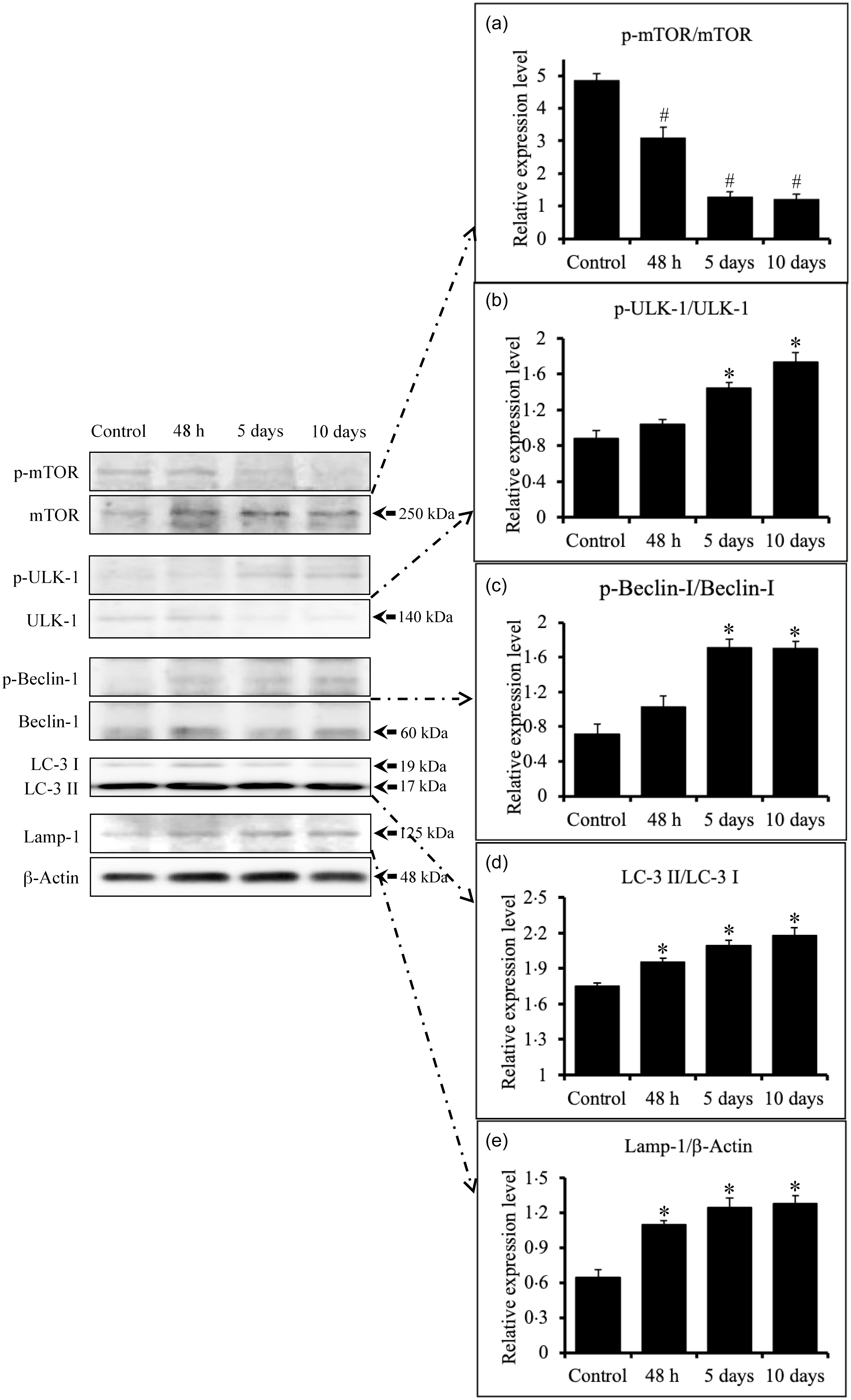
The mammalian target of rapamycin (mTOR) functions as an ATP as well as amino acid sensor and considered as inhibitor of autophagy. In the presence of sufficient nutrients, mTOR is phosphorylated at Ser2448 via the PI3 kinase/Akt signalling pathway and auto phosphorylated at Ser2481 that plays a key role in cell growth and homeostasis by preventing autophagy. On the other hand, food deprivation may decrease the p-mTOR/m-TOR ratio in order to initiate autophagy signalling. Data of the present study show that the food deprivation significantly (P < 0·05) inhibited p-mTOR/mTOR ratio in the ovary of female rats in a time-dependent manner (one-way ANOVA; P < 0·05; Figure 4(a)). One-way ANOVA followed by post hoc Tukey analysis further strengthened our observations that protein p-mTOR/mTOR ratio was significantly decreased in the ovary of female rats after 48 h of complete food deprivation or subjected to half diet for 5 and 10 d (Figure 4(a)).
Impact of dietary stress generated due to food deprivation or restriction on the autophagy marker proteins expression in the ovary. (a) p-mTOR/mTOR, (b) p-ULK-1/ULK-1, (c) p-Beclin-1/Beclin-1, (d) LC-3 II/LC-3 I and (e) Lamp-1. Data are mean (sem) of six replicates and analysed by one-way ANOVA followed by a post hoc Tukey test. ‘*’ denotes a significant increase, and ‘#’ denotes a significant decrease as compared with control.

The UNC51-like kinase-1 (ULK1) is the catalytic component of the autophagy preinitiation complex that plays a central role in the initiation stage of autophagy via phosphorylation of ATG14, Beclin1 and other autophagy proteins. Inhibition of p-mTOR/mTOR ratio in the ovary could increase p-ULK-1/ULK-1 phosphorylation ratio in order to induce autophagy signalling under dietary stress conditions. This is further strengthened by the observations that the food deprivation significantly increased p-ULK-1/ULK-1 phosphorylation ratio in the ovary of female rats in a time-dependent manner (one-way ANOVA; P < 0·05; Figure 4(b)). One-way ANOVA followed by post hoc Tukey analysis further supported our observations that the expression pattern of p-ULK-1/ULK-1 was significantly increased if the female rats were subjected to partial food deprivation (half diet) for 5 and 10 d as compared with control (Figure 4(b)).
The AMPK-mediated phosphorylation of Beclin1 is required for autophagy induction during starvation. Therefore, p-Beclin-1/Beclin-1 phosphorylation ratio was analysed in the present study. Data revealed that food deprivation elevated the expression pattern of autophagy markers such as p-Beclin-1/Beclin-1 in a time-dependent manner (one-way ANOVA; P < 0·05; Figure 4(c)). One-way ANOVA followed by post hoc Tukey analysis further strengthened our observations that the expression pattern of p-Beclin-1/Beclin-1 phosphorylation ratio was significantly increased if the female rats were subjected to partial food deprivation (half diet) for 5 and 10 d as compared with control (Figure 4(c)).
Microtubule-associated protein light chain 3 (LC3) is considered one of the definitive markers of autophagy. Initiation of autophagy causes the conversion of LC3-I to LC3-II that can be determined by increase of LC3-II/LC3-I ratio through western blotting. Therefore, the LC3-II/LC3-I ratio was analysed in the present study. Data indicate that food deprivation elevated the expression pattern of autophagy markers such as LC3-I to LC3-II in a time-dependent manner (one-way ANOVA; P < 0·05; Figure 4(d)). One-way ANOVA followed by post hoc Tukey analysis further strengthened our observations that the expression pattern of LC3-I to LC3-II ratio was significantly increased if the female rats were subjected to either complete food deprivation or partial food deprivation (half diet) for 5 and 10 d as compared with control (Figure 4(d)).
The lysosomal-associated membrane protein 1 (Lamp1) is a heavily glycosylated transmembrane protein. The newly synthesised Lamp1 enters the plasma membrane where they are in dynamic equilibrium between endosomes, lysosomes, amphisomes and autolysosomes to form degradative lysosomes that serve as hubs for autophagy. Therefore, the Lamp-1 was analysed in the present study. Data indicate that food deprivation elevated the expression pattern of autophagy markers such as Lamp-1 in a time-dependent manner (one-way ANOVA; P < 0·05; Figure 4(e)). One-way ANOVA followed by post hoc Tukey analysis further strengthened our observations that the expression pattern of Lamp-1 was significantly increased if the female rats were subjected to either complete food deprivation or partial food deprivation (half diet) for 5 and 10 d as compared with control (Figure 4(e)).
Dietary stress induces proapoptotic signalling in the ovary
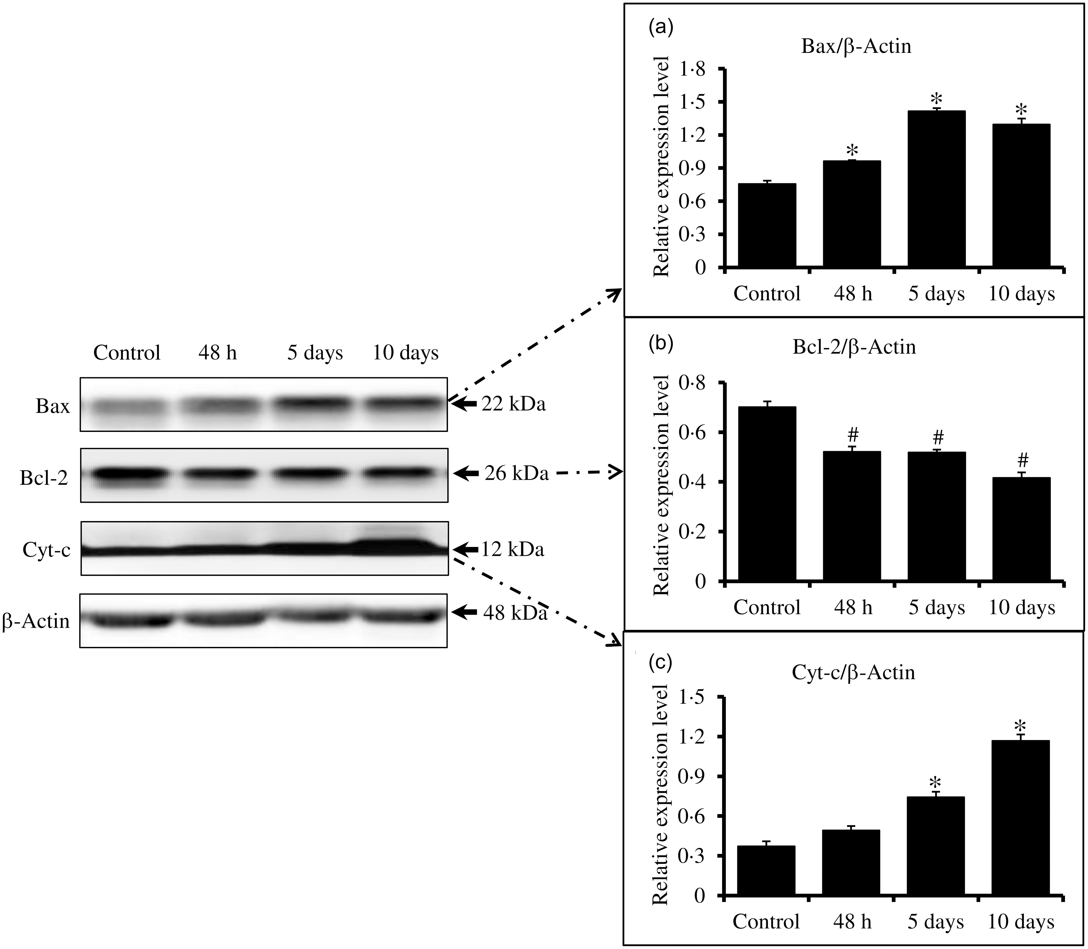
Dietary stress may induce proapoptotic signalling in the ovary. To confirm this possibility, the proapoptotic markers were analysed in the ovary of female rats. Data of the present study suggest that dietary stress caused the overexpression of Bax protein in a time-dependent manner (Figure 5(a)). One-way ANOVA followed by Tukey analysis further strengthened our observations that the expression pattern of Bax was significantly increased if the female rats were subjected to either complete food deprivation for 48 h or partial food deprivation (half diet) for 5 and 10 d as compared with control (Figure 5(a)). On the other hand, Bcl-2 expression was decreased in a time-dependent manner (Figure 5(b)). One-way ANOVA followed by Tukey analysis further strengthened our observations that the expression pattern of Bcl-2 was significantly reduced if the female rats were subjected to either complete food deprivation for 48 h or partial food deprivation (half diet) for 5 and 10 d as compared with control (Figure 5(b)).
Impact of dietary stress generated due to food deprivation or restriction on the apoptotic marker proteins expressions in the ovary. (a) Bax, (b) Bcl-2 and (c) Cyt-c expression. Data are mean (sem) of six replicates and analysed by one-way ANOVA followed by a post hoc Tukey test. ‘*’ denotes a significant increase, and ‘#’ denotes a significant decrease as compared to control.

The overexpression of Bax and underexpression of Bcl-2 might have altered Bax/Bcl-2 ratio and thereby mitochondrial outer membrane permeability. The altered mitochondrial outer membrane permeability allows the release of cytochrome-c from the mitochondrial intermembrane space to the cell cytoplasm that induce downstream apoptotic pathway. Therefore, cytochrome-c expression level was analysed in the present study. Results of the present study revealed that the dietary stress increased cytochrome c expression level in a time-dependent manner (Figure 5(c)). One-way ANOVA followed by Tukey analysis further confirms our observations that the expression pattern of cytochrome-c was significantly increased if the female rats were subjected to partial food deprivation (half diet) for 5 and 10 d as compared with control (Figure 5(c)).
Discussion
Nutrient metabolism homeostasis is critical for the maintenance of the integral health of mammals including human. Under nutrition, partial or complete starvation limits the daily supply of energy and nutrients required for the maintenance of overall health thus reducing body as well as various organs weight. Ovary is a dynamic organ that performs several functions and is very sensitive to dietary stress, especially during reproductive aging. This notion is supported by the observations that aged ovary exhibits increased sensitivity to stress and dysregulated cell death as well as survival pathways(Reference Sasaki, Hamatani and Kamijo32,Reference Yan, Zhao and Li33) . One-year-old female rats are normally considered towards advance reproductive aging that correspond to perimenopausal stage in humans and represent biologically relevant model to study the decline in ovarian function, altered hormonal milieu and increased susceptibility to cellular stress responses such as autophagy and apoptosis. Therefore, 1-year-old female rats were subjected to short dietary restriction in order to understand how aging ovaries respond to nutritional stress at the cellular and molecular levels, thereby broadening our understanding of fertility decline and ovarian pathophysiology during reproductive ageing.
Data of the present study further confirm that the short-term dietary stress generated due to complete food deprivation for 48 h and partial food deprivation (half diet for 5 d or 10 d that correspond to one or two estrous cycle) significantly decreased body weight by 3 %, 7 % and 12 %, respectively. Further, the dietary stress reduced ovary weight in a time-dependent manner (Figure 1). Dietary stress may induce a cascade of metabolic and hormonal changes that may be associated with reduced body weight and ovary weight in the present study. Since the body weight is one of the most noticeable and direct effect of dietary stress especially in female, a drop in the body weight is mainly due to the utilisation of fat and glycogen stores(Reference El-Zayat, Sibaii and El-Shamy34,Reference Harris and Martin35) . Similarly, dietary stress deprives the daily requirement of various nutrients to the body that negatively affects the growth, development and performance of various organs, reduces body weight and increases susceptibility towards various diseases in mammals, including humans(Reference Habib, Ali and Nazir1,Reference Bindari, Shrestha and Shrestha36) .
The nutritional investment in reproduction is much greater in female compared with male in mammals including human(Reference Rojas, Chávez-Castillo and Olivar37). Therefore, the females are more sensitive to dietary stress as compared with males and may cause reduction in gonadotrophin secretion as well as ovarian functions. This is supported by the data of the present study that dietary stress generated due to complete food deprivation or partial food deprivation not only significantly (P < 0·05) reduced serum levels of FSH (Figure 2(a)) as well as LH (Figure 2(b)) secretion but also the ovary weight (Figure 1) as well as estradiol-17 β in circulation in a time-dependent manner (Figure 2(c)). These data corroborate with the previous findings that the dietary stress caused due to food deprivation and food restriction significantly reduced gonadotrophin secretion, ovarian weight and thereby reproductive potential in mammals(Reference Cagampang, Maeda and Yokoyama38–Reference Subih, Salem and Al Omari40). The decrease in estradiol-17β level has been reported in experimental rats under starvation conditions(Reference Domazetovic, Fontani and Marcucci41). These data together with previous findings suggest that dietary stress not only reduces body weight but also gonadotrophins levels leading to reduced ovary weight as well as estradiol-17β level.
Dietary stress-mediated decline in gonadotropic hormones, ovary weight, as well as estradiol-17β level may adversely affect the histoarchitecture of the ovary. This possibility is further strengthened by the data of the present study that dietary stress caused due to complete food deprivation for 48 h increased the number of fragmented and irregularly shape oocytes (Figure 3(b)) as compared with control (Figure 3(a)). Further, the dietary stress generated due to partial food deprivation (half diet) for 5 d significantly reduced the number of primary follicles and altered the histoarchitecture of the ovary (Figure 3(c)). The severity of damage in the ovary was increased and the reduced the number of primary follicles, if the female rats were subjected to 10 d on half diet (Figure 3(d)). These data led us to hypothesise that the dietary stress generated in experimental female rats might have affected hypothalamo–pituitary–gonadal axis(Reference Bronson42,Reference Martin, Golden and Carlson43) , FSH and LH secretions as well as folliculogenesis in the ovary(Reference Cameron and Nosbisch44–Reference Wu, Van der Hoek and Ryan46). Similarly, dietary stress-induced structural alterations in the ovary have been reported in adult female rats and women(Reference de Morais Oliveira, Lupi and Silveira47,Reference Downs and Wise48) . Although the partial starvation is less severe, it can nevertheless decrease the size of the ovarian reserve and impede the growth of follicles(Reference Broughton and Moley49).
It is established that nutritional stress induces autophagy signalling through the major metabolic kinases such as AMP-PK and mTOR that are responsible for monitoring cellular energy and the concentration of amino acids in a cell(Reference He, Zhang and Zhao50). Since mTOR is a prominent autophagy inhibitor, dietary stress generated in the present study may decrease the p-mTOR/m-TOR ratio in order to initiate autophagy signalling. This possibility was further strengthened by the data of the present study that food deprivation inhibited p-mTOR/mTOR protein expression ratio in the ovary of female rats in a time-dependent manner (Figure 4(a)) suggesting the initiation of autophagy signalling in the rat ovary. Similarly, the inhibition of mTOR was associated with the dietary stress-mediated initiation of autophagy signalling in the mammalian ovary(Reference Shintani and Klionsky51).
Downstream to mTOR, ULK1 is essential for the initiation of autophagy, and the inhibition of p-mTOR/mTOR protein expression ratio might have allowed the initiation of autophagy signalling by inducing the p-ULK-1/ULK-1 phosphorylation ratio that provides valuable information regarding the activation state of autophagy(Reference Parzych and Klionsky52). Our results suggest that dietary stress increased p-ULK-1/ULK-1 phosphorylation ratio in the ovary of female rats in a time-dependent manner (Figure 4(b)). These data are in agreement with the previous observations that food deprivation induced autophagy in a lung cancer cell line(Reference Mejlvang, Olsvik and Svenning53). The AMPK is an energy sensor kinase and initiates autophagy by acting on the ULK complex and mTORC1(Reference Egan, Shackelford and Mihaylova54–Reference Kim, Baek and Lee56). The AMPK binding phosphorylates ULK1/2 complex that induces phosphorylation of other units of the same complex to activate the whole complex. The activated ULK1/2 complex recruits endoplasmic reticulum, mitochondria and plasma membrane for the formation of phagophore(Reference Kim, Baek and Lee56–Reference Ktistakis and Tooze58).
For phagophore formation, Class III phosphatidylinositol 3-kinase (Class III PI3K) complex is required that consist of factors like Beclin1(Reference Kang, Zeh and Lotze59,Reference Molejon, Ropolo and Re60) . Beclin1 is one of the important units of class III PI3K that undergo AMPK-mediated phosphorylation during starvation to induce autophagy(Reference Kang, Zeh and Lotze59). The Beclin-1 plays a crucial role in the autophagic process by acting as a pivotal regulator for the production of autophagosomes. The evaluation of Beclin-1 phosphorylation through the p-Beclin-1/Beclin-1 ratio provides insight into the regulatory action of Beclin-1 in autophagy(Reference Kaur and Changotra61). Therefore, p-Beclin-1/Beclin-1 phosphorylation ratio was analysed in the present study. Data revealed that food deprivation elevated the expression pattern of autophagy markers such as p-Beclin-1/Beclin-1 in a time-dependent manner (Figure 4(c)). These results are consistent with the previous observations that the p-Beclin-1/Beclin-1 ratio increased as an early autophagic response to dietary stress(Reference Maiuri, Zalckvar and Kimchi62).
The microtubule-associated protein 1 light chain 3 (LC3) in conjugation with Atg12-Atg5 is important for elongation and maturation of phagophore. The autophagy induction leads to an elevation LC3-II/LC3-I ratio and commonly employed to determine autophagy induction(Reference Kabeya, Mizushima and Ueno63) as well as autophagic activity(Reference Shintani and Klionsky51). Therefore, the LC3-II/LC3-I ratio was analysed in the present study, and the results indicate that food deprivation elevated the expression pattern of LC3-I to LC3-II in a time-dependent manner (Figure 4(d)) suggesting the involvement of LC3-II in phagophore formation during autophagy. Similarly, studies suggest the involvement of LC3-II to form autophagosome during the induction of autophagy(Reference Glick, Barth and Macleod55,Reference Kim, Baek and Lee56,Reference Eskelinen64) .
The lysosomal-associated membrane protein 1 (LAMP1) is a heavily glycosylated transmembrane protein. The examination of Lamp-1 expression under dietary stress conditions could offer the dynamics of lysosomes in the regulation of autophagy. Therefore, the Lamp-1 was analysed in the present study, and our results suggest that the dietary stress elevated the expression pattern of Lamp-1 in a time-dependent manner (Figure 4(e)). The newly synthesised LAMP1 enters in the plasma membrane where they are in dynamic equilibrium between endosomes, lysosomes, amphisomes and autolysosomes to form degradative lysosomes that serves as hubs for autophagy(Reference Eskelinen64). A positive association between increased Lamp-1 expression and lysosomal activity has already been documented(Reference Glick, Barth and Macleod55). Cells increase their autophagic capability, potentially as a means to recover vital metabolites and maintain cellular energy balance in situations where external nutrition availability is restricted(Reference Czaja, Ding and Donohue65). Our results together with previous observations suggest that the dietary stress might have increased Lamp-1 expression in the ovary to elevate lysosomal activity and thereby autophagy(Reference Saftig and Klumperman66).
The dietary stress not only induces autophagy signalling, and growing evidences suggest that it may also induce stress-mediated intrinsic apoptotic signalling pathway(Reference Yadav, Tiwari and Gupta8,Reference Pandey, Yadav and Premkumar12,Reference Yadav, Pandey and Premkumar13,Reference Yadav, Gupta and Sharma67) . For instance, the nutritional stress triggers overexpression of proapoptotic proteins such as Bax in the ovary(Reference Berens and Tyler68). The primary role of Bax is to facilitate the release of Cyt-c from the mitochondria that activates downstream caspases and thereby apoptotic cell death(Reference Wang and Youle69,Reference Wei, Zong and Cheng70) . Hence, proapoptotic markers such as Bax, Bcl-2 and Cyt-c levels were analysed in order to determine the onset of apoptotic markers in the ovary. Data of the present study suggest that dietary stress caused overexpression of Bax protein in a time-dependent manner (Figure 5(a)). On the other hand, Bcl-2 expression was decreased in a time-dependent manner (Figure 5(b)). The overexpression of Bax and under expression of Bcl-2 might have altered Bax/Bcl-2 ratio and thereby mitochondrial outer membrane permeability. The altered mitochondrial outer membrane permeability allows the release of cytochrome-c from the mitochondrial intermembrane space to the cell cytoplasm. This possibility was further strengthened by our results that the dietary stress increased cytochrome-c expression level in a time-dependent manner (Figure 5(c)). These findings are in agreement with the previous observations that under stress conditions, under expression of Bcl-2 and overexpressions of Bax as well as cytochrome-c initiate intrinsic apoptotic pathways in the ovary of female rats(Reference Tiwari, Prasad and Tripathi11,Reference Chaube, Prasad and Khillare71–Reference Tripathi, PremKumar and Pandey74) . Thus, the dietary stress generated due to complete food deprivation for 48 h and partial food deprivation for 5 and 10 d caused cellular stress sufficient enough to induce both autophagy as well as apoptotic signalling in parallel. The stress-mediated induction of both autophagy and apoptotic signalling has been reported in the rat ovary(Reference Escobar, Echeverría and Sánchez-Sánchez75–Reference Escobar Sánchez, Echeverría Martínez and Vázquez-Nin77).
Conclusion
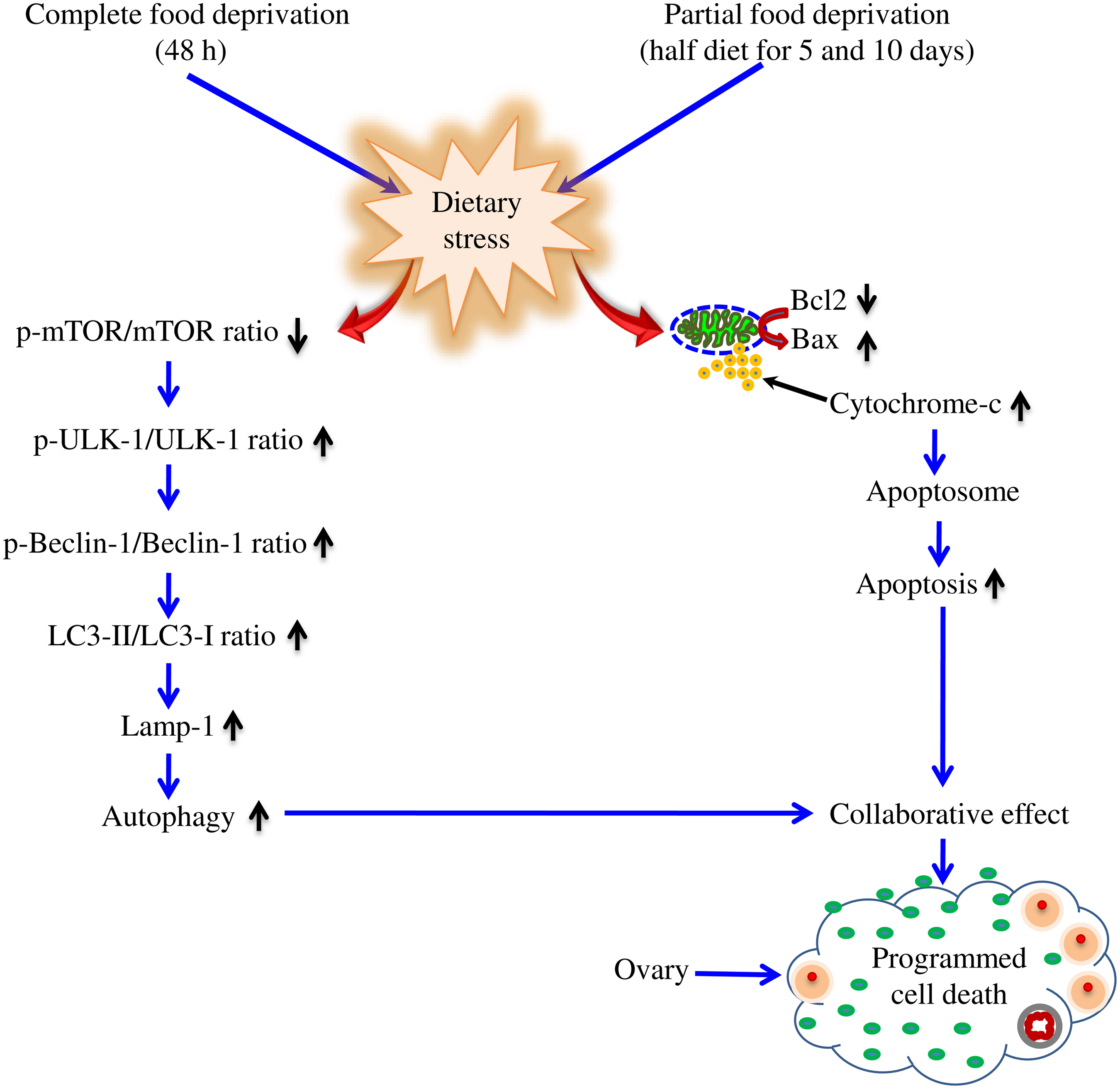
The dietary stress caused due to complete food deprivation for 48 h and partial food deprivation (half-diet) for 5 as well as 10 d decreased body weight, ovary weight, serum levels of FSH, LH and estradiol-17β. The decreased gonadotropins and steroid hormone levels affected histoarchitecture of the ovary, reduced number of follicles, deteriorated follicular somatic cells and oocyte architecture, initiated autophagy and apoptotic signalling in the ovary (Figure 6). Indeed, the dietary stress may initiate reproductive health disorders by altering the reproductive hormones level, structure and functions of the ovary. These findings could be useful in addressing the necessity for adequate precision lifestyle habits for women’s reproductive health.
Schematic diagram showing the effects of dietary stress on initiating autophagy and apoptotic signalling in the ovary. Dietary stress inhibited p-mTOR/mTOR ratio and elevated p-ULK-1/ULK-1 ratio. The phosphorylated ULK-1 activated Beclin-1/ LC3-II/ Lamp-1-mediated pathway to induce autophagy. The dietary stress increased Bax/Bcl2 ratio and cytochrome-c release that induce apoptosis. The collaborative effects of autophagy and apoptosis result in programmed cell death in the ovary.

Acknowledgements
The infrastructural facilities provided to Shail K. Chaube from the Department of Zoology, Institute of Science, BHU, Varanasi-221005, UP, India, are highly acknowledged. The research laboratory facilities at the Department of Physiology, Institute of Medical Sciences, Banaras Hindu University, Varanasi provided by Kumar Sarvottam and Bhupendra Singh Yadav are highly acknowledged.
The financial support in a part for the research reported herein has been provided to Shail K. Chaube from CAS, Department of Zoology, Institute of Science, BHU, Varanasi-221005, UP, India. The financial support provided to PKY by Council of Scientific & Industrial Research, New Delhi [File No. 09/013(0686)2017-EMR-I] is also acknowledged.
Author’s credit role: P. K. Y.: Writing – original draft, writing – review and editing, methodology, investigation, formal analysis, investigation, data curation, visualisation and validation. S. N. P.: Writing – review and editing. S. K. C.: Writing – review and editing, writing – original draft, visualisation, validation and supervision and conceptualisation.
The authors declare that they have no known competing financial interests or personal relationships that could have appeared to influence the work reported in this paper.
All procedures were performed in accordance with the guidelines of the Institutional Animal Ethical Committee of Institute of Science, Banaras Hindu University, Varanasi-221005, India (Approval number: BHU/DoZ/IAEC/2018–2019/015).